R E V I E W
Comparison of UVA Protection Factor
Measurement Protocols
This article was published in the following Dove Press journal: Clinical, Cosmetic and Investigational Dermatology
Kamand Hedayat 1
Saman Ahmad Nasrollahi 1,2
Alireza Firooz 1,2
Hossein Rastegar 2
Manouchehr Dadgarnejad3
1Center for Research and Training in Skin
Diseases and Leprosy (CRTSDL), Tehran University of Medical Sciences (TUMS),
Tehran, Iran;2Cosmetic Products
Research Center, Iranian Food and Drug Administration, Ministry of Health and
Medical Education, Tehran, Iran;3Iranian
Food and Drug Administration, Ministry of Health and Medical Education, Tehran, Iran
Background: In the past, it was taught that UVA wavelengths (320- 400nm) only plays a major role in skin aging but recently the scientific researches also show that UVA cause cancerous keratinocyte cells in deep layer of the epidermis. Therefore, the protective ability of the product against UVA is important in addition to protection against UVB rays. The UVA protective factor (UVA-PF) is used to evaluate the effectiveness of sunscreen products against UVA rays. This study aims to review and compare all outstanding protocols in the
field of UVA-PF measurement andfinally the introduction of the best method of measuring UVA-PF based on the further benefits.
Materials and Methods: Four standards including ISO 24443 (AS/NZS 2604: 2012 recommended approach), CEN 2006, FDA 2007 and FDA 2011 are selected.
Results:In order to measure UVA-PF with in vivo method, two standards of CEN 2006 and FDA 2007 recommended persistent pigment darkening (PPD) method. Although the general principle of both is similar, there are some differences in detail. For in vitro measurement of UVA-PF, CEN and FDA 2011 standards use critical wavelengths. FDA 2007 introduces the modified Diffey fraction, and ISO 24443 standard meets the UVA-PF measurement in a manner that is consistent with PPD.
Conclusion:Finally, this review discussed the comparison of all in vitro and in vivo UVA-PF measurement standards and provided information in the form of texts and tables to move towards the creation of an integrated standard. Since in vitro methods of UVA-PF measure-ment are not reproducible due to differences in test conditions, it may be concluded that the in vivo PPD method is a more suitable option.
Keywords:sunscreen, ultraviolet, UVA protection factor, in vivo, in vitro
Introduction
Part of the sunlight reaching the ground involves the ultraviolet spectrum consisting of three sections of UVC (200–290nm), UVB (290–320nm) and UVA (320–400nm). UVC is completely absorbed by the earth’s atmosphere. The amount of UVA reaching the ground is more than 20 times the UVB.1UVA plays a major role in premature aging and photocarcinogenesis. UVA increases the release of inflammatory factors, including IL10 from keratinocyte cells. Additionally, it increases the expression of inflammatory proteins such as TNFα, IL1α, IL6, and IL8.2,3UVA plays a considerable role in the creation of
mutations in mitochondrial DNA in human skin with the help of ROS (reactive oxygen spices). It also causes premature skin aging by creating a mutation infibroblast cells in the human skin.4The role of increased expression of the p53 protein in causing UVA-induced apoptosis is undeniable.58-hydroxy-2′-deoxyguanine (8-oxoG) is a valuable indicator to track the DNA oxidative damage caused by UVA, and it can lead to G→T transversions.6 Correspondence: Saman Ahmad Nasrollahi
Center for Research and Training in Skin Diseases and Leprosy (CRTSDL), Tehran, University of Medical Sciences (TUMS), No. 415 Taleqani Avenue, Tehran 1416613675, Iran
Tel/Fax +982188963804 Email [email protected]
Clinical, Cosmetic and Investigational Dermatology
Dove
press
open access to scientific and medical researchOpen Access Full Text Article
Clinical, Cosmetic and Investigational Dermatology downloaded from https://www.dovepress.com/ by 118.70.13.36 on 24-Aug-2020
Therefore, protection against UVA along with UVB is highly important.
The UVA protective factor (UVA-PF) is used to evaluate the effectiveness of sunscreen products against UVA rays.
Materials and Methods
In this study, global standards in the field of UVA-PF determination were reviewed and compared. Four stan-dards, namely ISO 24443 (provided by European union, at May 2013), CEN2006 (provided by European Union, South Africa, Japan in Brussels, at July 12, 2006), FDA 2007 (provided by United States, at August 27, 2007) and FDA 2011 (provided by United States, at June 17, 2011) have set up different protocols in thefield of UVA protec-tion factor. Some are dealt with using in vivo methods whereas others are dealt with using in vitro methods.7–11
Results
Due to the role of UVA in the occurrence of skin damages, it is important to use sunscreens which protect the skin from both UVA and UVB. In vitro methods of UVA-PF measurement are not reliable and reproducible due to differences in test conditions, such as distinction of sub-strates, variant amount of applied product and UV dose of irradiation.12 Among the FDA 2007 and CEN standards introducing the in vivo PPD approach, both have advan-tages and disadvanadvan-tages so that using a better approach in each case can yield better results.
Discussion
In vivo UVA-PF Determination Test
Assessment
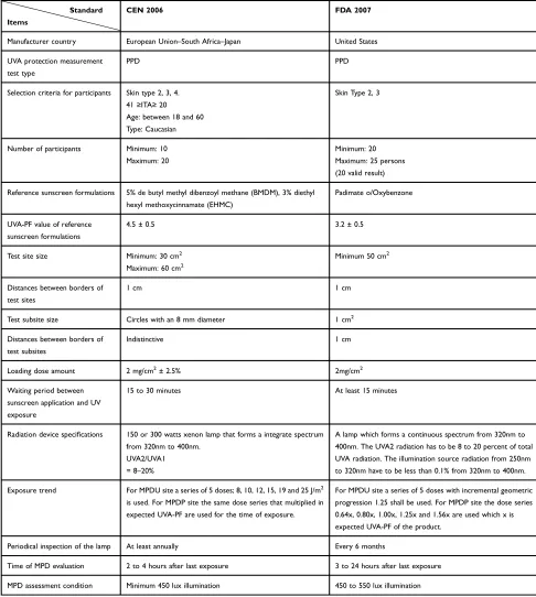
First, we will go through in vivo UVA-PF determination methods conducted using Immediate Pigment Darkening (IPD), Persistent Pigment Darkening (PPD) and Protection Factor A (PFA). However, in CEN and FDA 2007 stan-dards, the PPD method is accepted only in the in vivo UVA-PF determination method (Table 1).
In the PPD method, like the SPF (Sun Protection Factor) measurement method, people meeting the criteria for the test are selected with a specified number for participation. In the SPF test, the endpoint is an erythemal response versus the PPD test in which the endpoint is persistence pigment dar-kening of the test subsites. Test sites and subsites are designed in specific dimensions blindly on the participant bodies. The test and reference sunscreen products are weighed in the defined amount and are applied onto the
participant’s skin. Then, with a specific method, the product is spread over the skin followed by a waiting period for product to be dried. Individuals, based on a predetermined manner, are exposed to UVA radiation while sitting or lying position on the abdomen. After a certain amount of time from the last UV exposure, the level of permanent darkness of the pigment is evaluated as thefinal point of the test, and UVA-PF is calculated by a set of formulas (which will be discussed in“Statistics and Calculations”section ).
Selection Criteria for Participants
Despite the criteria mentioned inTable 1(FDA 2007 and CEN common criteria), CEN standards further their pre-cautions for entering the test.
The criteria for a healthy male and female to be con-sidered include:
● Skin type II, III and IV13
● Not being involved in any sun tests within the last two months (no remaining marks on the back).
● Not having sun exposure on the back area for at least 2 months prior to the study.
● Absence of scars, or active dermal lesions on the areas of the backtested.
● Test area must be uniform in color, without nevi, blemishes or solar lentigo without excessive hairs.
The CEN also set a series of exclusion test criteria;
● Subjects notfitting the previous inclusion criteria.
● Pregnant or lactating women,
● Past history of allergy, photoallergic, phototoxic, or other abnormal responses to sunlight or sensitivity to cosmetic products, toiletries, sunscreens, latex and/or topical drugs.
● Subjects with dermatological problems on the test area.
● Subjects having used self-tanning products on the back in the previous month.
Consequently, the CEN appears more suitable for indivi-duals selecting criterion owing to greater consistency.
Number of Participants
The FDA 2007 and CEN use 20 to 25 people and 10 to 20 people, respectively. Like other clinical studies, the higher number of the participants, brings higher test accuracy. On the other hand, the large sample size lead to some problems
Clinical, Cosmetic and Investigational Dermatology downloaded from https://www.dovepress.com/ by 118.70.13.36 on 24-Aug-2020
such as handling the protocol, providing enough samples and volunteer’s dropout.
Reference Sunscreen Formulations
It is recommended that a standard sample be used to verify the results of each test sample.
The test is valid if the average UVA-PF obtained for the reference sunscreen product is within the spe-cified range. According to Table 1, both reference sunscreens are good choices, since their UVA-PF results are close to each other, and their active ingre-dients are approved.
Table 1In vivo UVA-PF Determination Protocol Comparison
Standard
Items
CEN 2006 FDA 2007
Manufacturer country European Union–South Africa–Japan United States
UVA protection measurement
test type
PPD PPD
Selection criteria for participants Skin type 2, 3, 4.
41≥ITA≥20
Age: between 18 and 60
Type: Caucasian
Skin Type 2, 3
Number of participants Minimum: 10 Maximum: 20
Minimum: 20 Maximum: 25 persons
(20 valid result)
Reference sunscreen formulations 5% de butyl methyl dibenzoyl methane (BMDM), 3% diethyl
hexyl methoxycinnamate (EHMC)
Padimate o/Oxybenzone
UVA-PF value of reference sunscreen formulations
4.5 ± 0.5 3.2 ± 0.5
Test site size Minimum: 30 cm2 Maximum: 60 cm2
Minimum 50 cm2
Distances between borders of
test sites
1 cm 1 cm
Test subsite size Circles with an 8 mm diameter 1 cm2
Distances between borders of
test subsites
Indistinctive 1 cm
Loading dose amount 2 mg/cm2
± 2.5% 2mg/cm2
Waiting period between sunscreen application and UV
exposure
15 to 30 minutes At least 15 minutes
Radiation device specifications 150 or 300 watts xenon lamp that forms a integrate spectrum from 320nm to 400nm.
UVA2/UVA1 = 8–20%
A lamp which forms a continuous spectrum from 320nm to 400nm. The UVA2 radiation has to be 8 to 20 percent of total
UVA radiation. The illumination source radiation from 250nm to 320nm have to be less than 0.1% from 320nm to 400nm.
Exposure trend For MPDU site a series of 5 doses; 8, 10, 12, 15, 19 and 25 J/m2 is used. For MPDP site the same dose series that multiplied in
expected UVA-PF are used for the time of exposure.
For MPDU site a series of 5 doses with incremental geometric progression 1.25 shall be used. For MPDP site the dose series
0.64x, 0.80x, 1.00x, 1.25x and 1.56x are used which x is expected UVA-PF of the product.
Periodical inspection of the lamp At least annually Every 6 months
Time of MPD evaluation 2 to 4 hours after last exposure 3 to 24 hours after last exposure
MPD assessment condition Minimum 450 lux illumination 450 to 550 lux illumination
Clinical, Cosmetic and Investigational Dermatology downloaded from https://www.dovepress.com/ by 118.70.13.36 on 24-Aug-2020
Test Sites and Subsites Size
What is important in choosing the size of the sites and sub-sites is the possibility of using multi-source solar simulator. It means, choosing smaller sizes for the sites and sub-sites can result in multiple sub-sites being exposed to UV at the same time. Therefore, the CEN standard features (for site size 30 cm2and for subsites circles 8mm in diameter) are more appropriate in thisfield.
For distances between the two sub-sites, FDA 2007 suggests an interval of 1 cm. CEN does not specify any interval between subsites, making the FDA 2007 feature considerable for subsites distances.
Loading Dose Amount
The loading quantity of the test and reference product is determined in almost the same amount. CEN suggests the range 2mg/cm2±2.5%, and FDA 2007 proposes 2mg/cm2, which should be uniformly loaded and spread by a finger cot. The product should be placed in small droplets through-out the test area and spread with special movements.
The spreading time should take 20 to 50 s. Product loading areas should be selected randomly.
Waiting Period Between Sunscreen Application and UV Exposure
The waiting time suggested between the product loading and the UV exposure is at least 15 min and 15 to 30 min by both FDA 2007 and CEN, respectively.
Radiation Device and the Periodical Inspection of Its Lamp
Table 1presents the list of the radiation device specifi ca-tions for each of the two standards. Regarding the periodic inspection of the device, FDA 2007 and the CEN standard suggest that the inspection be conducted every 6 and 12 months, respectively.
According to the FDA 2011, calibration data of solar simulators and their UV lamps are stable for periods longer than 1 year; Thus, the periodic assessment of the device is better to consider once a year.
Exposure Trend
As shown for the exposure trend inTable 1, the concept of both methods is the same.
Time and Condition of MPD Assessment
MPD is the smallest amount of UVA radiation producing visible tangible pigmentation with visible borders. When the pigment darkness is fixed, the MPD is evaluated visually. For the MPD evaluation, CEN and FDA 2007 suggest 2 to 4
and 3 to 24 h after the last site exposure, respectively. Eye evaluation should be blindly by an experienced observer under adequate and uniform conditions of light. FDA notes that it appears the pigment darkening is most stable about 3 h or more after post-irradiation, and is thus proposing that this observation occur at 3 to 24-h post-irradiation. This time range provides increasedflexibility in the test method with-out sacrificing accuracy. Therefore, FDA 2007 criteria appear to be superior.
In vitro UVA-PF Determination Test
Assessment
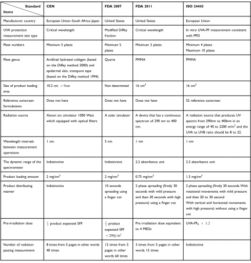
All in vitro methods are based on the evaluation of UV passing through a thin layer of sunscreen spreading over a rough plate so that the UV level is calculated before and after exposure to the controlled dose from the radiation source. However, at the stage of UVA protection level calcu-lation, each standard sets a different benchmark (Table 2).
The FDA 2007 standard uses the modified Diffey frac-tion where the area under the UVA1 absorpfrac-tion curve (340nm to 400nm) is divided into the total absorption region of UVA and UVB (290nm to 400nm).14,15 Nevertheless, FDA 2011 abolished the modified Diffey fraction for the following reasons:
1. This fraction provides considerable attention to the UVA1 region, while this area does not play a major role in producing harmful skin effects.
2. If one or more ingredients of the product produce, a high absorption in the short wavelength UVA2 region and low or no adsorption in the UVA1 region a large ratio is resulted, while this product does not produce a wide range of absorption.
3. This proposed ratio does not show a reliable degree of protection against UVA and does not apply any-where else in the world.
4. One way to increase this fraction is to reduce high-absorption compounds in the UVA2 and UVB regions, but this will reduce the level of protection in these areas of the spectrum (in other words, for increasing the Diffey fraction, SPF should decrease).
The ISO 24443 standard calculates the UVA protective factor in line with the PPD test results. The two standards CEN and FDA 2011 consider the critical wavelength method to be appropriate.
Clinical, Cosmetic and Investigational Dermatology downloaded from https://www.dovepress.com/ by 118.70.13.36 on 24-Aug-2020
òλc
290Að Þλdλ¼ 0:9ò 400 290Að Þλ dλ
A (λ) is the mean absorption at each wavelength, and d (λ) is the wavelength interval between each calculation.
The critical wavelength is the wavelength in which the region below the absorption curve comprises 90% of the total area under the UV absorption curve.
The FDA concluded that the accompanying critical wavelength test (to measure the extent of UVB and UVA
protection) and the SPF test (to measure the magnitude of UVB and UVA protection) provided a complete measure-ment of the wide spectrum capability. For a product to be labeled with a broad-spectrum statement, it should gener-ate a critical wavelength equal to or gregener-ater than 370 nm.
Test Plates
Plates, as the loading place, play a key role in the test. Among the plates of quartz, including PMMA Table 2In vitro UVA-PF Determination Protocol Comparison
Standard
Items
CEN FDA 2007 FDA 2011 ISO 24443
Manufacturer country European Union–South Africa–Japan United States United States European Union
UVA protection
measurement test type
Critical wavelength Modified Diffey
fraction
Critical wavelength In vitro UVA-PF measurement consistent
with PPD
Plate numbers Minimum 5 plates Minimum 5
plates
Minimum 3 plates Minimum 4 plates
Maximum 10 plates
Plate genus Artificial hydrated collagen (based on the Diffey method 2000) and
epidermal skin, transpore tape (based on the Diffey method 1994)
Quartz PMMA PMMA
Size of product loading area
10.2 cm 9cm Not determined 16 cm2
16 cm2
Reference sunscreen
formulations
Does not have Does not have Does not have S2 reference sunscreen
Radiation source Xenon arc simulator 1000 Watt
which equipped with opticalfilters
A solar simulator A device that has a continuous
spectrum of 290 nm to 400 nm.
A radiation source that produces UV
spectra from 290nm to 400nm in an energy range of 40 to 2200 w/m2
and the
UVA to UVB ratio should be 8 to 22.
Wavelength intervals between measurement
operations
1 nm 5 nm 1 nm 1 nm
The dynamic range of the
spectrometer
Indistinctive Indistinctive 2.2 absorbance unit 2.2 absorbance unit
Product loading amount 2 mg/cm2 2 mg/cm2 0.75 mg/cm2 1.3 mg/cm2
Product distributing
manner
Indistinctive 10 seconds
spreading using afinger cot
2 phase spreading (firstly 30
seconds with mild pressure and then 30 seconds with high
pressure) using afinger cot
2 phase spreading (firstly 30 seconds With
rotational movements with mild pressure and then 20 to 30 second
With vertical and horizontal movements with high pressure) without using afinger
cot
Pre-irradiation dose 1
3product expected SPF 23product expected SPF
200j=m2
Pre irradiation dose equivalent to 4 MEDs
UVA-PF0 1:2
Number of radiation
passing measurement
8 times from 5 pages in other words
40 times
12 times from 5
pages in other words 60 times
3 times from 5 pages in other
words 15 times
Indistinctive
Clinical, Cosmetic and Investigational Dermatology downloaded from https://www.dovepress.com/ by 118.70.13.36 on 24-Aug-2020
(polymethylmethacrylate), hydrated collagen, epider-mal skin and transpore tape, the PMMA plates are chosen for its newness. The following reasons are determining factors of preference to the other plate types:
1. It is cheaper than quartz.
2. It is ready to use and it does not need to be cleaned or re-roughened.
3. It is used for more than a decade.
As for the size of plates, when plates with dimensions of 16 cm2yield acceptable results, it is not necessary to use plates with larger dimensions (10.2cm9cm). Regarding the degree of roughness of these plates, the standard FDA 2011 proposes pages with a roughness of 2 to 7 µg, and the ISO 24443 offers 6-micron roughness. The choice of a constant roughness of 6 µm that is identical to all these tests seems reasonable.
Reference Sunscreen Formulation
According to Table 2, only ISO 24443 has a reference sunscreen product. The reference sunscreen S2 brings a UVA-PF of 12.7.
Radiation Source
Before measuring the radiation passage through the plates covered with the product, an irradiated phase for these products is performed to consider optical stability. FDA 2011 believes that the solar simulator (FDA 2007 pro-posed device) radiation is filtered such a way that its energy at wavelengths below 300nm is less than the spec-trophotometer sensitivity even when no sunscreen is loaded on to the plate (i.e. the passage rate is 100%).
Therefore, FDA 2011 proposes a radiation source pro-ducing continuous spectral distribution of UV radiation from 290 to 400 nanometers. Standard CEN proposes the solar simulator equipped withfilters.
Wavelength Intervals Between Measurement Operations
To measure the passage of radiated UV at each wave-length, the radiation intervals must be intermittent. These intervals are set in the intervals 1nm and 5nm according to
Table 2. It was mentioned in FDA 2011: The submissions stated that specifying a smaller interval would produce more accurate results and noted that current spectrometers are capable of making measurements at 1 nm intervals.
The Dynamic Range of the Spectrometer
Dynamic range is one of the indicators to calibrate the UV spectrophotometer. The dynamic range of spectrometer must be sufficient to accurately measure the passage of high-absorption sunscreens at all UV wavelengths (290nm to 400nm).
Product Loading Amount
The amount and process of loading the product on the rough section of the plate is specified in each standard. To ensure accuracy in the amount of product loading, we can use the pipette weight measurement method before and after loading or with the aid of density factor. The product is loaded in droplets of equal volume. The FDA 2007, CEN and ISO 24443 use a value of 2 and 2 mg/cm2 and 1.3 mg/cm2 respectively. The FDA believes that UV passage from a thick layer of 2mg/cm2is less, and leads to inconsistent responses. Therefore, the FDA set the loading amount at 0.75 mg/cm2to ensure that the UV passing level is within the dynamic range of UV detectors.
Product Distributing Manner
The smooth spreading of the sunscreen product on the plate considerably affects the accuracy of the test. As
Table 2 shows, the spreading process can be either one-phase or two-one-phase with use or non-use of a finger cot. The FDA 2011 considers the two-phase spreading a more effective way to achieve a solid and thick layer. This contributes to a better distribution for a wide range of drug forms than the 10-s soft distribution technique, and is also similar to the actual use of consumers. Therefore, the FDA 2011 and ISO 24443 opinion in a product-spreading manner seems the best options.
Pre-Irradiation Dose
This operation is performed to check the reduction of optical stability. The sunscreen is loaded onto the plate and then exposed to a source of radiation. According to FDA 2011, since at one time and place on the earth’s surface, the radiation of the sun is equal for products with high or low SPF and UVA-PF, then the pre-irradiation dose in the form of an SPF or UVA-PF fraction is not logical. Thus, FDA 2011 suggests that the pre-radiation dose is a constant value. The FDA received information, which showed that products containing Avobenzone, were completely destroyed in the face of exposure to a dose equivalent to 2–3 MED (Minimal Erythemal Dose).
This is the worst scenario for optical degradation, since Avobenzone is the most unstable sunscreen against light in
Clinical, Cosmetic and Investigational Dermatology downloaded from https://www.dovepress.com/ by 118.70.13.36 on 24-Aug-2020
sunscreens monographs. 1MED in skin type 2 is equiva-lent to 250 J/m2. Thus, in FDA 2011, the pre-irradiation dosage is set 4MED that is equivalent to 800J/m2.
Number of Radiation Passing Measurement
After exposure of products to pre-radiation dose, the plates covered with the product are placed in the spectrophot-ometer to measure the passage. According to FDA 2011 opinion (5 times measurement from 3 pages), when a satisfactory result is obtained with fewer computations, then no more time and cost are needed.
Statistics and Calculations
According to CEN, the critical wavelength is obtained from the following formula:
0:9¼ò
λc
290log10MPFλ ò400290log10MPFλ
whereλCis the critical wavelength, and MPF is the
mono-chromatic protection factor, which is the inverse of the transmittance at a given wavelength.
According to ISO 24443, initially, the individual UVA protection factor (UVA-PFI) on each base (at least 4 bases) is calculated according to the following formula:
UVAPFI ¼ ò
λ¼400
λ¼320Pð Þ λ Ið Þ λ dð Þλ òλλ¼400¼320Pð Þ λ Ið Þ λ 10Aeð Þλ cdð Þλ
where P(λ) is the PPD action spectrum.
I(λ) is the spectral irradiance received from the UVA source.
Ae(λ) is the mean monochromatic absorbance of the test product layer after UV exposure.
d(λ) is the wavelength step (1 nm).
C is the coefficient of adjustment. The initial absor-bance curve values are multiplied by a scalar value “C” until the in vitro calculated SPF values are equal to the in vivo measured SPF. This is accomplished in an iterative calculation process. The initial absorbance values multi-plied by this “C” value become the adjusted sunscreen absorbance curve that is used for determination of the initial UVA-PF0 value, and the exposure dose. The “C” value typically lies between 0.8 and 1.6 for valid inter-pretation. If it is outside this range, new samples should be prepared to validate the original observations.
Thus, according to the formula below, the average UVA-PF calculation is computed:
UVAPFn0¼∑UVAPFI n0
If this condition Confident interval (CI) n %½ ½ 17%UVAPF is met, the test is valid; otherwise, the number of bases should be increased as long as the condition is fulfilled.
If the mentioned condition is not met after using 10 bases, the test is declared invalid.
According to FDA 2011 and FDA 2007 standards, the UV passing level is calculated from the following formula:
Tð Þ ¼λ ∑
n
1Pð Þλ=n
∑n
1Cð Þλ =n
Cð Þλ :Measurments of spectral irradiance transmitted for each wavelength through control PMMA plates coated with 15 µl of glycerin.
Pð Þλ : Measurements of spectral irradiance transmitted for each wavelengthλ through the PMMA plate covered with the sunscreen product. The average absorption in
each wavelength (Að ÞÞλ is calculated from the negative logarithm of the mean UV passing as follows:
Að Þ¼ λ logTð Þλ
According to FDA 2011, the critical wavelength is calcu-lated from this formula;
òλc
290Að Þλdλ¼ 0:9ò 400 290Að Þλ dλ
According to FDA 2007, the Diffey fraction ratio is cal-culated as follows:
UVAI=UV¼UVA I area per unitλ UV area per unitλ
UVA I area per unitλ is given as:
aUVAI=λ¼5=3½A340þA400þ4 A345ð þ. . .þA395Þ
þ2 A350ð þA360þA370þ. . .þA390Þ=60
UV area per unitλis given as:
aUV=λ¼5=3½ðA290þA400
þ4 A295ð þA305þA315þ. . .þA395Þ þ2 A300ð þA310þ. . .þA390Þ=110
Conclusion
Due to the role of UVA in the occurrence of skin damages, the use of sunscreen products which protects the skin against UVA and UVB, seems indispensable. To assess UVA-PF, the in vivo method is preferred owing to its precision and repeatability, as well as simulation of the
Clinical, Cosmetic and Investigational Dermatology downloaded from https://www.dovepress.com/ by 118.70.13.36 on 24-Aug-2020
actual human use conditions. Among FDA 2007 and CEN standards introducing the in vivo PPD approach, both have advantages and disadvantages so that using a better approach in each case can yield better results.
Disclosure
The authors declare no conflict of interests.
References
1. Yadav N, Banerjee M. Molecular and genetic response of human skin under ultraviolet radiation. Photocarcinogenesis Photoprotection.
2018;15–27. doi:10.1007/978-981-10-5493-8_3
2. Levandovski R, Pfaffenseller B, Carissimi A, et al. The effect of sunlight exposure on interleukin-6 levels in depressive and non-depressive subjects. Biomed Central Psychiatry. 2013;13:75. doi:10.1186/1471-244X-13-75
3. Schneider LA, Raizner K, Wlaschek M, et al. UVA-1 exposure in vivo leads to an IL-6 surge within the skin. Exp Dermatol. 2017;26 (9):830–832. doi:10.1111/exd.13286
4. Brem R, Macpherson P, Guven M, et al. Oxidative stress induced by UVA photoactivation of the tryptophan UVB photoproduct 6-formylindolo[3,2-b]carbazole (FICZ) inhibits nucleotide excision repair in human cells.Sci Rep. 2017;7(1). doi:10.1038/s41598-017-04614-8
5. Hiramoto K, Yamate Y, Yokoyama S. Ultraviolet A eye irradiation ameliorates atopic dermatitis via p53 and Clock Gene Proteins in NC/ Nga Mice. Photochem Photobiol. 2018;94(2):378–383. doi:10.1111/ php.12853
6. Brash DE. UV signature mutations.Photochem Photobiol. 2015;91 (1):15–26. doi:10.1111/php.12377
7. Food and Drug Administration proposed rule: sunscreen drug pro-ducts for over-the-counter human use; proposed amendment offinal monograph 07-4131. C2007. [cited August 27, 2007]. Available from:https://www.federalregister.gov/d/07-4131/p-1. Accessed April 13, 2020.
8. Food and Drug Administration proposed rule: labeling and effective-ness testing; sunscreen drug products for over-the-counter human use 2011-14766. C2011. [cited June 17,2011]. Available from: https:// www.federalregister.gov/d/2011-14766/p-2. Accessed April 13, 2020. 9. Australian/New Zealand standard: sunscreen products –evaluation and classification. C2012. [cited May 30, 2012]. Available from:
https://shop.standards.govt.nz/catalog/2604%3A2012%28AS% 7CNZS%29/view?client=html5. Accessed April 13, 2020.
10. European Commission: standardisation mandate assigned to cen con-cerning methods for testing efficacy of sunscreen products. C2006. [Cited July 12, 2006]. Available from: ec.europa.eu/consumers/sec tors/cosmetics/files/doc/sunscreen_mandate_en.pdf. Accessed April 13, 2020.
11. International Organization for Standardization Determination of sunscreen UVA photoprotection in vitro C2012 [Cited June, 2012]. Available from: https://www.iso.org/standard/46522.html. Accessed April 13, 2020.
12. Moyal D. UVA protection labeling and in vitro testing methods. Photochem Photobiol Sci.2010;9:516–523. doi:10.1039/b9pp00139e 13. Fitzpatrick TB. The validity and practicability of sun-reactive skin types I through VI.Arch Dermatol.1988;124:869–871. doi:10.1001/ archderm.1988.01670060015008
14. Diffey BL. A method for broad-spectrum classification of sunscreens. Int J Cosmet Sci. 1994;16:47–52. doi:10.1111/j.1467-2494.1994. tb00082.x
15. Diffey BL, Matts PJ, Tanner PJ. In vitro assessment of the broad-spectrum ultraviolet protection of sunscreen products. J Am Acad Dermatol.2000;43:1024–1035. doi:10.1067/mjd.2000.109291
Clinical, Cosmetic and Investigational Dermatology
Dove
press
Publish your work in this journal
Clinical, Cosmetic and Investigational Dermatology is an interna-tional, peer-reviewed, open access, online journal that focuses on the latest clinical and experimental research in all aspects of skin disease and cosmetic interventions. This journal is indexed on CAS.
The manuscript management system is completely online and includes a very quick and fair peer-review system, which is all easy to use. Visit http://www.dovepress.com/testimonials.php to read real quotes from published authors.
Submit your manuscript here:https://www.dovepress.com/clinical-cosmetic-and-investigational-dermatology-journal
Clinical, Cosmetic and Investigational Dermatology downloaded from https://www.dovepress.com/ by 118.70.13.36 on 24-Aug-2020