International Journal of Nanomedicine
Dovepress
O r I g I N A L r e s e A r C h
open access to scientific and medical research
Open Access Full Text Article
The effect of magnetic nanoparticles of Fe
3
O
4
on immune function in normal ICr mice
Bao-An Chen1
Nan Jin1
Jun Wang1
Jiahua Ding1
Chong gao1
Jian Cheng1
guohua Xia1
Feng gao1
Yin Zhou1
Yue Chen1
guina Zhou1
Xiaomao Li2
Yu Zhang3
Men Tang3
Xuemei Wang3
1Department of hematology,
Zhongda hospital, Clinical Medical school, southeast University, Nanjing, People’s republic of China;
2Department of Physics, University
of saarland, D-266041 saarbruechen, germany; 3National Key Laboratory
of Bioelectronics (Chien-shiung Wu Laboratory), southeast University, Nanjing, People’s republic of China
Correspondence: Bao-An Chen Department of Hematology, The Affliated Zhongda hospital, southeast University, Nanjing 210009, People’s republic of China
Tel +86 25 8327 2006 Fax +86 25 8327 2011 email [email protected]
Abstract: We investigated the effect of magnetic nanoparticles of Fe3O4 (Fe3O4-MNPs) on the mice immune system. Imprinting control region (ICR) mice were assigned randomly into four groups and treated with normal saline or low, medium, or high doses of Fe3O4-MNPs, respectively. After intravenous administration of Fe3O4-MNPs for 72 hours, the peripheral T cells and the induction of primary immune responses in mice were investigated by flow cytometry and determined using enzyme-linked immunosorbent assay, respectively. The results showed that the ratio of spleen to body weight was not different between the experimental groups and control group (P . 0.05). The lymphocyte transformation rates in the suspension of spleen were higher in low-dose group than those in the control group (P , 0.05), while the proliferation of splenocytes was low in the medium and high groups when compared to the control group (P , 0.05). In peripheral blood, both the proportions of subset CD4+ and CD8+ T lymphocytes in the low-dose group were higher than those in the control group, whereas there was no difference in the number of CD4+ T cells between the medium- and low-dose groups. Interestingly, the Fe3O4-MNPs enhanced the production of interleukin-2 (IL-2), interferon-γ, and IL-10 but did not affect the production of IL-4 in peripheral blood. It is concluded that Fe3O4-MNPs could influence immune functions of normal ICR mice in a dose-dependent manner.
Keywords: magnetic nanoparticle of Fe3O4, immune function, splenocyte proliferation, cytokine
Nanotechnology offers an efficient alternative for cancer diagnostics and tumor target treatment due to the unique properties of nanostructures, such as large surface-to-volume ratio, porous structure, embedded effect, and size effect, which have been recognized as offering potential promising applications in biomedical engineering. Much effort has been extended to the development of novel nanocomposites and biomaterials for DNA
detection,1 intracellular labeling,2 drug carrier,3 cancer targeting,4 imaging,5 and so on.
Therefore, magnetic nanoparticles of Fe3O4 (Fe3O4-MNPs) as a kind of biocompatible
nanomaterial which is feasible to characterize and easily functionalize, may offer an excit-ing development toward developexcit-ing an effective drug delivery system while biocompatible superparamagnetic particles like magnetite could be utilized in tissue-specific release of
therapeutic agents and magnetic field assisted radionuclide therapy.6–9
As a novel material, though we have already proved that Fe3O4-MNPs have no
cytotoxicity, the exact function of Fe3O4-MNPs on immune function has not yet been
adequately clarified. In the present paper, we investigated the effects of Fe3O4-MNPs
on the immune system in imprinting control region (ICR) mice to elucidate the
inter-actions between Fe3O4-MNPs and immune system and to provide theoretic evidence
for the clinical applications.
International Journal of Nanomedicine downloaded from https://www.dovepress.com/ by 118.70.13.36 on 23-Aug-2020
For personal use only.
Number of times this article has been viewed
This article was published in the following Dove Press journal: International Journal of Nanomedicine
Dovepress
Chen et al
Materials and methods
experimental agents
Experimental agents were sourced from the following locations: RPMI1640 (Gibco Chemical Co., Carlsbad, CA, USA); Anti-CD3 (PE-Cy5), Anti-CD4, Anti-CD8 ( Pharmingen, San Diego, CA); Calf serum (Gibco Chemical Co); Enzyme-linked immunosorbent assay kit (Gibco, CA, USA); Con A (Sigma Chemical Co., St Louis, MO, USA).
Preparation of Fe
3O
4-MNPs
Based on our previous studies,10–11 the synthesis of Fe
3O4-MNPs
was prepared by the electrochemical deposition under oxidizing conditions. Before being applied in the present experiment, the magnetite nanoparticles were well-distributed in RPMI-1640 medium freshly added with 10% heated-inactivated fetal bovine serum (FBS) using ultrasound treatment in order to
obtain Fe3O4-MNPs colloidal suspension.
Animals and animal care
Female and male ICR mice, which were age-matched (eight weeks of age) and weight-matched (18–22 g), were purchased from Shanghai National Center for Laboratory Animals. Animals were kept with a 12-hour light/dark cycle and received water and food ad libitumina semi-barrier system. The experiments were performed in adherence to the guidelines for the Care and Use of Laboratory Animals of the National Institute of Health.
experimental groups and preparation
for blood samples
Mice were randomly assigned to one of four groups (n = 10
per group). The doses of 5.14 mg/kg (low dose group), 20.7 mg/kg (medium dose group), and 51.4 mg/kg (high dose
group) Fe3O4-MNPs, were dissolved in normal saline and
intravenously (iv) injected into mice once. Meanwhile, those injected with 0.2 mL normal saline alone formed the control group. After being monitored for 72 hours, the eyeballs of mice were extirpated for blood collecting at the end of this period and spleens were aseptically removed immediately, blotted, and weighed and then used for various analyses. Blood samples obtained from the mice were centrifuged
(1500 rpm) for 5 minutes at 4°C to separate plasma and
blood cells. The blood cells were used for analyzing surface
markers of lymphocytes and the plasma was stored at −80°C
for determination of cytokines.
Lymphocyte proliferation assay
Single-cell suspensions were prepared from the spleens in RPMI-1640 medium. Briefly, a cell suspension was produced
by puncturing the spleen with a 20-gauge needle gently
flushing the organ with ice-cooled (4°C) culture medium
solu-tion. The suspension was freed from debris by centrifugation
at 1000 rpm for 20 minutes at 4°C. And the remaining
splenocyte suspensions were washed twice and adjusted
to 2 × 10⁶ cells/mL with RPMI-1640. The splenocyte cell
suspension was placed in a 96-well microtiter plate in 200 µL
aliquots, and cultured in the presence or absence of T-cell
mitogen (concanavalin A [ConA], Sigma, USA) (5 µg/mL).
Meanwhile the wells receiving complete RPMI-1640 were
regarded as control. Cells were cultured for 68 hours at 37°C
in a 5% humidified CO2 atmosphere, following which 10 µL
MTT (0.5 mg/mL) were added to each well at 37°C in the dark for at least 4 hours, the formazan crystals were dissolved
in 150 µL dimethyl sulfoxide (Sigma Aldrich) and the
reduc-tion of MTT was quantified by absorbance at a wavelength of 570 nm using a microplate reader (Model-550; Bio-Rad Lab-oratories, Hercules, CA, USA). The results were expressed as
a mean differential optical density (ODmitogen-ODcontrol).
The proportions of lymphocyte subset
Phenotypic analyses of blood lymphocytes were performed using FCM. Briefly, the cells were incubated with PE or FITC-conjugated monoclonal antibodies [Anti-CD3 (PE-Cy5), Anti-CD4 (FITC), or Anti-CD8 (PE)] for 10 minutes, washed three times, and then resuspended in FACS permeabilizing solution before determination. At least 10,000 cells were analyzed for each MoAb staining using a FACScan flow cytometer (Becton Dickinson, Franklin Lakes, NJ, USA). Results were expressed as mean fluorescence intensity for a given molecule per cell.Assessment of cytokines
The levels of interleukin-2 (IL-2), interleukin-4 (IL-4),
interleukin-10 (IL-10), and interferon-γ (INF-γ) in serum were
measured in duplicate using enzyme-linked immunosorbent assay kit according to the manufacturer’s instruction. Briefly,
50 µL samples or standard control were added to 50 µL assay
diluents for each well, incubated at room temperature for 2 hours;
after thorough washing, 100 µL conjugate was added to each
well for incubation of 2 hours. Then 100 µL substrate solution
was added to each well and incubated for 30 minutes. Finally, a
100 µL stop solution was added to each well and the optical
den-sity was measured using ELISA reader (Bio-Rad Laboratories, Hercules, CA, USA) with dual wavelength of 450 nm.
statistical analysis
Data were analyzed using the Statistical Package for Social Science (version 13.0; SPSS Inc., Chicago, IL, USA). The
International Journal of Nanomedicine downloaded from https://www.dovepress.com/ by 118.70.13.36 on 23-Aug-2020
Dovepress Immune function in ICr mice
significant difference between groups was analyzed using
one-way ANOVA; P values ,0.05 were considered
statisti-cally significant.
Results
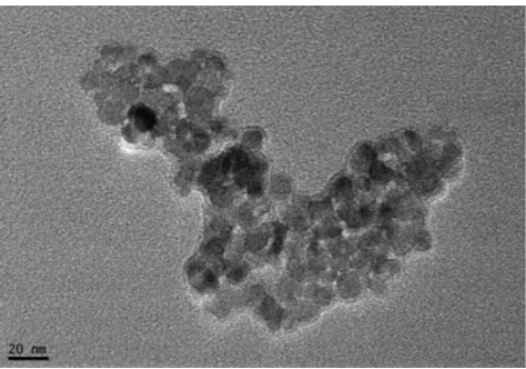
The characteristic of Fe
3O
4-MNPs
A colloidal suspension of Fe3O4-MNPs was achieved by
using ultrasound treatment and the magnetization and the
size of Fe3O4-MNPs were found to be 25.6 × 10⁻³ emu/mg
and 20 nm, respectively (Figure 1).
The changes of spleen weight in Fe
3O
4-MNPs-treated ICr mice
Mice treated with Fe3O4-MNPs appeared healthy and their
body weight gain patterns were similar to controls (data not
included). The spleens of Fe3O4-MNPs-treated mice showed
same appearance as the controls. Both the spleen weight and the ratio of spleen weight to body weight showed no significant difference between the experimental groups and
the controls (Table 1). It suggested that Fe3O4-MNPs did
not cause splenomegaly, which was due to the deposition of damaged erythrocytes or to recruitment and/or proliferation of splenic cells.
splenocyte proliferation
A significant increase of splenocyte proliferative capacity
was noticed after administration of Fe3O4-MNPs in low dose
(P , 0.05; low dose versus control). Both administration of
medium-dose and high-dose Fe3O4-MNPs affect splenocyte
proliferation and reduced the splenocyte proliferative capacity
compared with the control group (P , 0.05; control versus
medium-dose/high-dose Fe3O4-MNPs) (Table 2).12
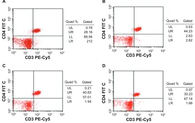
The proportions of lymphocyte subsets
in peripheral blood
There were no differences between the low-dose group and
the medium-dose group in the proportions of CD4+ T-cell
subset in peripheral blood of ICR mice, and both had more
CD4+ T lymphocytes than the control group. But the
high-dose group showed no difference compared with control
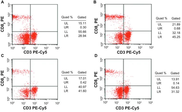
(P . 0.05) (Figures 2 and 3). Furthermore the proportions of
CD8+ T lymphocytes subset were slightly lower after the
com-mencement of high dose. Though both low- and medium-dose
groups indicated that they have more CD8+ T lymphocytes
than those of control group, and the low-dose group showed significantly more when compared to the medium-dose group
(P , 0.05) (Figures 4 and 5).
Cytokine release
To detect whether Fe3O4-MNPs can alter cytokine production
patterns in peripheral blood, enzyme-linked immunosorbent
assay was conducted. Fe3O4-MNPs altered the production
of IL-2, INF-γ, and IL-10. Interestingly, low dose of
Fe3O4-MNPs significantly increased the levels of IFN-γ,
IL-2, IL-10 and IL-4 (P , 0.05) when compared to medium
or high doses of Fe3O4-MNPs (P , 0.05), and there were
no significant differences between medium and high doses
of Fe3O4-MNPs (P . 0.05), suggesting that low dose of
Fe3O4-MNPs significantly increased the ability of splenocytes
to release cytokines. But no significant changes of IL-4 were observed between the experiment groups and the control group (Figure 6).
Discussion
Close attention has been paid to current nanoparticle techniques. It is well known that nanoparticles could present a versatile nanoscale surface for biomolecular recognition because of the numerous potential benefits in merging biomacromolecules and nanoparticles. Meanwhile, much effort has been explored to the development of new nano-composites and their application in many research fields such as DNA detection, intracellular labeling, drug carrier, and so on.
The magnetic nanoparticle of Fe3O4 we tested has good
biocompatibility and no cytotoxicity.13,14 Our group have
proved that Fe3O4-MNPs could increase the intracellular
effective concentration of chemotherapeutic drugs in vitro to reverse MDR. The focus of this study was to investigate
whether Fe3O4-MNPs have effects on mice immune
system, especially the T cell functions after given different dose.
Figure 1 TeM Image of magnetic nanoparticles of Fe3O4, Bar = 20 nm (X40000
times).
International Journal of Nanomedicine downloaded from https://www.dovepress.com/ by 118.70.13.36 on 23-Aug-2020
Dovepress
Chen et al
The total body weights of mice were not changed signif icantly, and the spleen/body weight ratio was unchanged. Actually, quantification of body weight and organ weight forms an integral part of any toxicological study providing an initial assessment of overall animal health status, as well as potential pathology. Descriptions of the tier approach to immunotoxicity evaluation should incorporate measurements of body weight, weights of spleen, thymus, kidney, and liver, as well as organ/body weight ratios in an
initial screen.15
Proliferation of lymphocytes following exposure to mitogenic stimuli is an important methodology for the assessment of cell-mediated immunity. This assay has enjoyed frequent use in immunotoxicology studies for
its ease of performance and relative reproducibility.16
Therefore, we have investigated the effects of Fe3O4-MNPs
on lymphoproliferatior in spleen following exposure to Con-A. The results showed that the proliferation of lymphocytes was significantly increased when mice were
injected with 5.14 mg/kg Fe3O4-MNPs compared with the
controls. However, the other two groups did not act in a similar way.
To detect the levels of proportion of CD4+ or CD8+ T cells,
which are markers for T cell lymphocytes. Fe3O4-MNPs
influenced the function of helper T cells or suppresser T cells,
when the less the Fe3O4-MNPs were given, the more of an
increase in T cell numbers was seen, suggesting that low
concentration of Fe3O4-MNPs can regulate T cell functions
in ICR mice.
The Th1 cytokines promote cellular immunity by activating
macrophages, cytotoxic CD8+ T lymphocytes, and so on,
while the Th2 cytokines enhance humoral immunity, including activation and class switching of antibody
producing B cells.17 Th1 cells are defined by their ability to
secrete the inflammatory cytokines IL-2 and INF-γ and are
involved in cellular immunity, some autoimmune disease, and in chronic inflammatory disorders. The Th2-biased cells preferentially produce IL-4, IL-5, IL-10, and IL-13 and participate in humoral response and antibody
produc-tion.18 In the present study, low dose of Fe
3O4-MNPs was
found to strongly affect the production of Th1 cytokine, and also affect some Th2 cytokine release, suggesting that
Fe3O4-MNPs might be involved in inflammations associated
with infections.
Conclusion
In conclusion, our results indicate Fe3O4-MNPs can influence
immune functions of mice in a dose-dependent manner.
Further study indicated that a high dose of Fe3O4-MNPs
has no significant influence on the immune systems of the mice. These data could be useful for improving biomedical
applications of Fe3O4-MNPs, but these immunological effects
of Fe3O4-MNPs should be further conducted both in vivo
and in vitro.
Acknowledgments
This work was supported by 973 National Key Fundamental Research Project of China (No. 2006CB933205), 863 Project of People’s Republic of China (No. 2007AA022007), National Nature Science Foundation of People’s Republic of China (No. 30740062, 30872970), and Special-Purpose Science Research Foundation for High School (No. 20070286042).
Table 1 effects of Fe3O4-MNPs on the spleen weight and the ratio of spleen weight to body weight (n = 10, mean ± sD)
Groups Spleen weight (g) The ratio of spleen weight
to body weight (×10-3)
Control (saline) 0.0940 ± 0.0152 3.3102 ± 0.4478
Low dose of Fe3O4-MNPs (5.14 mg/kg) 0.0720 ± 0.0286 2.3793 ± 0.8027*
Medium dose of Fe3O4-MNPs (20.7 mg/kg) 0.0900 ± 0.0187 3.1488 ± 0.5225*
high dose of Fe3O4-MNPs (51.4 mg/kg) 0.1120 ± 0.0084 4.0481 ± 0.8744*
Note: *P . 0.05, when compared to the control.
Table 2 Influence of Fe3O4-MNPs on the rate of lymphocyte transformation in spleen suspension (n = 10, mean ± sD)
Groups The rate of lymphocyte
transformation
Control (saline) 0.0193 ± 0.001027 Low dose of Fe3O4-MNPs (5.14 mg/kg) 0.0398 ± 0.005155* Medium dose of Fe3O4-MNPs (20.7 mg/kg) 0.0111 ± 0.003029*
high dose of Fe3O4-MNPs (51.4 mg/kg) 0.0046 ± 0.001517*
Note: *P, 0.05, when compared to the control.
International Journal of Nanomedicine downloaded from https://www.dovepress.com/ by 118.70.13.36 on 23-Aug-2020
Dovepress Immune function in ICr mice
10
4
10
3
10
2
10
1
10
0
10
4
10
3
10
2
10
1
10
0
104
103
102
101
100
10
4
10
3
10
2
10
1
10
0
104
103
102
101
100
104
103
102
101
100
10
4
10
3
10
2
10
1
10
0
104
103
102
101
100
CD4 FIT
C
CD3 PE-Cy5 CD3 PE-Cy5
CD3 PE-Cy5 CD3 PE-Cy5
CD4 FIT
C
CD4 FIT
C
CD4 FIT
C
Quad % Gated
UL UR LL
LR 212
68.94 28.16 0.78
Quad % Gated
UL UR LL
LR 2.62
2.63 44.23 0.53
Quad % Gated
UL UR LL
LR 1.66
67.14 30.23 0.97
Quad % Gated
UL UR LL
LR 1.94
57.01 40.83 0.21 A
C D
B
Figure 3 effect of Fe3O4-MNPs on the proportion of CD4+ T lymphocyte subset in peripheral blood by FCM.
Notes: A) 0.2 mL saline; B) low dose of Fe3O4-MNPs (5.14 mg/kg); C) medium dose of Fe3O4-MNPs (20.7 mg/kg); D) high dose of Fe3O4-MNPs (51.4 mg/kg).
0.8
0.7
0.6 0.5
0.4
0.3
0.2
0.1
0
Control
The proportions of CD8
+ cells (%)
Low dose Medium dose High dose
CD8
Figure 4 effect of Fe3O4-MNPs on the proportion of CD8+ T lymphocyte subset in peripheral blood.
Notes: Control:0.2 mL saline; Low dose: low dose of Fe3O4-MNPs (5.14 mg/kg); Medium dose: medium dose of Fe3O4-MNPs (20.7 mg/kg); high dose: high dose of Fe3O4
-MNPs (51.4 mg/kg). CD4+, CD8+represent CD4, CD8-positive T cells.
50 45 40 35 30 25 20 15 10 5 0
Control
The proportions of CD4
+ cells (%)
Low dose Medium dose High dose
CD4
Figure 2 The effects of Fe3O4-MNPs on the proportion of CD4+ T lymphocyte subset in peripheral blood.
Notes: Control:0.2 mL saline; Low dose: low dose of Fe3O4-MNPs (5.14 mg/kg); Medium dose: medium dose of Fe3O4-MNPs (20.7 mg/kg); high dose: high dose of Fe3O4
-MNPs (51.4 mg/kg). CD4+, CD8+ represent CD4, CD8-positive T cells.
International Journal of Nanomedicine downloaded from https://www.dovepress.com/ by 118.70.13.36 on 23-Aug-2020
Dovepress
Chen et al
10 4 10 3 10 2 10 1 10 0 10 4 10 3 10 2 10 1 10 0 104 103 102 101 100 10 4 10 3 10 2 10 1 10 0 104 103 102 101 100 104 103 102 101 100 10 4 10 3 10 2 10 1 10 0 104 103 102 101 100 CD8 β PE CD8 β PE CD8 β PE CD8 β PE
CD3 PE-Cy5 CD3 PE-Cy5
CD3 PE-Cy5 CD3 PE-Cy5
Quad % Gated UL UR LL LR 28.94 55.66 0.30 15.11
Quad % Gated UL UR LL LR 45.25 32.18 0.68 21.89
Quad % Gated UL UR LL LR 31.32 54.63 0.14 13.91 Quad % Gated
UL UR LL LR 41.60 40.97 0.42 17.01 A C D B
Figure 5 effect of Fe3O4-MNPs on the proportions of CD8+ T lymphocyte subset in peripheral blood by FCM.
Notes: A) 0.2 mL saline; B) low dose of Fe3O4-MNPs (5.14 mg/kg); C) medium dose of Fe3O4-MNPs (20.7 mg/kg); D) high dose of Fe3O4-MNPs (51.4 mg/kg).
80 70 60 50 40 30 20 10 0 Control
The level of cytokine (pg/mL)
Low dose Medium dose High dose
IFN−γ IL−2 IL−4 IL−10
Figure 6 effect of Fe3O4-MNPs on the production of cytokine in peripheral blood.
Notes: Control:0.2 mL saline; Low dose: represents low dose of Fe3O4-MNPs (5.14 mg/kg); Medium dose: represents medium dose of Fe3O4-MNPs (20.7 mg/kg); high dose:
represents high dose of Fe3O4-MNPs (51.4 mg/kg).
Disclosure
The authors confirm no conflicts of interest in this work.
References
1. Weizmann Y, Patolsky F, Katz E, Willner I. Amplified DNA sensing and immunosensing by the rotation of functional magnetic particles. J Am Chem Soc. 2003;125(12):3452–3454.
2. Song HT, Choi JS, Huh YM, et al. Surface modulation of magnetic nano-crystals in the development of highly efficient magnetic resonance probes for intracellular labeling. J Am Chem Soc. 2005;127(28):9992–9993. 3. Gao XH, Cui YY, Levenson RM, Chung LW, Nie S. In vivo cancer
tar-geting and imaging with semiconductor quantum dots. Nat Biotechnol. 2004;22(8):959–960.
4. Chen Y, Yang L, Feng C, Wen LP. Nano neodymium oxide induces mas-sive vacuolization and autophagic cell death in non-small cell lung cancer NCI-H460 cells. Biochem Biophys Res Commun. 2005; 337(1): 52–60. 5. Zhang L, Zhang K, Prändl R, Schöffl F. Detecting DNA-binding of
proteins in vivo by UV-crosslinking and immunoprecipitation. Biochem Biophys Res Commun. 2004;322(3):705–711.
6. Cho CS, Cho KY, Park IK, et al. Receptor-mediated delivery of trans-retinoic acid to hepatocyte using poly (L-lactic acid) nanopar-ticles coated with galactose-carrying polystyrene. J Control Release. 2001;77:7–15.
7. Jain TK, Morales MA, Sahoo SK, Leslie-Pelecky DL, Labhasetwar V. Iron oxide nanoparticles for sustained delivery of anticancer agents. Mol Pharm. 2005;2(3):194–205.
8. Alexiou C, Arnold W, Klein RJ, et al. Locoregional cancer treatment with magnetic drug targeting. Cancer Res. 2000;60:6641–6648. 9. Tiefenauer LX, Kuhne G, Andres RY. Antibody-magnetic nanoparticles:
In vitro characterization of potential tumor-specific contrast agent for magnetic resonance imaging. Bioconjug Chem. 1993;4(5): 347–352.
10. Chen BA, Sun Q, Wang XM, et al. Reversal in multidrug resistance by magnetic nanoparticle of Fe3O4 loaded with adriamycin and tetrandrine in K562/AO2 leukemic cells. Int J Nanomedicine. 2008;3: 277–286.
11. Jiang Z, Chen B A, Xia G H, et al. The reversal effect of Fe3O4-magnetic nanoparticles loaded with cisplatin on the SKOV3/DDP ovarian carcinoma cells. Int J Nanomedicine. 2009;4:1–8.
International Journal of Nanomedicine downloaded from https://www.dovepress.com/ by 118.70.13.36 on 23-Aug-2020
International Journal of Nanomedicine
Publish your work in this journal
Submit your manuscript here: http://www.dovepress.com/international-journal-of-nanomedicine-journal
The International Journal of Nanomedicine is an international, peer-reviewed journal focusing on the application of nanotechnology in diagnostics, therapeutics, and drug delivery systems throughout the biomedical field. This journal is indexed on PubMed Central, MedLine, CAS, SciSearch®, Current Contents®/Clinical Medicine,
Journal Citation Reports/Science Edition, EMBase, Scopus and the Elsevier Bibliographic databases. The manuscript management system is completely online and includes a very quick and fair peer-review system, which is all easy to use. Visit http://www.dovepress.com/ testimonials.php to read real quotes from published authors. Dovepress
Dovepress
Immune function in ICr mice
12. Müller K, Skepper JN, Posfai M, et al. Effect of ultrasmall super-paramagnetic iron oxide nanoparticles (Ferumoxtran-10) on human monocyte-macrophages in vitro. Biomicrofluidics. 2007;1(4):44104. 13. Cheng FY, Su CH, Yang YS, et al. Characterization of aqueous
disper-sions of Fe(3)O(4) nanoparticles and their biomedical applications. Biomaterials. 2005;26(7):729–738.
14. Zhang R, Wang X, Wu C, et al. Synergistic enhancement effect of magnetic nanoparticles on anticancer drug accumulation in cancer cells. Nanotechnology. 2006;17(14):3622–3626.
15. Luster MI, Portier C, Pait DG, et al. Risk assessment in immunotoxi-cology Sensitivity and predictab ility of immune tests. Fundam Appl Toxicol. 1992;18(2):200–210.
16. Snyder C, Valle CD. Lymphocyte proliferation assays as potential biomarkers for toxicant exposures. J Toxicol Environ Health. 1991; 34(1):127–139.
17. Charlton B, Lafferty KJ. The Th1/Th2 balance in autoimmunity. Curr Opin Immunol. 1995;7(6):793–798.
18. Del Prete GF, De Carli M, Almerigogna F, et al. Human IL-10 is pro-duced by both type 1 helper (Th1) and type 2 helper (Th2) T cell clones and inhibits their antigen-specific proliferation and cytokine production. J Immunol. 1993;150(2):353–360.
International Journal of Nanomedicine downloaded from https://www.dovepress.com/ by 118.70.13.36 on 23-Aug-2020