International Journal of Nanomedicine
Concepts and practices used to develop
functional PLGA-based nanoparticulate systems
Hongkee Sah1,2 Laura A Thoma2 Hari R Desu2 Edel Sah3
George C Wood2
1College of Pharmacy, Ewha Womans University, Sedaemun-gu, Seoul, South Korea; 2College of Pharmacy, The University of Tennessee Health Science Center, Memphis, TN, USA; 3College of Science, University of Notre Dame, Notre Dame, IN, USA
Correspondence: Hongkee Sah College of Pharmacy, Ewha Womans University, 52 Ewhayeodae-gil, Sedaemun-gu, Seoul 120-750, South Korea
Tel +82 2 3277 4367 Fax +82 2 3277 2851 Email [email protected]
Abstract: The functionality of bare polylactide-co-glycolide (PLGA) nanoparticles is limited to drug depot or drug solubilization in their hard cores. They have inherent weaknesses as a drug-delivery system. For instance, when administered intravenously, the nanoparticles undergo rapid clearance from systemic circulation before reaching the site of action. Furthermore, plain PLGA nanoparticles cannot distinguish between different cell types. Recent research shows that surface functionalization of nanoparticles and development of new nanoparticulate dosage forms help overcome these delivery challenges and improve in vivo performance. Immense research efforts have propelled the development of diverse functional PLGA-based nanoparticulate delivery systems. Representative examples include PEGylated micelles/nanoparticles (PEG, polyethylene glycol), polyplexes, polymersomes, core-shell– type lipid-PLGA hybrids, cell-PLGA hybrids, receptor-specific ligand-PLGA conjugates, and theranostics. Each PLGA-based nanoparticulate dosage form has specific features that distinguish it from other nanoparticulate systems. This review focuses on fundamental concepts and practices that are used in the development of various functional nanoparticulate dosage forms. We describe how the attributes of these functional nanoparticulate forms might contribute to achievement of desired therapeutic effects that are not attainable using conventional therapies. Functional PLGA-based nanoparticulate systems are expected to deliver chemotherapeutic, diagnostic, and imaging agents in a highly selective and effective manner.
Keywords: nanoparticulate dosage forms, nanoparticles, polylactide-co-glycolide, functionality
Introduction
Biodegradable polylactide-co-glycolide (PLGA) nanoparticles have been used as carriers for drugs, peptides, proteins, vaccines, and nucleotides.1 Nanoparticles can protect drug moieties from degradation and provide sustained drug release. Nanoparticles are sometimes effective in facilitating intracellular delivery of bioactive materials. Parenteral PLGA nanoparticles have been used for various applications in vaccination, cancer therapy, and the treatment of cerebral disorders.2 In particular, nanoparticles have been widely investigated for use in cancer therapy. Nanoparticles, if given intravenously, can extravasate into and accumulate within tumor tissues that have defective blood vessels and impaired lymphatic drainage.3 This enhanced permeability and retention (EPR) effect helps direct nanoparticles to tumor sites.4 Even though PLGA nanoparticles have emerged as a promising carrier, they have a number of intrinsic drawbacks including the following:
Dove
press
R E v I E W
open access to scientific and medical research
Open Access Full Text Article
International Journal of Nanomedicine downloaded from https://www.dovepress.com/ by 118.70.13.36 on 23-Aug-2020
For personal use only.
Number of times this article has been viewed
This article was published in the following Dove Press journal: International Journal of Nanomedicine
• Prolonged systemic circulation of nanoparticles is a key prerequisite for maximizing the EPR effect. However, if administered intravenously, bare PLGA nanoparticles are rapidly removed from circulation in the blood, due to opsonization.
• There is increasing demand for the development of PLGA-based formulations other than microspheres and nanoparticles. For example, polymeric micelles and polyplexes are receiving increasing attention as nanocarriers. However, PLGA alone does not permit such formulations.
• PLGA nanoparticles cannot distinguish different cell types. Although cell-recognition capabilities can be conferred to PLGA nanoparticles through conjugation with an appropriate ligand, PLGA itself does not have versatile functional groups that could be used for surface derivatization.
• In vivo blood circulation time of nanoparticles and the extent to which they are taken up by cells are affected by various factors, such as surface charge. For example, cationic nanoparticles enhance cellular uptake and open tight junctions. It is sometimes necessary to change the negative charge of bare PLGA nanoparticles.
• The passive diffusion of PLGA nanoparticles into tumor tissues, driven by the EPR mechanism, may not elicit an efficacious clinical response. Transferring more drug payload to cancer cells is required.
• Bare PLGA nanoparticles do not easily enter cells, and they cannot pass through the blood–brain barrier (BBB). Innovative measures should be taken to improve their uptake by cells or to use them as a drug carrier to the brain.
Versatile strategies are being adopted to overcome the limitations of bare PLGA nanoparticles. The objectives of these strategies are to improve their functionality and in vivo performance. Functionalization of PLGA nanoparticles includes surface modification by PEGylation (PEG, polyethylene glycol), lipid- or surfactant-coating, polyion complexation, and conjugation with cell-targeting ligands. Functional PLGA-based nanoparticulate systems offer various beneficial outcomes, such as: minimization of opsonin adsorption, prolongation of blood residence time, reduction of drug side effects, elimination of the use of toxic adjuvants for drug solubilization, improved targeted drug delivery, efficient cellular translocation and subcellular trafficking, and synergic drug combination therapy.5,6 Broadly, functional PLGA-based nanoparticulate forms may be classified as PEGylated micelles, polyplexes, PEGylated nanoparticles,
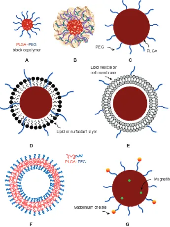
polymersomes, core-shell type hybrid nanoparticles/ nanocells, cell-mimicking nanoparticles, targeting ligand– PLGA conjugates, and theranostics (Figure 1). This review focuses on the fundamentals and strategies that are used to develop diverse functional PLGA nanoparticulate carriers. Representative practices for each topic are discussed, rather than specific details.
Why are PLGA and PEG preferred
over other polymers?
The US Food and Drug Administration (FDA) approved the use of PLGA and polylactide (PLA) polymers in parenteral microspheres, implants, and periodontal drug-delivery systems (eg, Lupron Depot, Sandostatin LAR Depot, Zoladex, Vivitrol, Risperdal Consta, OsteoScaf, Arestin, and Atridox). The long approval history of PLA/PLGA-based drug products by worldwide regulatory agencies is a major reason for the preferential use of these biodegradable polymers over others. For convenience, PLGA is used hereinafter to represent both PLA and PLGA. The characteristics of PLGA are controlled by the stereochemistry of lactic acid (D, L, or DL), degree of crystallinity, lactic acid/glycolic acid ratio, and molecular weight. PLGA is available with different end groups, namely a free carboxylic end group and an ester-terminated group.7 End-capped PLGA usually has a lauryl or a methyl ester, and is more stable than uncapped PLGA.
PEGylation technology contributes to (a) increasing the aqueous solubility and stability, (b) reducing intermolecular aggregation, (c) decreasing immunogenicity, and (d) prolonging the systemic circulation time of a compound. PEG is often linked to PLGA to achieve similar beneficial effects.8 PEG conjugation to PLGA also enables the preparation of self-assembling micelles. In addition, the end groups of PEG can be modified to aldehyde, amino, carboxyl, or methyl groups to alter the surface charge of a nanoparticulate carrier. There are several kinds of PEGs: linear monofunctional PEG derivatives having a reactive group (eg, carboxyl or amine group) on one side and a methoxy group or a sugar group on the other side, homobifunctional PEG derivatives, heterobifunctional PEG derivatives, Y-shape branched PEG derivatives, fork-shaped PEG derivatives, and multi-arm PEG derivatives.9 These PEG derivatives are used for conjugation with ligands that have amine, thiol, or carboxyl groups.
PEGylated PLGA micelles
PEG-PLGA block copolymers are utilized to prepare micelles.10 Although various kinds of block copolymers can be synthesized, the most commonly synthesized block Dovepress
Sah et al
International Journal of Nanomedicine downloaded from https://www.dovepress.com/ by 118.70.13.36 on 23-Aug-2020
copolymers have AB, BAB, or ABA block structures, where A and B stand for PEG and PLGA blocks, respectively.11,12 Synthetic methods for producing these copolymers are well established.13–15 Currently, a number of block copolymers are commercially available from companies such as Akina (http://www.akinainc.com) and Polysciences, Inc (http://www.polysciences.com). Block copolymers with low molecular weights and/or high PEG/PLGA ratios are water-soluble, whereas those with high molecular weights
and/or low PEG/PLGA ratios are water-insoluble. Block copolymers, which are more hydrophilic than bare PLGA, are considered to be more suitable for the delivery of hydrophilic macromolecules such as proteins.
Due to their amphiphilic nature, when PEG-PLGA block copolymers are dispersed in an aqueous medium, they self-assemble into micellar forms. PEG acts as a hydrophilic corona, while PLGA serves as a hydrophobic core. A polymeric micelle can incorporate aqueous
A B C
D E
F G
PLGA–PEG block copolymer
Lipid or surfactant layer
Lipid vesicle or cell membrane
Gadolinium chelate
Magnetite PEG
PLGA
PLGA–PEG
Figure 1 Representative PLGA-based nanoparticulate dosage forms. (A) PEGylated micelle (eg, PEG-PLGA block copolymeric micelle), (B) polyplex (eg, DNA-PEI-PLGA), (C) PEGylated PLGA nanoparticle, (D) core-shell type nanoparticle (eg, PEGylated lipid-PLGA hybrid nanoparticle), (E) cell membrane-PLGA hybrid nanoparticle (eg, PLGA nanoparticles encased by red blood cell), (F) polymersome (eg, PEG-PLGA block copolymeric vesicle), and (G) magnetic PLGA particle either conjugated with gadolinium-chelate or laden with magnetite (eg, theranostics).
Note: Allof these nanoparticulate carriers can be further derivatized by cell-recognizable ligands.
Abbreviations: PLGA,polylactide-co-glycolide; PEG, polyethylene glycol; DNA, deoxyribonucleic acid; PEI, polyethyleneimine.
Dovepress Functional PLGA-based nanoparticulate delivery systems
International Journal of Nanomedicine downloaded from https://www.dovepress.com/ by 118.70.13.36 on 23-Aug-2020
hydrophobic drugs such as paclitaxel. Polymeric micelles can prolong the blood residence time of drugs, lessen systemic toxicity, and direct drugs to the site of action.16 One example of PEG-PLGA micellar technology is the Genexol-PM formulation of Samyang Corporation, Seoul, Korea.17 This formulation was approved by the Korea FDA in 2007. Genexol-PM is a Cremophor EL-free PEG-PLGA micelle formulation of paclitaxel that is intended to treat breast cancer and non-small cell lung cancer. This formulation allows administration of higher doses of paclitaxel than the Cremophor EL-based formulation. The maximum tolerated doses of the Genexol-PM and Cremophor EL-containing Taxol formulation were 390 and 230 mg/m2, respectively.18 In addition, specialized intravenous infusion sets, in-line filtration, and premedication are not required for the administration of the Genexol-PM. In preclinical models, the Genexol-PM achieved higher paclitaxel tumor concentrations than the Cremophor EL-based formulation.13 These results indicate that polymeric micelles may passively target tumors, stemming from the EPR effect and the prolonged systemic circulation time. It may be of interest to the readers to note that Davis et al summarized the pharmacokinetic parameters of nanoparticulate carriers including Genexol-PM, Taxol, Abraxane, Doxil, and other polymer nanoparticles.5 Recently, Shin et al carried out an interesting pharmacokinetics study of individual and concurrent delivery of paclitaxel, demethoxygeldanamycin, and rapamycin using aqueous PEG-PLGA micellar solutions.19 After single drug-loaded, dual drug-loaded, and triple drug-loaded micelles were separately injected via the tail vein of mice, their pharmacokinetic patterns were characterized. The combination of drugs and their dosage affected the extent of pharmacokinetic differences among 1-, 2-, and 3-drug-loaded micelles. Shin et al speculated that these differences might have arisen from alterations of drug–drug interactions, drug metabolism, and micellar stability.
There are many reports of the use of PLGA-based diblock or triblock micelles for solubilizing hydrophobic drugs.20–22 The encapsulation of photosensitizers in PEG-PLGA micelles is an example of this solubilization. In photodynamic cancer therapy, photosensitizers react to a specific wavelength of light and produce an activated oxygen form that kills nearby cells.23,24 However, photosensitizers also enter healthy tissues, leading to their photosensitization. In addition, many photosensitizers are too hydrophobic for development into an aqueous injectable formulation. As a potential solution for these problems, a photosensitizer (mTHPP; 5,10,15,20-tetrakis (meso hydroxyphenyl)porphyrin) was
loaded into PEG-PLGA micelles.25,26 The 30 nm micelles, which contained 2% mTHPP, entered HN5 head and neck cancer cells and H2009 lung cancer cells. Upon illumination with a 532 nm laser, the micelles killed the cancer cells.
An interesting approach to controlling the release of drugs from micelles is the use of a mechanical technique.27 Hydrophobic nile red was used as a model drug to be encapsulated into PEGylated PLGA micelles. After micellar solubilization, dye release was triggered by high intensity focused ultrasound (HIFU). The strong HIFU-induced cavitation led to the degradation of the diblock copolymer into its constituents, thereby prompting dye release. The authors suggested that the HIFU technique could be utilized as a noncontact and remote control approach to manipulating drug release from polymeric micelles.
The hydrophobicity of PLGA cores can be increased by chemical derivatization. For example, mono-hexyl and di-hexyl substituted lactide monomers have been used to produce hexyl-PLGA via ring-opening polymerization.28,29 This technology, owned by SurModics Pharmaceutical Inc, makes it possible to prepare 20–80 nm micelles. PEG-hexyl-PLGA micelles, due to their elevated hydrophobicity, have greater capacity for loading hydrophobic drugs than do typical PEG-PLGA micelles. For example, the aqueous solubility of cyclosporine A is 0.01 mM, but its solubility in PEG-hexyl-PLGA micelles increases to 1.10 mM.30
Non-PEGylated PLGA micelles
Polyethyleneimine (PEI) has been widely used as a surface-modified adsorbent, complex-forming polyelectrolyte, colloidal particle, gene-delivery vehicle, and the like.31,32 Attempts have been made to apply PEI to PLGA nanotechnology.33 In one study, PEI-PLGA diblock copolymers were prepared according to the conventional dicyclohexylcarbodiimide/ N-hydroxysuccinimide (DCC/NHS) conjugation chemistry. The PEI-PLGA micelles showed better translocation into human keratinocytes than plain PLGA nanoparticles. It is likely that such improvement in cellular uptake arose from the cationic surface of PEI-PLGA micelles and the strong affinity of PEI to lipopolysaccharides and cell membranes, as has been suggested elsewhere.32
A poly(L-cysteine)-b-PLGA copolymer, linked through a disulfide bond, was formulated into a micellar dosage form.34 The hydrophilic polyamino acid domain formed a corona around the hydrophobic PLGA core. The chain lengths of block polymers and the redox status of the disulfide bond affected the size of the polymeric micelles. Other polypeptides made of aspartic acid, glutamic acid, and Dovepress
Sah et al
International Journal of Nanomedicine downloaded from https://www.dovepress.com/ by 118.70.13.36 on 23-Aug-2020
lysine have also been used as coronary segments of PLGA-based micelles.35–37 For example, carboxylic end groups of PLGA chains were reacted with primary ε-amino groups of a cationic poly(L-lysine) (PLL), to produce PLGA-g-PLL copolymers of various grafting percentages (3.6%–8.0%).37 These conjugates self-assembled into 70–80 nm micelles in water. These polymeric micelles are more thermodynamically stable and have lower critical micelle concentrations than typical surfactant micelles.
Another interesting micellar template consists of hydrophilic siRNA conjugated to PLGA via a disulfide bond.38 The amphiphilic conjugate self-assembles in an aqueous environment to form 22 nm micelles. The net negative charge of the siRNA-PLGA conjugate micelles is nullified by coating with a cationic PEI. The PEI coated siRNA-PLGA micelles exhibited better cellular translocation than typical siRNA-PEI complexes. Another notable feature of siRNA-PLGA conjugate is the disulfide bond between the siRNA and PLGA components. It is expected that the glutathione-laden cells reduce the disulfide bond and release the siRNA from the conjugate.39
Because antisense oligonucleotides do not easily enter target cells, a delivery system is required to facilitate cellular uptake. Jeong and Park conjugated a c-myc antisense 15-mer to a carboxyl end group of PLGA via DCC/NHS chemistry.40 The resultant amphiphilic oligonucleotide-PLGA conjugate self-assembled into 65 nm micelles. The endocytosis efficiency of the conjugate was studied in NIH 3T3 mouse fibroblast cells. The oligonucleotide sparsely entered the cells (,0.1%). In sharp contrast, the cellular uptake of the polymeric micelle was significantly greater (68.3%). This suggests that these micelles have potential as an antisense carrier.
Polyplexes
An innovative strategy was used to prepare PEI-PLGA micelle-based polyplexes for gene delivery.41 PLGA and branched PEI (bPEI) were linked together to produce
PLGA-b-bPEI-b-PLGA by DCC/NHS conjugation chemistry. This polymer was made into 50–60 nm micelles that had a positive surface charge. The micelles were then condensed with a firefly luciferase receptor gene-encoding plasmid deoxyribonucleic acid (pDNA). In the MCF7 breast cancer cell model, the transfection efficiency of the resulting polyplexes was 103–104 times lower than that of bPEI/pDNA complexes. The authors introduced a lower molecular weight bPEI to the micelle/pDNA complexes to induce tighter layer-by-layer complexation and augment the cationic surface charge of the polyplexes. The transfection efficiency of
the final polyplexes was 50–100 times higher than that of the primary polyplexes. It is also noteworthy that the final polyplexes were lyophilized to powder, whereas typical bPEI/pDNA complexes became sticky after lyophilization.
PLGA-PEG-PLL triblock copolymers have been used to prepare nanoparticulate carriers. PEG imparts a stealth effect upon the PLGA core, whereas PLL confers a cationic charge to the nanoparticles.42 The primary ε-amine groups in the lysine moieties of PLL can form polyionic complexes with negatively charged compounds. Such a system was explored as a nonviral vector for DNA transfection. In vitro gene transfection studies using HeLa and HepG2 cells showed that better transfection efficiency was attained with PLGA-PEG-PLL/DNA polyplexes than with typical PEI/DNA complexes. Such a system might serve as a safer nonviral gene delivery vector than typical PEI/DNA complexes.
Acid-responsive micelles
As nanoparticles enter cells by endocytosis, proton pumping into endocytic vesicles is accompanied by a vesicular pH decrease to 5. If nanoparticles are responsive to pH change, this property could be used for intracellular drug targeting.43 Based on this strategy, PLGA-b-poly(2-ethyl-2-oxazoline)-b -PLGA triblock copolymer micelles were prepared to deliver doxorubicin into cancer cells.44 The micelles self-assemble into a flower-like morphology – the hydrophilic poly(2-ethyl-2-oxazoline) domains form a shell layer over the hydrophobic PLGA core. When poly(2-ethyl-2-oxazoline) (pKa = 7.1) is in an acidic environment, the protonated nitrogen interacts with a carbonyl group via hydrogen-bonding. This triggers the deformation of the polymeric micelles. The pH-dependent destabilization of the micelle structure causes changes in size, zeta potential, fluorescence pattern, and doxorubicin release. A related study on human cervix HeLa cells showed that these polymeric micelles significantly suppressed tumor cells.
Preparation of functional
PLGA-based micelles
Typical methods for preparing polymeric micelles include: (1) the solid dispersion method, (2) the emulsion-based solvent evaporation/extraction method, (3) nanoprecipitation, and (4) the solvent dialysis method.45 Suitable manufacturing methods are selected based on the aqueous solubility of the amphiphilic block copolymer, the molecular weight of each block constituent, and the proportion of each block constituent. In the solid dispersion method, an amphiphilic polymer and a hydrophobic drug are dissolved in an organic solvent. The solvent is evaporated under reduced pressure to form a Dovepress Functional PLGA-based nanoparticulate delivery systems
International Journal of Nanomedicine downloaded from https://www.dovepress.com/ by 118.70.13.36 on 23-Aug-2020
gel-like polymeric matrix. Adding water into the gel-like matrix followed by dispersing the mixture at elevated temperature lead to the formation of polymeric micelles (Figure 2A). In the emulsion-based method, a polymeric dispersed phase is emulsified in an aqueous phase. Solvent removal by evaporation and/or extraction causes the rearrangement of polymeric chains to form micelles (Figure 2B). Nanoprecipitation involves the use of a water-miscible solvent (eg, tetrahydrofuran, acetonitrile, acetone, and dimethyl formamide) as a dispersed solvent. Addition of the dispersed phase to an aqueous phase triggers instant solvent diffusion, leading to spontaneous formation of polymeric micelles. Finally, in the solvent dialysis method, a water-miscible solvent is removed by dialyzing with an excessive amount of water.
PEGylated PLGA nanoparticles
When administered intravenously, bare PLGA nanoparticles are rapidly cleared from the blood. This phenomenon is attributed to the fact that hydrophobic nanoparticles with a negative surface charge are vulnerable to opsonization.46,47 To overcome this limitation, the surface of PLGA nanoparticles is layered with PEG that functions as a hydrophilic corona. Improved systemic circulation time of PEGylated PLGA nanoparticles is well documented.48–50 PEGylation of PLGA nanoparticles can be conducted in two ways. In one method, nanoparticles are prepared using PEGylated PLGAs such as PEG-PLGA, PLGA-PEG-PLGA, and PEG-PLGA-PEG block copolymers. The other method involves a chemical reaction that links PEG to the surface
of preformed PLGA nanoparticles. For example,
1-ethyl-3-(3′-dimethylaminopropyl) carbodiimide (EDC)/NHS
conjugation results in an amide bond between an amine-terminated PEG derivative and a free carboxyl end group of PLGA. Physicochemical properties of PEG-PLGA nanoparticles (size, surface charge, surface hydrophilicity, and surface PEG content) affect their biodistribution and cellular uptake.51,52 Li et al have correlated these nanoparticle properties to biodistribution patterns using a physiologically based pharmacokinetic model.52
A comprehensive review of current advances in the stealth coating of nanoparticulate drug-delivery systems was recently published.53 The article particularly cautions against the undesirable effects of PEGylation on the in vivo performance of nanoparticulate carriers. The major concerns are the interference of the PEG corona with nanoparticle–cell interactions and the endosomal escape of nanoparticles after extravasation. Another major concern is the immune response of the host to repeated administration of a PEGylated nanoparticulate carrier. It is also important to consider the dependence of stealth properties on the density and morphology of PEG corona. The corona morphology of PEG is often described by mushroom (random coil, low density), brush (elongated coil, high density), or mushroom/ brush transition models.54–56 PEG coronas in brush-like and intermediate configurations inhibit phagocytosis and complement activation, whereas mushroom-like PEG morphology triggers complement activation and phagocytosis. Therefore, the molecular weight and shape
PLGA PEG Drug
Water Organic solvent
B
o/w nanoemulsion A
Gel
Figure 2 Schematic representation of typical nanoencapsulation techniques used to prepare PLGA-based micelles: (A) the solid dispersion method and (B) the emulsion-based solvent evaporation/extraction method.
Abbreviations: PLGA,polylactide-co-glycolide; PEG, polyethylene glycol; o/w, oil-in-water.
Dovepress
Sah et al
International Journal of Nanomedicine downloaded from https://www.dovepress.com/ by 118.70.13.36 on 23-Aug-2020
of PEG, as well as the PEG/PLGA molar ratio, play critical roles in the behavior of PEGylated nanoparticles.
Even though there are a number of nanoencapsulation technologies, the most commonly used technique to prepare PLGA nanoparticles is an emulsion-based solvent evaporation/ extraction technique.57,58 The manufacturing principles of this technique are very similar to those used in the production of commercial PLGA microspheres. A water-in-oil-in-water double emulsion technique is used for nanoencapsulation of hydrophilic drugs, whereas an oil-in-water emulsion method is chosen for encapsulation of hydrophobic drugs. Briefly, an organic solvent with substantial volatility (eg, methylene chloride, chloroform, or ethyl acetate) is used to dissolve nanoparticle-forming materials. The dispersed phase is then emulsified in an aqueous phase using a propeller stirrer, a turbine impeller mixer, a rotor–stator homogenizer, a high pressure microfluidizer, a porous membrane mixer, or a sonicator.59,60 Solvent removal from nanoemulsion droplets brings about their transformation into solid nanoparticles. Although nanoprecipitation follows the same manufacturing principle as the emulsion-based procedure, the former favors the use of a water-miscible semipolar solvent (eg, acetone, acetonitrile, or tetrahydrofuran) over water-immiscible solvents. As a result, mixing of the polymeric dispersed phase with an aqueous continuous phase leads to instant generation of solid nanoparticles.
Polymersomes
There are often critical problems with typical liposomes, such as low drug loading efficiency, storage instability, drug leakage during storage, or burst drug release. Polymersomes have therefore been proposed as an alternative to liposomes.61,62 Polymersomes are self-assembled bilayer vesicles made from synthetic amphiphilic copolymers such as PEG-PLGA (Figure 1). Polymersomes can be further engineered to deliver, target, and release bioactive materials.63,64 Although polymersomes and liposomes are both bilayered vesicles, they differ from one another in several ways. For example, PEGylated lipid content of stealth liposomal formulations is limited to 5%–10%, because greater concentrations result in curved micelles.65 In contrast, polymersomes accommodate much higher concentrations of a PEGylated ingredient, and can be stable for months in aqueous solutions.66 The more densely packed PEG brush of polymersomes might confer better resistance toward opsonization than the less densely packed PEG brush of other nanoparticles, thereby extending their systemic circulation time. Both hydrophilic drugs and hydrophobic drugs can
be loaded into polymersomes. Drug cocktail therapy using polymersomes has been suggested as a method for directing multiple drugs to multiple cellular targets; for example, taxol to microtubules and doxorubicin to DNA.67,68
The self-assembling dynamics of an amphiphile are determined by the characteristics of its constituents and their proportions. The geometry of a self-assembled amphiphile is described by a core packing parameter (p) as follows:
p = νH/(a0 × lc), where νH is the volume of a compactly packed hydrophobic core, a0 is the effective cross-sectional area occupied by a hydrophilic group at a nanoparticulate-solution interface, and lc is the length of a hydrophobic core.69 Spherical micelles have P value of less than 1/3, and the P value of cylindrical micelles falls between 1/3 and 1/2. Lamellar vesicles have P values ranging from 1/2 to 1. Many reports prefer to use the f value (the fraction of the hydrophilic block in an amphiphile), rather than the P value. The relationship between f and p is expressed by the following equation: ln f ≈ −p/β where β = 0.66.61 This correlation is helpful to those who are interested in manipulating the morphology of nanoparticulate matters. When a PEG-PLGA block copolymer is used to prepare polymersomes, f values of 0.35 ± 0.1 are recommended as a starting point. A copolymer with f . 0.45 is likely to produce micelles, whereas a copolymer with f, 0.25 gives rise to solid-like particles. To produce polymersomes, the constituents of a polymersome are dissolved in an organic solvent. After solvent removal, the resultant thin film is hydrated with the aid of heat and/ or a cosolvent. The formation of a bilayered vesicle is favored thermodynamically. Sometimes, polymersomes are sonicated or extruded to tailor their size. They can be further labeled with targeting ligands or fluorescent dyes.
Ester linkages in the backbone of PLGA are subject to random hydrolytic chain scission. This hydrolytic progress influences the vesicular integrity of PEG-PLGA polymersomes and drug release. PLGA hydrolysis transforms the bilayer-forming PEG-PLGA into PEG-abundant segments. These degradation byproducts either segregate to generate hydrophilic pores in the polymersome membrane, or congregate to form detergent-like micelles.70 Continual PLGA degradation eventually leads to the rupture of the polymersomic membrane. Accordingly, drug release is degradation-dependent and can range from days to months, depending on polymer characteristics.
Core-shell type hybrid nanoparticles
Core-shell type lipid-PLGA hybrid nanoparticles usually consist of three major components: (1) drug-loaded PLGA Dovepress Functional PLGA-based nanoparticulate delivery systems
International Journal of Nanomedicine downloaded from https://www.dovepress.com/ by 118.70.13.36 on 23-Aug-2020
nanoparticles, (2) a lipid layer/membrane surrounding the surface of PLGA nanoparticles, and (3) a hydrophilic stealth material such as PEG.71–73 PEG is conjugated either to PLGA, or to a lipidic component such as distearoyl phosphatidylethanolamine (DSPE). In addition, a targeting material may be linked to the lipidic material such as lecithin, or conjugated to the surface of nanoparticles. In general, the thickness of the lipid layer on PLGA nanoparticles ranges from 5 to 12 nm. Relevant variables (eg, lipid/PLGA ratio, PLGA property, and lipid type) are changed to prepare core-shell nanoparticles with desired in vivo behavior.74 A core-shell type nanoparticle provides better drug encapsulation efficiency than PEG-PLGA nanoparticles or bare PLGA nanoparticles.75 It is likely that the lipidic layer acts as a molecular fence that helps to entrap drug molecules inside the hard core of hybrid nanoparticles.
Lipid-PLGA hybrid nanoparticles are prepared by either a single-step procedure or a two-step procedure.76–78 In the typical single-step process, PLGA is dissolved in an organic solvent (eg, acetonitrile). Separately, a preheated aqueous solution containing 4%–6% ethanol is used to dissolve lecithin and functionalized lipids (eg, PEG-DSPE). The polymeric solution is added dropwise into the aqueous phase with vigorous mixing to generate lipid-coated nanoparticles. The hydrophobic domains of the lipidic materials adsorb to the surface of PLGA cores, and their hydrophilic domains project toward the aqueous phase. The organic solvent diffuses into the aqueous phase and is removed by dialysis, evaporation, or extraction. In the preparation of lipid-PLGA hybrid nanoparticles, the lipid/PLGA ratio is critical. At a lipid concentration above its critical micelle concentration value, micelles and/or liposomes might coexist with core-shell nanoparticles. In the two-step manufacturing process, PLGA nanoparticles and lipid vesicles are prepared separately. They are then merged together to produce lipid-PLGA hybrid nanoparticles. When a hydrophilic drug is loaded into hybrid nanoparticles, counter ionic excipients (eg, PEG-PE and dextran sulfate) can be included in their formulation. Ionic excipients induce electrostatic interactions with drugs, thereby improving their incorporation efficiency.79–81
Lipid-PLGA hybrid nanoparticles enable multiple drug combination therapy and temporal release of more than two drugs. An interesting nanoparticulate delivery system was proposed to target tumor cells and neovasculature at the same time.77 The “nanocell” architecture consisted of a nuclear nanoparticle and an extranuclear lecithin/PEGylated lipid envelope, which could be categorized as a core-shell template. More specifically, a doxorubicin-PLGA conjugate was used
as a nanoparticle-forming matrix. In a separate experiment, combretastatin A4, which could damage tumor blood vessels, was loaded into liposomes consisting of DSPE-PEG, phosphatidylcholine, and cholesterol. After doxorubicin-PLGA nanoparticles were added to the liposome suspension, the mixture was extruded to produce nanocells. In a murine tumor model, combretastatin A4 was released quickly to inhibit the growth of tumor blood vessels. Following the breakdown of the doxorubicin-PLGA conjugate, doxorubicin was slowly released to kill tumor cells.
In the preparation of other core-shell type hybrid nanoparticles, the lipidic shell component can be replaced with other materials, as in a surfactant-PLGA hybrid nanoparticulate system.82,83 After surfactant-coated nanoparticles that contained doxorubicin HCl were injected intravenously into tumor-bearing rats, their survival time was monitored. Animal survival time was considerably longer when rats were injected with nanoparticles coated with polysorbate 80, TPGS (D-α-tocopheryl polyethylene glycol 1000 succinate) or poloxamer 188 than when they were injected with bare doxorubicin-loaded nanoparticles. Another example of core-shell type nanoparticles is a chitosan-PLGA hybrid. Chitosan is cationic and interacts with the anionic components (eg, sialic acid) of glycoproteins on cell membranes. Chitosan is regarded as less toxic than other cationic polymers such as PLL and PEI. It can condense with negatively charged nucleotides, thus improving their transfection efficacy.84,85 Based on these properties of chitosan, it has been used to modify the surface of PLGA nanoparticles in order to improve their binding capacity to negatively charged nucleotides and to enhance cellular internalization.86,87 Zhao et al used PEGylated octadecyl-quaternized lysine-modified chitosan as a lipid shell for PLGA nanoparticles containing paclitaxel.88 The surface of the nanoparticles was further functionalized by the conjugation with folic acid for targeted delivery of paclitaxel. In a study of mice bearing cervix tumors, the functional PLGA nanoparticles provided better antitumor efficacy than Taxol. The use of functional PLGA nanoparticles also resulted in a drug concentration at the tumor site that was ten times that of a paclitaxel solution. The EPR effect and receptor-mediated endocytosis might explain these beneficial outcomes.
Cell-PLGA hybrid nanoparticles
Encapsulating PLGA nanoparticles into erythrocyte membranes has been studied as a new means of constructing a biomimetic PLGA nanoparticulate delivery system.89 Fresh Dovepress
Sah et al
International Journal of Nanomedicine downloaded from https://www.dovepress.com/ by 118.70.13.36 on 23-Aug-2020
red blood cells (RBCs) were incubated in a hypotonic medium to induce hemolysis. This procedure led to RBC ghosts that were devoid of cytoplasmic content. These ghosts were sonicated and extruded through a polycarbonate membrane to produce RBC membrane-derived vesicles. Preformed PLGA nanoparticles were put together with the RBC membrane-based vesicles, and the mixture was extruded to fuse them together. The PLGA core was approximately 70 nm in diameter, and the outer membrane shell was 7–8 nm thick. A related in vitro study demonstrated that the cell-PLGA hybrid nanoparticles entered HeLa cells without being disrupted. A pharmacokinetic study using mice showed that the hybrid nanoparticles had a superior blood retention effect, compared with PEG2000-functionalized lipid-PLGA hybrid nanoparticles (their corresponding elimination half-lives were 39.6 and 15.8 hours, respectively). This beneficial effect might have arisen from the higher structural rigidity and better biomimetic property of the RBC membrane, in comparison to PEG2000-functionalized lipid-PLGA hybrid nanoparticles.
Surface derivatization of PLGA
nanoparticles
Functionalization of PLGA nanoparticles can also be achieved by modifying the nanoparticle surface with cell-targeting ligands. Attaching the desired ligand to the surface of nanoparticles can be performed by simple physical associations or conjugation reactions (Figure 3). Physical associations are driven by electrostatic interactions and hydrophobic associations, as seen in polyplexes and core-shell type lipid-PLGA hybrid nanoparticles. Specific binding affinity is responsible for the interactions between ligands and biotin-functional PLGA nanoparticles.90 For example, preformed PLGA nanoparticles with carboxyl end groups can be conjugated with biotin-PEG-NH2. Avidins (avidin and its homologues such as Streptavidin and NeutrAvidin) show very high affinity to biotin. Therefore, biotinylated PEG-PLGA nanoparticles serve as a platform for further noncovalent binding with avidin-ligand conjugates (Figure 3A). Similarly, biotin-ligand conjugates can be attached to avidin-functionalized PLGA nanoparticles. Targeting ligands (eg, antibodies and Fab fragments) can be attached to PLGA nanoparticles using these simple methods. Such approaches help avoid harsh conjugation chemistry conditions, thereby guaranteeing ligand stability.
The formation of an amide using a carbodiimide is often used to conjugate a ligand to nanoparticles (Figure 3B–E). For example, a water soluble carbodiimide reagent such as
EDC reacts with a carboxyl group in PLGA, forming an amine-reactive O-acylisourea intermediate. Although the intermediate reacts with an amine group in the ligand, it is also prone to other reactions. Therefore, NHS is added to transform the intermediate into its NHS ester derivative. The rapid reaction of the ester derivative with any primary amine group and the liberation of NHS lead to the formation of a PLGA-ligand conjugate that is linked through an amide bond. This EDC/NHS conjugation reaction occurs in an aqueous environment. Because some portions of free carboxyl end groups in PLGA are embedded inside PLGA nanoparticulates, their availability for direct conjugation might be limited. To avoid this situation, PLGA is dissolved in an organic solvent such as dimethyl formamide, prior to the conjugation reaction. In this case, EDC is replaced with a hydrophobic carbodiimide reagent (eg, DCC).
The surface of PLGA is often derivatized by various PEG types with different end group functionalities for PEGylation effects and reactions with diverse ligands. At present, amine-, aldehyde-, carboxyl-, maleimide-, succinimidyl ester-, and sulfhydryl-functionalized PEG-PLGA polymers are available. Representative examples of the conjugation methods used for surface derivatization of PLGA nanoparticulate systems are discussed in the relevant sections of this manuscript.
Bisphosphonate-functional PLGA
nanoparticles
Bisphosphonates cause osteoclast apoptosis and are used to increase bone mass and to treat bone metastases. Non–nitrogen-containing bisphosphonates (eg, clodronate and etidronate) are metabolized to nonhydrolyzable analogs of adenosine triphosphate and diadenosine tetraphosphate. In contrast, nitrogen-containing bisphosphonates (eg, zoledronate, pamidronate, ibandronate, and alendronate) are inhibitors of the mevalonate pathway. Bisphosphonates have strong affinity to calcium and tend to accumulate in bones. This property has been utilized to synthesize biphosphonate-PLGA conjugates that can be used as a nanoparticle-forming matrix.91 Alendronate and ibandronate have primary amine groups that can be conjugated to the carboxyl end groups of PLGA. For example, an alendronate-PLGA conjugate was used to prepare 200–300 nm nanoparticles that could be directed to the bones.92 Alendronate-PLGA conjugate nanoparticles do not have cytotoxic effects on endothelial cells or trabecular osteoblasts. These nanoparticles adsorb onto hydroxyapatite to a greater extent than bare PLGA nanoparticles.
Bisphosphonates are cytotoxic to tumor cells and macrophages.93,94 For example, clodronate encapsulated Dovepress Functional PLGA-based nanoparticulate delivery systems
International Journal of Nanomedicine downloaded from https://www.dovepress.com/ by 118.70.13.36 on 23-Aug-2020
in liposomes is reported to be toxic to macrophages, and liposomal alendronate is more potent against cancer cells than free alendronate. Tumor associated macrophages (TAMs) support tumor growth and help existing tumors evade the host immune system. Therefore, the targeting of TAMs is perceived as a promising cancer therapy. Liposomal clodronate is an example of this approach.95,96 Similarly, clodronate-containing PLGA nanoparticles have been proposed as a method for reducing macrophage density in tumor patients.97 To improve tumor targeting, the surface of the nanoparticles is functionalized by the addition of a tumor-specific peptide, LyP-1 (CGNKRTRGC). An in vivo study using BALB/C mice bearing 4T1 tumors
demonstrated that clodronate-loaded, LyP-1-functionalized PLGA nanoparticles specifically targeted and reduced TAMs. Such studies suggest that bisphosphonate-containing PLGA nanoparticles have potential as a drug carrier for the treatment of bone diseases and cancer immunotherapy.
Lectin-functional PLGA
nanoparticles
Because lectins are capable of recognizing sugar molecules, they show specific binding affinity toward glycosylated membrane components. For example, wheat germ agglutinin, a lectin from Triticum vulgare, is capable of binding to N-acetyl-D-glucosamine- and sialic acid-containing
C
OH O
H2N C
O
NH
HS N
O O
S
SH HS S S
H2N CH2
O
CH C O
O N
O O
H2N C
O
NH
N
O O
+
+
+
+
+
NH2 C
HO
O
+
NH C O
+
Biotinylated nanoparticle avidin-ligand conjugate
A
B
C
D
E
F
G NH
Figure 3 Derivatization of the surface of PLGA-based nanoparticulate carriers with cell-recognizable ligands: (A) physical association driven by the specific avidin-biotin binding affinity; (B–D) amide coupling reactions using carbodiimide reagents; (E) maleimide-thiol reaction; (F) thiol-thiol reaction; and (G) aldehyde-amine reaction. In most conjugation reactions, the surface of PLGA is first derivatized by PEGs with different end groups to produce amine-, aldehyde-, maleimide-, succinimidyl ester-, or sulfhydryl-functional PEG-PLGA conjugates.
Abbreviations: PLGA,polylactide-co-glycolide; PEG, polyethylene glycol.
Dovepress
Sah et al
International Journal of Nanomedicine downloaded from https://www.dovepress.com/ by 118.70.13.36 on 23-Aug-2020
glycoproteins which are abundant in tumors, the intestine, and the nasal cavity. Several lectins have been conjugated with PEG-PLGA nanoparticles to promote cellular uptake.98,99 In most cases, lectins were conjugated to maleimide-derivatized PEG-PLGA through a maleimide-thiol coupling reaction. Lectin-functional PEG-PLGA nanoparticles bind to various model cells and become endocytosed by clathrin- and caveolae-mediated pathways.100 In addition, lectin-conjugated PEG-PLGA nanoparticles are reported to have potential as a drug carrier for brain targeting.101,102
Mannan-decorated PLGA
nanoparticles
C-type lectin receptors (CLRs) that recognize exogenous and endogenous carbohydrates are examples of dendritic cell endocytic receptors. More than 60 human CLRs have been identified.103 A mannose receptor that belongs to the CLR family is expressed in subsets of dendritic cells and in macrophages. Mannan-decorated PLGA nanoparticles have therefore been designed to achieve targeted antigen delivery to dendritic cells.104 Mannan was either physically adsorbed on the surface of PLGA nanoparticles (139 ± 21 µg per mg PLGA nanoparticles) or covalently bound via DCC/ NHS conjugation chemistry (365 ± 36 µg per mg PLGA nanoparticles). A study using bone marrow-derived dendritic cells demonstrated that the cellular uptake efficiency of mannan-decorated PLGA nanoparticles was about twice that of bare PLGA nanoparticles. This result indicates the potential of Mannan-decorated PLGA nanoparticles as vaccine carriers to dendritic cells.
Sialic acid-functional PLGA
nanoparticles
PLGA nanoparticles conjugated with BBB receptor-specific ligands have been used to target the brain. Tosi et al modified the surface of PLGA nanoparticles with a BBB-penetrating peptide (simil-opioid peptide, 7 mer) and sialic acid.105,106 Sialic acid was used because of its abundance in the brain parenchyma. While bare PLGA nanoparticles were unable to cross the BBB, the bifunctional PLGA nanoparticles not only provided a targeted brain delivery but also prolonged drug residence time within the brain parenchyma. Although loperamide alone could not penetrate the BBB, the drug-loaded bifunctional PLGA nanoparticles entered the brain and elicited central pharmacological efficacy for more than 24 hours. The duration of drug action attained with the bifunctional PLGA nanoparticles was longer than that attained using other nanoparticulate systems.107,108 Biodistribution studies
of the bifunctional PLGA nanoparticles revealed that 6% of the total injected dose of loperamide was localized inside the brain parenchyma of rats. This phenomenon could be attributed to interactions between the bifunctional PLGA nanoparticles and sialic acid receptors in the brain. These exciting preliminary findings indicate that bifunctional nanoparticles can enter the brain and release cargo over an extended period of time.
Biotin-functional PLGA
nanoparticles
Because biotin receptors are overexpressed in many types of cancer cells, it is assumed that decoration of PLGA nanoparticles with biotin enables tumor targeting. Paclitaxel and tariquidar have been coencapsulated into biotin-conjugated PEG-PLGA nanoparticles to target breast cancer cells.109 In comparison to biotin-free nanoparticles, the resultant nanoparticles improved the degree of tumor reduction and the survival rate of tumor-bearing mice. Recently, Patil et al have proposed an interesting strategy to overcome multidrug resistance.110 Both paclitaxel and a P-glycoprotein-targeted siRNA were coencapsulated into biotin-functional PLGA nanoparticles to maximize antitumor efficacy. An in vivo study of BALB/c mice bearing tumors led to the conclusion that dual drugs containing functional nanoparticles were more effective in reducing tumor volume in mice than control treatments. Concurrent delivery of chemosensitizers and chemotherapeutics via tumor-targeted nanoparticles may be an effective practice in solving multidrug resistance of tumors.
Folate-functional PLGA
nanoparticulates
Many types of cancer cells overexpress folate receptors by as much as 100–300 times the rate of normal cells.111 Yoo and Park used a folate receptor for targeting nanoparticulate doxorubicin micelles.112 In their study, PLGA was reacted
with PEG-bisamine to produce a PLGA-PEG-NH2 diblock
copolymer. Folate was conjugated to this diblock copolymer via carbodiimide chemistry, and the conjugate was used to prepare doxorubicin-containing micelles. The folate-functional micelles were more cytotoxic to the human squamous carcinoma cell line of oral cavity (KB cells) than controls. The half-maximal inhibitory concentration values of the folate-functional micelles, doxorubicin micelles, and free doxorubicin were about 50, 70, and 75 µM, respectively. In an in vivo mouse study, the folate-functional micelles caused more significant suppression of tumor growth among Dovepress Functional PLGA-based nanoparticulate delivery systems
International Journal of Nanomedicine downloaded from https://www.dovepress.com/ by 118.70.13.36 on 23-Aug-2020
various doxorubicin preparations. The increase of cellular uptake may have been due to the folate receptor-mediated endocytosis mechanism. Another recent study resulted in the same conclusion.113 Wang and Hsiue linked folate and PLGA to PEI diamine, to produce a folate-PEI-b-PLGA conjugate.114 This amphiphilic block copolymer was able to condense a plasmid DNA (luciferase-encoding pUHC 13-3) to form micellar polyplexes. The folate-PEI-b-PLGA polyplexes reduced the degree of luciferase expression. Another folate-receptor targeted nanoparticle was more readily translocated into folate receptor positive cancer cells (SKOV3) than bare nanoparticles.115 Polymeric nanoparticles with surface-exposed folic acid have the potential to be rapidly cleared from systemic circulation due to their immediate interactions with receptors that are present in both cancer and normal cells. To minimize their premature elimination, folate-poly(amidoamine) dendrimer conjugates were encapsulated into PEG-PLGA nanoparticles.116 The EPR mechanism helps these nanoparticles accumulate at the targeted tumor. Once released at the tumor site, the dendrimers (12.1–19.6 nm) are likely to enter the tumor cells more effectively than larger nanoparticles (97.6–146.9 nm). The release kinetics of the dendrimers may be further controlled by altering the PEG-PLGA properties.
Transferrin-functional PLGA
nanoparticles
Transferrin is a glycoprotein that has a molecular weight of 80 kDa. Transferrin receptors are dimeric transmembrane glycoproteins that are overexpressed in many types of tumor cells.117 Many research groups have therefore anchored transferrin to PLGA-based nanoparticles.118,119 As an alternative to direct linking of transferrin to PLGA nanoparticles, Zheng et al prepared a transferrin-DOPE (1,2-dioleoyl-sn-glycero-3-phosphoethanolamine) conjugate.120 They used the conjugate as a component of core-shell type lipid-PLGA hybrid nanoparticles that contained an aromatase inhibitor. An in vitro aromatase inhibition test on SKBR-3 cells showed that the activity of transferrin receptor-targeted nanoparticles was two times that of transferrin-free nanoparticles. Other studies have also demonstrated the receptor-mediated improved cellular uptake of transferrin-coupled liposomes and lipid nanoparticles.121,122
Peptide-functional PLGA
nanoparticulates
Integrins are heterodimeric transmembrane subunits of α
and β that have specific affinities toward peptides with an
arginine-glycine-aspartate (RGD) sequence. Among the numerous kinds of RGD, cyclic RGD (cRGD) peptides have better cell attachment than the corresponding linear RGD peptides.123 A number of cRGD peptides are currently available for drug-delivery systems and imaging tools for tumors. For example, PLGA-four-arm-PEG branched nanoparticles conjugated to cRGD were developed to target pancreatic tumors.124 These functional nanoparticles entered U87MG glioma cells that overexpressed the αvβ3 integrin. An in vivo study demonstrated that more cRGD-functional nanoparticles accumulated at the pancreatic tumor site than did other controls. Toti et al conjugated a cRGD peptide to maleimide-derivatized PLGA nanoparticles to target αvβ3 integrins that were overexpressed in tumor vasculature and tumor cells.125 A mouse 4T1 tumor model study demonstrated that the cRGD-based functionalization contributed to a nearly twofold increase in the accumulation and retention of the functional nanoparticles in the tumor tissue, as compared with bare nanoparticles. Other RGD-based approaches include creation of new copolymeric materials such as poly(lactide-co-lysine) and simple coating of polymeric surfaces with PLL-RGD peptide conjugates.126,127
Cell-penetrating peptides (CPPs) have high translo-cational activity and facilitate cellular uptakes of large molecules and nanoparticulates.128,129 Their membrane translocation is mediated by different pathways including direct penetration, endocytosis, and formation of a transitory structure. Trans-activating transcriptional activators (TATs) are examples of CPPs. Several TATs were conjugated to PLGA nanoparticulate systems to direct anticancer agents to tumors and to achieve brain drug delivery.130,131 For example, Yu et al synthesized the aldehyde-PEG-PLGA block copo-lymer, which was used to prepare nanoparticles loaded with paclitaxel.130 Using the N-terminal PEGylation technique, a K237 peptide (HTMYYHHYQHHL) was linked to the aldehyde group of PLGA-PEG nanoparticles. (The K237 peptide binds specifically to the kinase insert domain recep-tor [KDR] receprecep-tors that were predominantly expressed on the surface of tumor neovasculature, thereby suppressing the vascular endothelial growth factor [VEGF]-KDR angio-genic signal pathway.) The K237-conjugated nanoparticles displayed improved translocation into human umbilical vein endothelial cells (HUVEC) through K237-KDR interaction, resulting in enhanced paclitaxel activity. Nam et al reported a similar result with a TAT peptide that was conjugated to PLGA nanoparticles.132
A bifunctional PEG-PLGA nanoparticulate system containing vincristine sulfate has been recently proposed Dovepress
Sah et al
International Journal of Nanomedicine downloaded from https://www.dovepress.com/ by 118.70.13.36 on 23-Aug-2020
for use in place of monofunctional CPP-conjugated PLGA nanoparticles.133 In this approach, both folic acid and a CPP (R7, RRRRRRR-NH2) were linked to PEG-PLGA nanoparticles. However, there was only a marginal increase in the cellular uptake of these nanoparticles by MCF-7 cancer cells, compared with PEG-PLGA nanoparticles decorated with either folic acid or R7 alone.
It is worth describing an example of the utilization of the pathophysiology of tumors or ischemic cardiac cells for drug targeting. Their hypoxic conditions lead to the accumulation of lactic acid, thereby generating an acidic environment. Acid-sensitive PLGA-based nanoparticulate drug-delivery systems have been designed based on this idea. One example of such systems is a multifunctional micellar system that consists of TAT-PEG-PLGA and a pH-sensitive diblock copolymer, poly(L-cystine bisamide-g-sulfadiazine)-b-PEG (PCBS-b-PEG).134 At normal pH, the negatively charged sulfadiazine in the PCBS-b-PEG associates with the cationic TAT that is linked to the PEG-PLGA. This electrostatic interaction triggers the shielding of TAT domains. Because only PEG is present on the surface of the micellar polyplexes, their systemic circulation is extended. At an acidic tumor site, the nanoparticles are subject to a change in the charge of sulfonamide, which in turn destroys the physical association between sulfonamide and TAT. This deshielding leads to TAT-mediated translocation of the TAT-PEG-PLGA micelles into tumor cells. On the other hand, the detached PCBS-b-PEG is degraded by glutathione.
A different kind of peptide has also been used for PLGA functionalization. Intracellular cell-adhesion molecules (ICAM) are examples of cell adhesion molecules that are involved in cell trafficking and proliferation of various metastatic cancer cells. For example, ICAM-1 is overexpressed in many diseases such as atherosclerosis, ischemia, asthma, arthritis, and cancer metastasis.135 Linear LABL and cyclic LABL (cLABL) peptides (eg, ITDGEATDSG, cyclo-(1,12)-PenITDGEATDSGC) bind effectively to ICAM-1 and inhibit its activity. Furthermore, these peptides undergo cellular translocation that is mediated by ICAM-1. Chittasupho et al made use of this mechanism to target doxorubicin-loaded PLGA nanoparticles to lung epithelial cells.136 The surface of the doxorubicin-loaded PLGA nanoparticles was coated with carboxyl group-modified Pluronic F127 using a simple adsorption process. The amine group of cLABL was reacted with the carboxyl group of Pluronic F127 via EDC/NHS conjugation chemistry. Cellular uptake studies using A549 lung epithelial cells showed that the binding and uptake of the cLABL-PLGA conjugate nanoparticles were considerably
more rapid than those of bare nanoparticles. Zhang et al showed that LABL-functionalized PLGA nanoparticles were more selective for HUVEC cells than plain PLGA nanoparticles.137
Antibody-directed PLGA
immunonanoparticles
To improve the targeting efficiency of PEGylated PLGA nanoparticles to a tumor site, the surface of the nanoparticles is often conjugated with monoclonal antibodies (mAb) against overexpressed or lineage-restricted tumor-specific antigens. Examples of mAb linked to PLGA nanoparticles include anti-Her2, anti-ICAM-1, anti-epithelial cell adhesion molecule, anti-DR5, and anti-cytokeratin.138–144 For example, DR5 is overexpressed in colorectal tumor cells.144 Conatumumab is a mAb that specifically binds to the extracellular domains of DR5 and agonizes the DR5 receptor to trigger apoptotic cell death. Fay et al linked conatumumab to camptothecin-loaded PLGA nanoparticles, in order to target DR5-expressing HCT116 cells.142 In parallel, the authors explored the chemotherapeutic effect of camptothecin and the apoptosis-inducing capability of immunonanoparticles. Using EDC/NHS conjugation, mAb was linked to preformed camptothecin-containing nanoparticles. An in vitro HCT116 cell study indicated that the immunonanoparticles exhibited specific cell-surface binding and receptor-induced internalization. The immunonanoparticles provoked the DR5-caspase 8 apoptotic process and potentiated the chemotherapeutic effect of camptothecin to a greater extent than mAb-free PLGA nanoparticles. Similarly, a cell-specific and targeted delivery was achieved using anti-Fas targeted camptothecin-containing PLGA nanoparticles.145
The success of PLGA immunonanoparticles often depends on how well an mAb is engineered onto the surface of nanoparticles. For example, Obemajer et al loaded cystatin into PLGA nanoparticles and modified the surface of the nanoparticles with anti-cytokeratin mAb to target cytokeratin on the membrane of breast tumor cells.143 The mAb was attached to the nanoparticles by either physical adsorption or EDC conjugation. The mAb adsorbed nanoparticles entered MCF neoT tumor cells and released cystatin, whereas the same nanoparticles did not pass through noncancerous Caco-2 cells. In contrast, the mAb-nanoparticle conjugates produced via EDC conjugation displayed poor cellular uptake efficiency. It is probable that EDC conjugation damaged the structural integrity of the mAb. Earlier, Kocbek et al reported negative effects of EDC conjugation chemistry on the binding affinity of a mAb.146 Similarly, it is suggested elsewhere Dovepress Functional PLGA-based nanoparticulate delivery systems
International Journal of Nanomedicine downloaded from https://www.dovepress.com/ by 118.70.13.36 on 23-Aug-2020
that direct coupling of a targeting moiety to the surface of nanoparticles results in poor binding to the intended receptor and, thus, a poor therapeutic outcome.147
The applications of PLGA immunonanoparticles are not limited to cancer therapy. For example, transferrin and insulin receptors are abundant in the brain capillary endothelium.148 Brain-targeting PEGylated PLGA immunonanoparticles have therefore been proposed. In one study based on this rationale, the ends of PEG strands in PEG-PLGA block copolymers were functionalized with maleimide, which was used in the fabrication of nanoparticles.149 Thiolated mouse OX26 anti-rat transferrin receptor mAb was then conjugated to the reactive nanoparticles. A transmission electron microscopy evaluation of the immunonanoparticles labeled with anti-mouse IgG antibody-gold conjugate demonstrated that 67 antibodies were conjugated to one nanoparticle. In Alzheimer’s disease, amyloid β (Aβ) proteins accumulate in the cerebral vasculature, and cause cerebral angiopathy. This pathophysiological feature was considered in the development of a carrier that could permeate the BBB and target Aβ proteins. Chitosan-coated PLGA nanoparticles conjugated with an anti-Aβ antibody were developed to meet this challenge.150 The immunonanoparticles penetrated the BBB and improved targeting to the amyloid proteins. Another application of PLGA immunonanoparticles is the targeting of dendritic cells that are involved in the activation of T-cell-mediated immunity.151,152 To accomplish this, PLGA nanoparticles were linked to dendritic cell-specific antibodies including anti-CD11c, anti-CD40, Fcγ, ανβ3, and ανβ5 integrin receptors. Compared with bare PLGA particles, these immunonanoparticles more effectively induce the maturation of dendritic cells.
Nucleotide-functional PLGA
nanoparticles
Nucleotide-based therapies usually involve three kinds of technologies that make use of antinucleotides, RNA interference, and aptamers. In particular, aptamers are widely used for cancer targets including extracellular ligands and cell-specific proteins.153 Aptamers are relatively short DNA- or RNA-based oligonucleotides that have 25–40 nucleotides. They possess sequence-specific three-dimensional structures that bind to and inhibit specific target molecules. Macugen, a synthetic PEGylated oligonucleotide that specifically binds to VEGF, is the first such product approved for use in the United States.154 However, nucleic acids have several weaknesses, including short half-lives and poor cellular translocation efficiency. To overcome these challenges,
aptamers are frequently used to functionalize PLGA nanoparticles for targeted cancer therapy. For example, AS1411 (a 28-bp DNA aptamer) binds to nucleolin, which is highly expressed in the plasma membranes of cancer cells and endothelial cells in angiogenic blood vessels.155 Nucleolin is involved in endocytosis and/or macropinocytosis, and it could serve as a targeting ligand for tracking C6 glioma cells.156,157 Guo et al therefore evaluated the potential of the AS1411-nucleolin interaction for effective drug delivery to gliomas.158 Specifically, the AS1411 DNA aptamer was conjugated to the carboxyl end groups of PEG-PLGA nanoparticles that contained paclitaxel. In a rat intracranial tumor model, treatment with the functional nanoparticles increased the animal survival time by 4, 7, and 13 days over treatment with bare nanoparticles, Taxol, and saline, respectively. These results demonstrate the potential of AS1411-nucleolin mediated recognition and internalization of aptamer-functional nanoparticles into C6 glioma cells. AS1411 anti-nucleolin aptamers have also been conjugated to PEGylated lipid-PLGA hybrid nanoparticles containing paclitaxel to target tumor cells that overexpressed nucleolin receptors.159 These functional lipid-PLGA hybrid nanoparticles displayed greater cytotoxic effects toward cancer cell lines such as MCF-7 cells than did the aptamer-free nanoparticles. Additional notable features were that this approach enabled better drug encapsulation efficiency and superior sustained drug release than was possible with bare paclitaxel-loaded PLGA nanoparticles. Another example of the use of aptamers for nucleotide-based therapy is the A10 2′-fluoropyrimidine RNA aptamer which displays specific binding affinity to the extracellular domain of a prostate-specific membrane antigen (PSMA).160,161 The aptamer-PEG-PLGA conjugate nanoparticles provide a targeted drug delivery (eg, docetaxel) to the prostate cancer cells, thereby effectively inhibiting the proliferation of tumor cells. Similarly, cellular uptake of PLGA-PEG-COOH nanoparticles that were conjugated with a 3′-NH2-modified 5′-FITC-labeled PSMA aptamer was seven times that of bare PEGylated PLGA nanoparticles.162
Magnetic nanoparticles
A magnetic resonance imaging (MRI)-assisted strategy can improve image resolution at a local site of interest and enable drug delivery to the targeted site. Paramagnetic gadolinium chelates and superparamagnetic iron oxide particles are two main classes of clinically useful MRI con-trast agents. Wang et al fabricated 100 nm poly(D,L-lactic acid-co-α,β-malic acid) nanoparticles that contained about 8% magnetite.163 The superparamagnetic nanoparticles Dovepress
Sah et al
International Journal of Nanomedicine downloaded from https://www.dovepress.com/ by 118.70.13.36 on 23-Aug-2020