JAK/STAT SIGNALLING
Hayaatun Is’hare
Thesis presented in partial fulfilment of the degree of Doctor of Philosophy to the University
of London
2002
Biochemical Regulatory Mechanisms Laboratory
Cancer Research UK
44 Lincoln’s Inn Fields
London
WC2A 3PX
University College London
Gower Street
London
ProQuest Number: U642500
All rights reserved
INFORMATION TO ALL USERS
The quality of this reproduction is dependent upon the quality of the copy submitted.
In the unlikely event that the author did not send a complete manuscript and there are missing pages, these will be noted. Also, if material had to be removed,
a note will indicate the deletion.
uest.
ProQuest U642500
Published by ProQuest LLC(2015). Copyright of the Dissertation is held by the Author.
All rights reserved.
This work is protected against unauthorized copying under Title 17, United States Code. Microform Edition © ProQuest LLC.
ProQuest LLC
789 East Eisenhower Parkway P.O. Box 1346
ABSTRACT
JAK-STAT signalling pathways play a critical role in transducing responses from Interferon
(IFN) and cytokine receptors, but additional pathways are also engaged. Our objective was to
characterise further JAK-receptor complexes, firstly through analysis of the structural basis of
JAK-cytokine receptor interaction and secondly by identification and delineation of novel
signals emanating from activated receptor complexes.
The protein tyrosine kinase, JAKl, non-covalently associates with the intracellular domain of
a subunit of many cytokine receptors, including the signal-transducing subunit of the receptor
for the IL-6 family of cytokines, gpl30. The N-terminal domains of JAKl translated in vitro
were sufficient for binding to a biotinylated peptide encompassing the JAK-interaction motif
of gpl30. The region of JAKl concerned is predicted to adopt a (3-grasp fold. Mutation of
key residues within the putative P-grasp fold rendered JAKl incapable of reconstituting IFN,
as well as IL-6, responses in JAKl-deficient U4C cells, suggesting a similar mode of
association between JAKl and both class I and class II cytokine receptors.
To identify 'novel' signalling molecules not previously implicated in JAK-mediated
signalling, phosphotyrosine profiles of lysates from unstimulated and cytokine-treated cell
lines were analysed. Recombinant SH2 domains, biotinylated receptor peptides, anti-
phosphotyrosine antibodies, anion exchange and sizing columns and 2D gel electrophoresis
were used to select molecules tyrosine phosphorylated in response to cytokine. Ligand
inducible differences were observed and purification of inducibly tyrosine phosphorylated
species to provide material for identification by mass spectrometry has been attempted.
Substantial phosphorylation of the IFNy receptor 1 (IFNGRl) subunit is observed in response
( C r i C D S ^ h ^
to low level stimulation of the IL-6 and OSM receptors. It is also mediated to significant
levels through EFOI^gpl30 chimeras and to a lesser extent through the IFNa receptor. As
yet no absolute requirement for IFNGRl in the responses through the endogenous IL-6, OSM
or IFN a receptors, or the chimeric receptors, has been detected. Similarly, cross
phosphorylation of IFNGRl following OSM stimulation does not markedly prime or
modulate the IFNy response. The mechanistic basis for and biological significance of this
ACKNOWLEDGEMENTS
I would like to thank past and present members of the Kerr Lab, who each in their own way
contributed towards my time in the BRM: Neil Rogers; Uta Schwidetzky; Tim Williams;
Gesan Arulampalam, Joerg Schlaak, Bjom Lillemeier; Birgit Strobl; Diane Watling; Sally
Newman; Catherien Hilkens and Ana Costa-Pereira. I particularly thank Neil for getting me
started, Diane and Sally for their help in the lab and with experiments, Birgit for her
invaluable scientific input and friendship and Ana for her great kindness and continual
support, both scientific and social. I also have to give a special mention to Gemma Smith for
her solidarity and sympathy during the past four years and to thank her for always being so
good-natured and cheerful. I wish them all every success in the future.
In addition, I thank Peter Heinrich, Iris Behrmann, Heike Hermanns and Claude Haan in
Aachen for an enjoyable and productive collaboration. I would also like to express my
appreciation to the staff of the Equipment Park and Peptide Synthesis/2D gel Laboratory for
their cooperation and assistance on numerous occasions. I especially wish to thank Tolla,
Pooja and David for their help and good company. In addition, I thank the washroom staff,
Dolly, Olga and Tutu, for looking after us so well and generally making the fifth floor a more
pleasant place to be. Thanks also to Jo and Anita for their much-appreciated secretarial
assistance.
Above all, I would like to convey my deep gratitude and respect to Ian Kerr and thank him
for his exceptional supervision, concern and support. It has been a rare privilege to work in
the BRM.
I also thank my brother, Laeeth, and my friends for their care and companionship during
these four years. Finally I would like to say thank you from the bottom of my heart to my
parents, who should really take the credit for everything I have been able to accomplish so
TABLE OF CONTENTS
page number
ABSTRACT...3
ACKNOWLEDGEMENTS... 4
TABLE OF CONTENTS... 5
TABLE OF FIGU RES... 11
TABLE OF TABLES... 14
ABBREVIATIONS...15
CHAPTER 1: INTRODUCTION...20
1.1 The Interferons... 20
1.2 Biological responses to the interferons...21
1.2.1 Antiviral responses... 21
1.2.2 Antiproliferative/apoptotic responses... 22
1.2.3. Immunomodulatory responses... 23
1.3.1 The IL-6 family of cytokines...24
1.3.2 IL-6... 24
1.3.3 OncostatinM... 25
1.4 The JAK-STAT signalling pathway... 26
1.4.1 Overview of JAK-STAT signalling...26
1.4.2 Cytokine receptor complexes...29
1.4.3 Receptor cross talk... 30
1.4.4 The JAKs... 31
1.4.5 STATs...35
1.4.6 STAT serine phosphorylation... 38
1.4.7 Signalling through the IFNa/p Receptor... .39
1.4.8 Signalling through thelFNy Receptor...40
1.4.10 Signalling through IL-6 family cytokine receptors... .42
1.5 Transcriptional responses to cytokines...45
1.5.1 Transcriptional responses to IFNs...45
1.5.2 Transcriptional responses to IL-6 family cytokines...47
1.6 Negative-regulation of the JAK-STAT pathway...47
1.6.1 SOCS...48
1.6.2 PIAS... 49
1.6.3 Phosphatases... .50
1.6.4 Proteosome...51
1.6.5 Other... .51
1.7 Additional signalling pathways... 52
CHAPTER 2: MATERIALS AND METHODS...61
2.1 Chemicals... 61
2.2 Tissue Culture... 61
2.3 Transient and stable transfections... 62
2.4 Generation of stable populations by retroviral infection...62
2.4.1 Production of viral stock... .62
2.4.2 Infection of target cells... .63
2.5 Cellular assays... 66
2.5.1 Fluorescence Activated Cell Scanner (FACS) analysis... .66
2.5.2 Cytopathic antiviral assay...66
2.6 Molecular biology...67
2.6.1 DNA manipulation, preparation and sequencing... .67
2.6.2 Agarose gel electophoresis...68
2.7 Biochemistry... 72
2.7.1 Preparation of cell lysates...72
2.7.2 Immunoprécipitation (IP) and coimmunoprecipitation... .72
2.7.3 Cross phosphorylation assay... .73
2.7.5 Anti-phosphotyrosine immunoprécipitation...73
2.7.6 Gel electrophoresis of proteins...74
2.7.7 Silver and Coomassie staining of SDS-PAGE gels... 74
2.7.8 Electroblotting of proteins for Western analysis...75
2.7.9 Western blot analysis...75
2.7.10 Acetone precipitation of proteins... .75
2.7.11 Trichloroacetic acid precipitation of proteins...76
2.8 in vitro translation of JAKl constructs...79
2.9 gpl30 boxl/box2 receptor peptide pull-down assay... 79
2.10 Purification of HIS-T7-boxl/box2 peptide... 81
2.10.1 Bacterial expression... 81
2.10.2 Bacterial lysis and peptide purification... 81
2.11 Purification of GST-fusion proteins... 82
2.11.1 Bacterial expression... 82
2.11.2 Bacterial lysis and protein purification... 83
2.12 Coupling of GST fusion proteins to Affigel-10...83
2.13 GST fusion protein pull downs... 83
2.14 2D gel electrophoresis... 83
2.15 SMART chromatography... 84
2.16 EMSA analysis... 85
2.16.1 EMSA probe labelling...85
2.16.2 EMSA...86
2.17 RNase protection analysis and expression profiling... 86
2.17.1 RNA preparation...86
2.17.2 Probe labelling...87
2.17.3 Hybridisation and mapping... 87
2.17.4 Expression profiling; macroarray analysis...89
2.18 Acknowledgements...89
CHAPTER 3: PHOSPHOTYROSINE PROFILING...90
3.1 Introduction... 90
3.2 Summary...91
3.3 Results...92
peptide... 92
3.3.2 Affinity purification using recombinant GST-SH2 domain fusion proteins... 92
3.3.3 Immunoprécipitation with anti-phosphotyrosine antibodies... .94
3.3.4 2D phosphotyrosine profiling...96
3.3.5 SMART column chromatography... .99
3.4 Discussion...101
3.4.1 Confirmation of novel participants in JAK-STAT signalling...101
3.4.2 Impact of buffer composition... 101
3.4.3 Lack of reproducibility of phosphotyrosine profiles...102
3.4.4 Low phosphotyrosine signalmoise ratio... 103
3.4.5 Comparison of profiling methods...103
3.4.6 Future developments in profiling technology... 104
3.4.7 Profiling of other post-translational modifications... 105
CHAPTER 4: JAK-RECEPTOR INTERACTIONS... 124
4.1 Introduction...124
4.2 Summary... 125
4.3 Results... 126
4.3.1 Establishment of a specific JAK-receptor peptide binding assay... 126
4.3.2 JH6-7 interacts with boxl/box2 of gpl30...128
4.3.3 The putative JAKl PERM Domain...128
4.3.4 Loop 4 of the predicted JAKl (3-grasp is implicated in interactions with cytokine receptors... 129
4.4 Discussion...131
4.4.1 A gp 130 receptor peptide pull down assay was established...132
4.4.2 The receptor peptide pull down assay could not be extended to class I cytokine receptors... 132
4.4.3 The JAKl N-terminus was predicted to adopt a PERM domain fold... 133
4.4.4 Loop 4 in the predicted JAKl p-grasp is implicated in receptor association... 133
4.4.5 Loop 4 does not impart specificity to cytokine receptor
4.4.6 N-terminal JAKl polypeptides interact with the gpl30 boxl/box2
peptide... 134
4.4.7 Other requirements for JAK association with cytokine receptors...! 35
4.4.8 Additional functions for JAK FERM domains...136
CHAPTER 5: CROSS TALK TO IFNGRl, THE SIGNAL TRANSDUCING SUBUNIT OF THE IFNy RECEPTOR...154
5.1 Introduction... 154
5.2 Summary... 155
5.3 Results... 157
5.3.1 IFNGRl is cross phosphorylated in response to signalling through chimeric receptors... 157
5.3.2 IFNGRl is cross phosphorylated through endogenous cytokine receptors... 159
5.3.3 Cross phosphorylation of IFNGRl is not essential for the responses through transfected EgABY440 or endogenous OSM and IFNa receptors... 162
5.3.4 The influence of OSM on the IFNy response... 163
5.3.5 Cross phosphorylated IFNGRl is capable of signalling...165
5.3.6 Complementation of naturally IFNGRl-deficient human fibroblasts... 166
5.3.7 F440 is capable of transducing signals in response to IFNy... 169
5.4 Discussion... 171
5.4.1 Site of cross phosphorylation...171
5.4.2 Mechanism of cross phosphorylation...171
5.4.3 Role of cross phosphorylation in OSM response... 173
5.4.4 Role of cross phosphorylation in responses through chimeric receptors... 174
5.4.5 Effect of cross phosphorylation on the IFNy response...175
5.4.6 Prevalence of cross phosphorylation...178
5.4.7 Role of IFNGRl Y440 in the IFNy response... 179
CHAPTER 6: FUTURE DIRECTIONS...226
6.1 Profiling of post-translational modifications...226
6.2 JAK-receptor interactions...228
6.3 Cross phosphorylation of IFNGRl...231
6.4 Role of IFNGRl Y440 in the IFNy response...234
6.5 Potential positive regulation of IL-6 family cytokine signalling by PTPs 235 CHAPTER 7; REFERENCES... 237
APPENDICES...262
Appendix 1 : Phosphotyrosine profiling to identify novel components of interferon and
interleukin 6-family cytokine signalling
Appendix 2: Mapping a Region within the N Terminus of Jakl Involved in Cytokine
Receptor Interaction
TABLE OF FIGURES
Fig. 1.1: Overview of JAK-STAT signalling pathways downstream of IFNy and
IL-6 family cytokines... .54
Fig. 1.2: The general structure of JAK family proteins... 56
Fig. 1.3: The general structure of STAT family proteins...56
Fig. 1.4: Model for the IFNy receptor complex...58
Fig. 1.5: Model for the IFNo/p receptor complex... 58
Fig. 1.6: Model for the IL-6 receptor complex...60
Fig. 1.7: Model for the OSM receptor complex...60
Fig. 3.1: Profiling strategy, showing methods under investigation...107
Fig. 3.2: Affinity purification using cytokine receptor biotinylated peptide: inducible phosphotyrosine profiles... 109
Fig. 3.3: Affinity purification using GST-conjugated Grb2-SH2 domains: OSM-inducible phosphotyrosine profiles... 111
Fig. 3.4: Screening different anti-phosphotyrosine monoclonal antibodies for immunoprécipitation... 113
Fig. 3.5: Immunoprécipitation of high molecular weight species using the anti-phosphotyrosine mAb, 4G10: IFNy- and OSM-inducible anti-phosphotyrosine profiles...115
Fig. 3.6: Specific elution of high molecular weight species from a PY20 antibody column...117
Fig. 3.7: 2D Phosphotyrosine Profiling...119
Fig. 3.8: SMART anion exchange chromatography...121
Fig. 3.9: SMART size fractionation chromatography... 123
Fig. 4.1 A: A schematic representation of JAKl JH domains and putative FERM and p-grasp regions...139
Fig. 4. IB: A schematic representation of the synthetic, biotinylated gpl30 boxl/box2 peptide and ‘mutant boxl/box2’ peptide... 139
Fig. 4. 2: Specific interaction of JAKs from cell lysates with the biotinylated gpl30 boxl/box2 peptide...141
A. Sequence alignment of ubiquitin and JAK N-termini... 145
B. Ribbon diagram of the JAKl N-terminal (3-grasp domain... 145
Fig. 4.5: Loop 4 mutants of JAKl are defective in IFNy as well as IL-6 responses... 147
Fig. 4.6: Loop 4 mutants of JAKl retain autokinase activity... 149
Fig. 4.7: Mutation of loop 4 impairs the interaction of JAKl with the boxl/box2 region of g p l3 0 ... 151
Fig. 4.8: Loop 4 mutants of JAKl that do not complement U4C cells do not associate with IL-5R(3/gpl30...153
Fig. 5.1: Schematic representation of the EPOR/gp 130-based chimeras... 183
Fig. 5.2: STAT activation through the EgAB and EgABY440 receptors... 185
Fig. 5.3: ISG transcription through EgAB and EgABY440 chimeric receptors... 185
Fig. 5.4: Specificity of cross phosphorylation: IFNGRl is cross phosphorylated in response to EPO and IL-6 in 293T.EgABY440 cells, but the EgABY440 chimera is not phosphorylated in response to IFNy or IL-6... 187
Fig. 5.5: Cross phosphorylation of IFNGRl and STAT activation through transiently expressed GCSFR-gpl30 chimeras... 189
Fig. 5.6: Kinetics of IFNGRl cross phosphorylation... 191
Fig. 5.7: Cross phosphorylation of IFNGRl but not gpl30 in HeLa cells...193
Fig. 5.8: Dose responses of IFNGRl cross phosphorylation... 195
Fig. 5.9: High levels of JAKl activation without correspondingly elevated levels of IFNGRl phosphorylation...197
Fig. 5.10: IFNGRl/JAKl/gpl30 coimmunoprecipitation assays in 2fTGH cells...199
Fig. 5.11: No cross phosphorylation of gpl30 is detected following IFN treatment... 201
Fig. 5.12: Effect of pharmacological inhibitors of signalling on IFNGRl cross phosphorylation... .203
Fig. 5.13: The antiviral response mediated through the EgABY440 receptor is not dependent on IFNGRl... .205
Fig. 5.14: OSM does not detectably modulate STATl activation by IFNy ... 207
Fig. 5.15: Priming with OSM does not significantly influence induction of ISGs by IFNy... .209
Fig. 5.16: Modulation of IFNGRl cell surface expression by IFNy and OSM... 211
Fig. 5.17: Transient overexpression of IFNGRl modestly enhances STATl, but
not STAT3,activation following OSM and EPO treatment of
2fTGH.EgAB cells...213
Fig. 5.18: Kinetics of STAT activation in response to IFNy, IFNa and OSM in Uz
derivatives...215
Fig. 5.19: Cross phosphorylation of wild type and Y440F mutant IFNGRl in Uz
derivatives...217
Fig. 5.20: Kinetics of STATl S727 phosphorylation and upregulation of STATl
protein levels in response to IFNy, IFNa and OSM in Uz derivatives...219
Fig. 5.21: Kinetics of Akt S473 phosphorylation in response to IFNy, IFNa and
OSM in Uz derivatives... .221
Fig. 5.22: Kinetics of MEKl/2 (S217/S221) phosphorylation in response to IFNy,
IFNa and OSM in Uz derivatives... .223
Fig. 5.23: Induction of gene transcription in response to IFNa and IFNy in Uz
TABLE OF TABLES
Table 1.1: Major biological responses to IL-6 family cytokines...25
Table 1.2: Activation of JAKs and STATs by cytokines and growth factors... 28
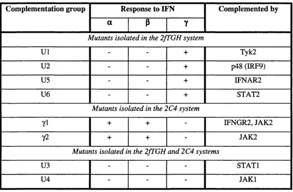
Table 1.3: Complementation groups of mutants in IFN signalling pathways... 42
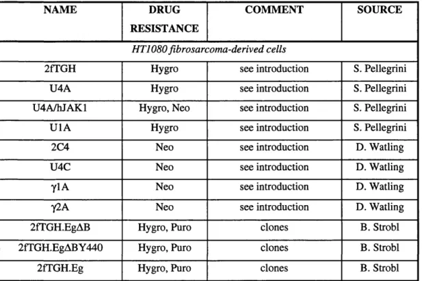
Table 2.1: Stable cell lines... 63
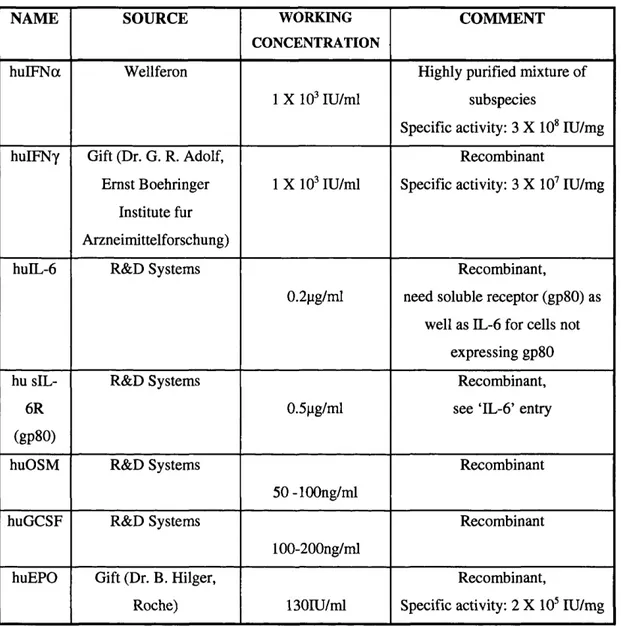
Table 2.2: Cytokines...65
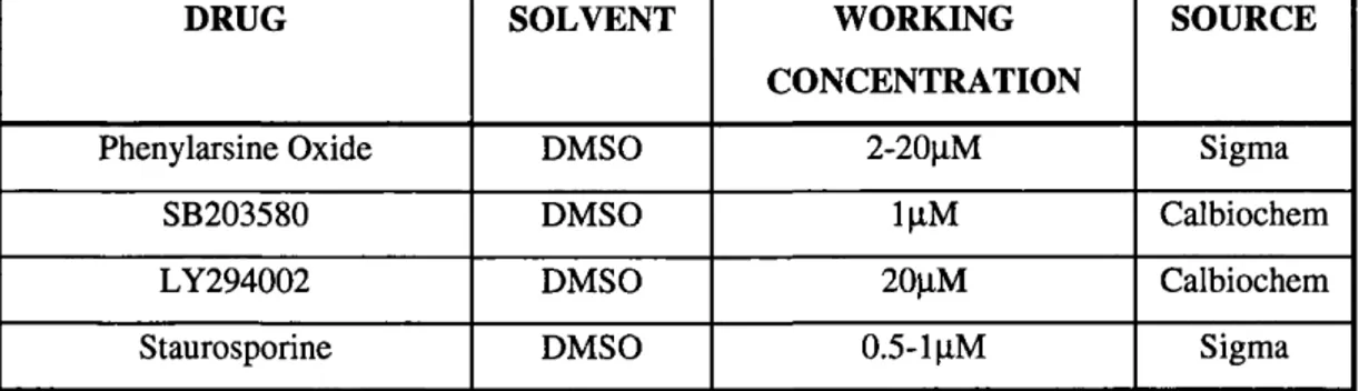
Table 2.3: Drugs... 66
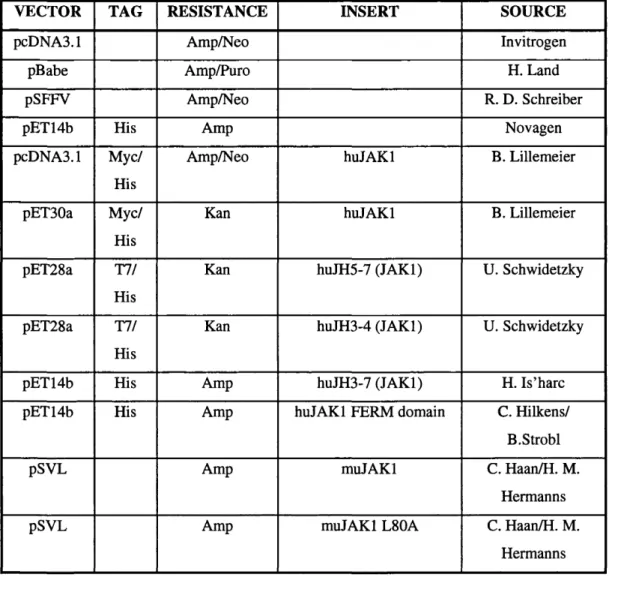
Table 2.4: Constructs...68
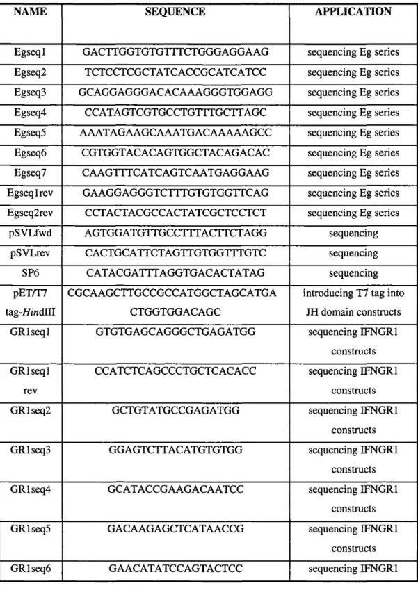
Table 2.5: Oligonucleotides and primers ...71
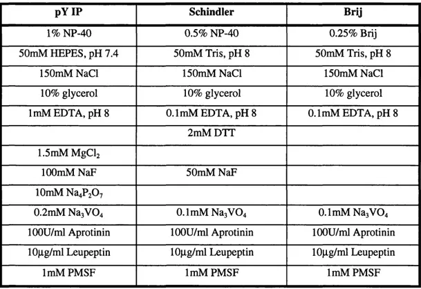
Table 2.6: Commonly used lysis buffers ...76
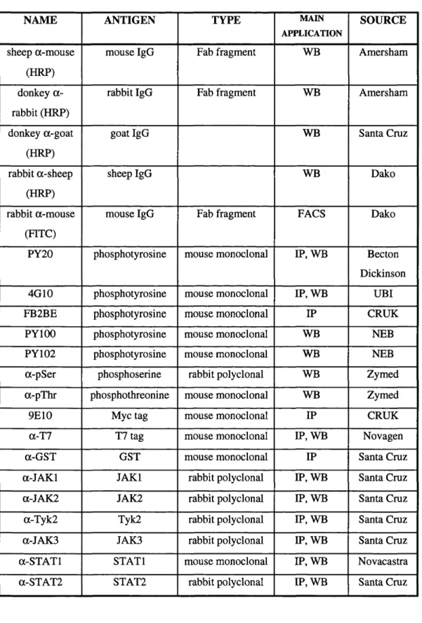
Table 2.7: Antibodies... 77
Table 2.8: Synthetic Peptides...80
Table 2.9: EMSA Probes,... 85
Table 2.10: RNase protection probes... 88
Table 3.1: GSM-responsive phosphotyrosine proteins affinity purified using GST-Grb2 SH2 domain, detected by anti-pY Western blot...93
Table 3.2: GSM-responsive high molecular weight proteins immunoprecipitated with anti-phosphotyrosine antibodies, detected by silver staining. ... 96
Table 3.3: IFNy-responsive tyrosine phosphorylated proteins identified by SMART profiling and anti-phosphotyrosine Western blot... 100
Table 4.1: Effect of mutations in the putative JAKl P-grasp region...131
Table 5.1: Modulation of IFNGRl cell surface expression by IFNy and GSM...165
ABBREVIATIONS
ID one dimensional
2D two dimensional
6TG 6-thioguanine
amp ampicillin
ATP adenosine triphosphate
bp base pair(s)
ESA bovine semm albumin
CHAPS 3-[3-Cholamidopropyl)-dimethylammonio]- 1-propanesulfonate
GUTA Class II transactivator
C/EBP CCAAT enhancer-binding protein
CEP CREE-binding protein
CIS cytokine inducible SH2-domain containing protein
CNTF ciliary neurotrophic factor
cpm counts per minute
CRUK Cancer Research UK
CT-1 cardiotrophin 1 ,
i^c tP d io tt^ c y n d iA c S DEPC diethyl pyrocarbonate
DMSG dimethyl sulphoxide
DOC deoxycholate
dsRNA double stranded RNA
DTT dithiothreitol
DMEM Dulbecco’s modified Eagle’s medium
e.c. extracellular
ECL enhanced chemiluminescence
EDTA ethylenediamine tetra-acetic acid
EOF epidermal growth factor
EGTA ethylene glycol-bis(p-aminoethylether)N,N,N’,N’-tetra-acetic acid
EMCV encephalomyocarditis virus
EMSA electrophoretic mobility shift assay
EPO erythropoietin
E S T
EVHl
FACS
PCS
FERM
F ix e
GAS GATE GFP GH G(M)-CSF gpl 30 gpt GR GTP GST h HAT HEPES HLA HPRT HRP hu hygro i.e. ICRF IFF IFN IFNAR IFNGR IL IP IPTG IRF 1RS 1/2
Enabled/VASP homology 1
fluorescence activated cell scanner
fetal calf serum
band 4.1, ezrin, radixin, moesin
fluorescein isothiocyanate
IFNy-activated sequence
fFNy-activated transcriptional element
green fluorescent protein
growth hormone
granulocyte (macrophage) colony stimulating factor
glycoprotein 130
guanine phosphoribosyltransferase
glucocorticoid receptor
guanosine triphosphate
glutathione-S-transferase
hour(s)
hypoxanthine, aminopterin, thymidine
N-2-hydroxyethypiperazine-N’ -2-ethanesulphonic acid
human leukocyte antigen
hypoxanthine phosphoribosyltransferase
horseradish peroxidase
human
hygromycin
intracellular
Imperial Cancer Research Fund
isoelectric focusing
interferon
IFNot/p receptor (subunit)
IFNy receptor (subunit)
interleukin
immunoprécipitation
isopropyl-p-D-thiogalactopyranoside
interferon regulatory factor
insulin receptor substrate 1/2
ISG interferon stimulated gene
ISGF3 interferon stimulated gene factor 3
ISRE interferon stimulated response element
lU international units
IVT in vitro translated
JAK Janus kinase/just another kinase
JH JAK homology
kan kanamycin
kb kilobase(s)
kD kilodalton(s)
LB Luria broth
LIF leukemia inhibitory factor
LTR long terminal repeat
LPS lipopolysaccharide
mAh monoclonal antibody
MAPK mitogen activated protein kinase
MHC major histocompatibility complex
min minute(s)
mu murine
neo neomycin/G418
NFS nuclear export sequence
NLS nuclear localisation sequence
NMR nuclear magnetic resonance
NP-40 Nonidet P40
OAc acetate
0-GlcNAc N-acetylglucosamine
OSM oncostatin M
PAGE polyacrylamide gel electrophoresis
PAO phenylarsine oxide
PBS phosphate buffered saline
PGR polymerase chain reaction
PDGF platelet derived growth factor
pfu plaque forming units
PI3K phosphatidylinositol 3-kinase
PIAS protein inhibitor of activated STAT
PIPES 1,4-piperazinediethanesulphonic acid
PMA phorbol 12-myristate 13-acetate
PMSF phenylmethylsulphonylfluoride
PRL prolactin
PTB phosphotyrosine binding domain
PTP protein tyrosine phosphatase
pY phosphotyrosine
puro puromycin
PVDF polyvinylidene difluoride
R receptor
RNAi RNA interference
rpm revolutions per minute
s second(s)
s e n ) severe combined immunodeficiency
SDS sodium dodecyl sulphate
SH2/3 Src homology (domain) 2/3
SHP//2- SH2 domain-containing tyrosine phosphatase //Z_
SIE c-Sis inducible element
sIL-6R soluble IL-6 receptor (gp80)
siRNA short interfering RNA
JC/fSihVC
SOCS suppressor of cytokine signalling ^
ssRNA single stranded RNA
STAT signal transducer and activator of transcription
STS staurosporine
TAE Tris-acetate/EDTA buffer
TBE Tris-borate/EDTA buffer
TBS Tris buffered saline
TEST Tris buffered saline with 0.1% Tween 20
TCA Trichloroacetic acid
TE lOmM Tris/HCl, ImM EDTA
TGE Tris-glycine/EDTA buffer
TGF transforming growth factor
TNF tumor necrosis factor
TPO thrombopoietin
Tris 2-amino-2(hydroxymethyl)-1,3-propandiol
tRNA transfer RNA
UTP uridine 5’-triphosphate
v/v volume for volume
w/v weight for volume
WB Western blot
CHAPTER 1: Introduction
CHAPTER 1: INTRODUCTION
Cytokines comprise a large family of soluble proteins that regulate cell growth,
differentiation and survival. Their production is generally transient and strictly controlled.
Individual cytokines often stimulate production of multiple other cytokines, which then act in
an autocrine or paracrine manner. Different cytokines may interact additively,
synergistically, or antagonistically. Thus, a complex network of intercellular mediators is
established and responses of cells and tissues are likely to always reflect the integration of
signals generated by exposure to combinations of cytokines. Cytokines initiate these signals
by binding to receptors at the cell surface, thereby engaging phosphotyrosine-dependent
signalling pathways that result in modulation of gene transcription. For the vast majority of
cytokines, signalling critically depends on components of the Janus Kinase (JAK)-Signal
Transducer and Activator of Transcription (STAT) pathway.
1.1 The Interferons
Interferons (IFNs) were originally identified as soluble factors possessing antiviral properties,
secreted by cells in response to viral infection (L indenm ann 1982). Two classes of
interferon exist: type I (IFNa, P, to) and type II (IFNy), each of which exerts its action
through a distinct receptor complex at the cell surface, resulting in the transcription of
e-hAl-overlapping sets of genes (reviewed in (Stark^l998)). In both cases, the biological effects
are mediated via the essential JAK-STAT pathway, in conjunction with additional signalling
pathways (Stark^l998). A range of stimuli, including challenge with virus or double
stranded RNA, bacterial lipopolysaccharide (LPS) and immunostimulatory cytokines can
elicit the production of different IFN subtypes in a cell type-specific manner (reviewed in
(Sen 2001)). IFNa is predominantly secreted by lymphocytes, monocytes and macrophages,
whilst IFNP is mainly produced by fibroblasts, as well as certain epithelial cells (S tark
1998). Major sources of IFNy are T-cells and natural killer (NK) cells (reviewed in (Boehm efed,
1997))
CHAPTER 1: Introduction
Type I IFNs constitute a family of highly related and evolutionarily conserved proteins,
encoded by a cluster of intronless genes located on the short arm of chromosome 9 in
humans. It is widely believed that all type I IFN genes derive from a common ancestral gene
Str\^
(reviewed in (V ilcek^996). Three subfamilies of type I IFN have been defined. In humans,
the al-IFNs consist of 14 functional genes and one pseudogene. The all-IFN group refers to
a single functional gene, IFNto, together with five pseudogenes. The third group comprises
IFNP alone. With the exception of IFNco, which has 172 amino acid residues, all secreted
forms of type I IFN are proteins of 165-166 amino acids. All type I IFNs interact with the
same receptor complex as monomers (Sen 2001), however, IFNP engages the receptor in a
different manner to IFNa, presumably accounting for the distinct biological properties of
IFNP (Abramovich^ 9 9 6 ) (Platanias, U ddin et al. 1996) (Rani 1996). Quantitative and
possibly qualitative differences in the responses stimulated by the various IFNa subtypes
have been noted (Hilkens, CMU., and Schlaak, JF., manuscript in preparation) although the
biological significance of the multiple IFNa subtypes remains enigmatic.
IFNy is the only known ligand of the type II receptor complex and interacts with the receptor
as a homodimer. Despite the functional similarity, IFNy bears little structural resemblance to
type I IFN. The IFNy protein comprises 143 amino acids and is encoded by a single copy
gene that contains three introns and localises to chromosome 12 in humans (reviewed in
(B oehn^l997) and (V ilcek^996)).
1.2 Biological responses to the interferons
1.2.1 Antiviral responses
Exposure to IFN confers upon cells an antiviral state in which the replication of a wide
spectrum of viruses is inhibited. Indeed, the interferon system is considered to be the first
line of defence against viral infection in mammals. Type I IFN is rapidly induced in response
to infection by virus and can act to potently limit the development and spread of viral
infection. This is reflected by the extreme susceptibility of the type I IFN receptor mouse
knockout to infection by viral pathogens (van den B ro ek ^ 9 9 5 ). The antiviral response is
CHAPTER 1: Introduction
protection to cells against viral infection, it is not directly induced by virus and is likely to
play a more fundamental immunomodulatory role (van den B ro e^ l9 9 5 ).
Multiple proteins contribute towards establishment of the IFN-induced antiviral state and the
molecular mechanisms involved are not yet fully understood. However, major players in the
antiviral response include the dsRNA-dependent protein kinase (PKR), dsRNA-dependent
2’-5’ oligoadenylate synthetase (2-5 OAS) and Mx proteins (reviewed in (Starl^998)). PKR is
a serine/threonine kinase and likely exerts its antiviral effects via phosphorylation and
consequent inactivation of the translation initiation factor, eIF2a. In addition, apoptosis may
cJ'ul.
be involved in the antiviral activity of PKR ( J a g u s ^ 9 9 9 ) . The 2-5 OAS family are
stimulated by dsRNA to catalyse the production of 2’-5’ oligoadenylates that activate RNase
L, leading to cleavage of ssRNA and inhibition of protein synthesis (S ilv erm an^997). Mx
proteins are GTPases and may interfere with intracellular trafficking and/or transcriptional
activity of viral ribonucleotide proteins (S trand en^9 93 ) although the basis of their antiviral
activity remains poorly understood. The existence of additional IFN-induced antiviral
pathways has been confirmed through studies in triple knockout mice deficient in PKR, (A 4/
RNase L and Mx (Zhoi^l999).
1.2.2 Antiproliferative/apoptotic responses
Inhibition of cell proliferation and modulation of apoptosis are related but distinct properties
of IFNs: IFN-inducible proteins have been identified that are selectively involved in
apoptosis with no influence on proliferation (D eiss^l995). These IFN-mediated responses
are obviously of considerable significance for the suppression of both tumours and infections
(Sangfelt^O O O). IFNs have long been known to inhibit the growth of many cell types
(Balkwill.1'978), (Lin 1986) and a requirement for fully transcriptionally active STATl for
^ (A /,
the mediation of this effect has now been demonstrated (B rom berg ^1996). For IFNa,
growth inhibition may involve down regulation of cyclin D and cdc25A (Tiefenbrin^l 996),
whilst increased levels of the cyclin-dependent kinase inhibitor, correlate with
the inhibition of^rowth of gliobj^om a and epidermal carcinoma cell lines in response to
IFNy (Kom insky^ 1998) (Chin^ 9 9 6 ) . However, regulation of p21 expression by p53 is
CHAPTER 1: Introduction
dominant over the effects of IFNy such that p21 is only induced by IFNy in tumour cell lines
deficient for wild type p53. Both IFNa/p and IFNy may also mediate anti-growth properties
through the suppression of c-myc, a key regulator of proliferation (Resnitzsky 1991)
(Ram ana 2000). It should also be noted that IFNy has been reported to exert a growth
stimulatory effect in certain cell systems (Caux 1992).
1.2.3. Immunomodulatory responses
IFNs are critical regulators of the immune system. Whilst the role of type I IFN in
immunomodulation seems largely restricted to enhancing the adaptive immune response
against viral infection, type II IFN has various profound immunoregulatory effects (reviewed
in (B o e h m ^ 1 9 9 7 )). Both type I and II IFN promote surface expression of major
histocompatability complex (MHC) class I molecules which stimulates development of CD8+
T-cell responses (reviewed in (S tark^998)). IFNy can also induce expression of MHC class
II at the cell surface via induction of the transcription factor, CIITA, and indeed MHC class II
induction is considered to be one of the defining characteristics of an IFNy-like response
(Boehm^ 9 7 ) (Stark^l998). IFNy also contributes to promotion of antigen processing by
upregulating subunits of the proteosome, a multienzyme complex which generates peptides
^ A '• th
for presentation by MHC class I proteins (Boehm^ 9 9 7 ) ( S ta r ÿ 9 9 8 ) . IFNy, together with
IL-12, drives naïve T-cells towards a Thi phenotype, promoting an inflammatory response
(B oehm ^ 9 9 7 ) (S ta rk ^ 9 9 8 ) . IFNy also plays a central role in generating activated
macrophages, inducing the expression of genes such as indoleamine 2,3-dioxygenase (IDO)
and inducible nitric oxide synthase (iNOS; induced by IFNy in murine but not human
eJ~A(.
macrophages) (S tark ^ 9 9 8 ). The critical function of IFNy in the response to intracellular
parasites is demonstrated by the fact that patients with naturally occurring IFNy receptor
deficiencies are exquisitely sensitive to infection by a number of microbial pathogens,
CHAPTER 1: Introduction
1.3.1 The IL-6 family of cytokines
The IL-6 cytokine family comprises a group of ligands that share the common property of
utilising the gpl30 receptor subunit for signalling, either alone or combination with other
receptor subunits. As a result, they elicit an array of overlapping responses that collectively
play vital roles in the regulation of a broad spectrum of biological processes in vivo. There are currently nine members of the family: IL-6; IL-11; Oncostatin M (OSM); Leukemia
Inhibitory Factor (LIF); Ciliary Neurotrophic factor (CNTF); Cardiotrophin-1 (CT-1);
Cardiotrophin-Like Cytokine/Novel Neurotrophin-1 (CLC/NN-1) and two viral IL-6
homologues. All a ^ ] ^ 0 - 2 0 0 residues long but overall possess only -20% amino acid
homology (Bravo^ 0 0 0 ) . Gene ablation studies in mice have revealed unique phenotypes
for each member of the family, indicative of the specificity of responses induced by each of
the IL-6 type cytokines, IL-6 deficient mice are impaired in acute-phase and antiviral
responses (K opf 1994) (Poli 1994), Deletion of the LIF gene produces female infertility as
a consequence of embryo implantation defects (S te w a rt^ 992), It is possible that the
unexpectedly mild phenotypes of mice lacking a single IL-6-type cytokine may reflect the
ability of other family members to compensate, at least partially, for absence of a particular
family member. By contrast, knockout mice foi^aiy of the major components of IL-6 family
cytokine signal transduction (gpl30 (Y oshida 1996), LIFR (W are 1995), JAKl (Rodig
1998), STAT3 (Takeda^l997)) have embryonic or perinatal lethal phenotypes.
1.3.2 IL-6
IL-6 was initially identified as an inducer of immunoglobulin production in activated B cells
(reviewed in (Taga^ 9 9 7 )), It is now apparent that IL-6 is a pleiotropic cytokine with a wide
range of biological functions in cells of many lineages. The mature IL-6 protein varies /At
between 21 and 28kD in size, depending or^lycosylation and phosphorylation state (Hirano
1991), IL-6 production can be induced in many cell types by a range of stimuli that include
ehA.1-bacterial LPS, IL-1, TNFa, TGFp and OSM (A k ira ^ 9 9 3 ), The ability of such stimuli to
elicit IL-6 production reflects the fact that one of the key roles of IL-6 is to mediate an acute
CHAPTER 1: Introduction
phase response to tissue damage and infection. The biological activities of IL-6
are summarised in Table 1.1.
1.3.3 Oncostatin M
OSM was originally purified from conditioned media of PMA-treated U937 cells and derives
its name from its capacity to inhibit proliferation of several cancer cell lines whilst
stimulating growth of normal fibroblasts
(Zarling^l986).
The protein is secreted as a 28kDmonomeric glycoprotein and shows 27% sequence identity with LIF (reviewed in
(Gomez-Lechon 1999)).
The similarities in gene structure, location and expression of these twocytokines suggest that OSM and LIF arose through a gene duplication; they can also signal
through the same receptor complex
(Ros^l991) (Mosley^ 996).
OSM mRNA is expressedin various cell types, including activated T cells, endothelial cells, hepatocytes, macrophages
and the embryonic aorta-gonad-mesenephros (AGM) region (site of primitive hematopoietic
development) and is induced as an early response gene in myeloid and lymphoid cells by IL-
2, IL-3 or GM-CSF treatment
(Yoshimura^l996).
OSM has multiple, cell type-specificeffects, summarised in Table 1.1, including activities in hematopoiesis, bone remodelling,
inflammation, neuronal differentiation and regulation of cell growth (reviewed in
(Gomez-Lechon 1999)
and(Gran^909)).
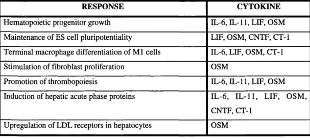
Table 1.1: Major biological responses to IL-6 family cytokines
RESPONSE CYTOKINE
Hematopoietic progenitor growth IL-6, IL-11,LIF, OSM
Maintenance of ES cell pluripotentiality LIF, OSM, CNTF, CT-1
Terminal macrophage differentiation of M l cells IL-6, LIF, OSM, CT-1
Stimulation of fibroblast proliferation OSM
Promotion of thrombopoiesis IL-6, IL-11,LIF, OSM
Induction of hepatic acute phase proteins IL-6, IL-11, LIF, OSM,
CNTF, CT-1
CHAPTER 1: Introduction
RESPONSE CYTOKINE
Upregulation of adhesion molecules OSM, IL-6, IL-11,-LIF
Effects on extracellular matrix OSM
Inhibition of lipoprotein lipase, resulting in fat depletion IL-6, IL-11, LIF, OSM
Effects on bone remodelling and osteoclast development IL-6, IL-11, LIF, OSM
Increase proliferation/differentiation of T cells 11-6, LIF, OSM
Increase proliferation/differentiation of vascular endothelial
cells
OSM
Induction of neuronal survival/differentiation IL-6, IL-11, LIF, OSM,
CNTF, CT-1
Induction of cytokines (IL-6, G-CSF, GM-CSF, LIF) OSM
1.4 The JAK-STAT signalling pathway
1.4.1 Overview of JAK-STAT signalling
A generalised scheme for JAK-STAT signalling in response to IFNy and IL-6 family
cytokine stimulation is depicted in Figure 1. JAKs are protein tyrosine kinases that
constitutively associate with the intracellular domains of cytokine receptors (reviewed in
(R ane and R eddy 2000)). Ligand binding results in a conformational change at the
receptor, bringing the associated JAKs into juxtaposition and permitting their activation,
presumably by auto- and transphosphorylation (reviewed in (Stark^998)). Activated JAKs
phosphorylate tyrosine residues in the receptor cytoplasmic tail, which then serve as docking
sites for STAT molecules. STATs, bound to the receptor phosphotyrosines via their SH2
domains, themselves become substrates for the JAKs, Tyrosine phosphorylated STATs
dissociate from the receptor and form dimers through reciprocal phosphotyrosine-SH2
e-i-al-domain interactions in an affinity-driven process (G reen lu n d ^ l9 9 5 ). Activated STAT
dimers translocate to the nucleus where they bind DNA and modulate transcription, either
alone or in concert with other transcription factors and co-activators (reviewed in
(C h atterje e-K ish o re ^ 0 0 ^ ) . The activation of the JAK-STAT pathway is generally
CHAPTER 1: Introduction
transient and STATs appear to be dephosphorylated prior to export from the nucleus. This is
discussed further in section 1.15.
This represents the simplest model of JAK-STAT signalling - there are of course a number of
permutations on this theme. As well as being activated by most cytokines, there are several
instances of JAKs and STATs participating in signalling through growth factor receptors and
examples are given in Table 1.2. In addition, studies in the lower eukaryote, Dictyostelium,
identified cAMP, which signals through a serpentine receptor, as an activator of Dd-STAT.
AchruHn
The paradigm w a^,« /f % mammalian cells with the observation that angiotensin II
binding to the AT, serpentine receptor rapidly activates JAK2, STATl, STAT3 and STAT5 in
an apparently heterotrimeric G protein-independent manner (reviewed in (W illiam s 1999)).
There is controversy over whether non-activated STATs ever exist as monomers in a
physiological context and some studies suggest that prior to activation, STATs are
sequestered in high molecular weight ‘statosome’ complexes (N d ub uisi^l99 9). There are
also data implying the existence of alternative mechanisms of STAT recruitment to and
activation by, ligand-bound receptor complexes. For example, STAT5 is reported to
associate with JAK2 and can thus be activated directly by JAK2 following GH treatment,
ey
*it-without binding to receptor phosphotyrosines ( F u jita n i^ 9 9 7 ) . STAT2, meanwhile, is
preassociated with the IFN a/p receptor and STATl activation in response to type I IFN
appears dependent both on prior activation of STAT2 and a constitutive interaction with the
adaptor protein, RACKl (U sacheva, Sm ith et al. 2001). For IFNy, the Fanconi anemia
protein, FANCC, is reported to bind non-phosphorylated STATl and mediate its recruitment
ihal.
to the receptor and consequent phosphorylation (Pang^2000). JAKs are not necessarily
required for activation of STATs by growth factor receptors and may phosphorylate
alternative substrates in these scenarios. For both EGF and PDGF, the receptor itself and/or
■e/- nl• A Src family tyrosine kinases may effect STAT phosphorylation (reviewed in (B ris c o e ^ 9 9 ^ ).
Furthermore, growth factor-induced tyrosine and serine phosphorylation of STAT3 was
recently demonstrated to occur through direct binding of STAT3 to the Racl GTPase (S im o n ^
CHAPTER 1: Introduction
oxygen species generated, for example, in response to growth factors (Simon, Rai et al.
1998).
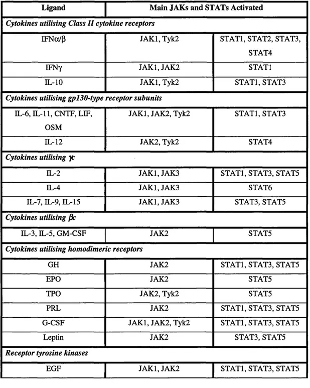
Table 1.2: Activation of JAKs and STATs by cytokines and growth factors
Ligand Main JAKs and STATs Activated
Cytokines utilising Class II cytokine receptors
IFNo(/p JAKl, Tyk2 STATl, STAT2, STAT3,
STAT4
IFNy JAKl, JAK2 STATl
IL-10 JAKl, Tyk2 STATl, STAT3
Cytokines utilising gpI30-type receptor subunits
IL-6, XL-11, CNTF, LIF,
OSM
JAKl, JAK2, Tyk2 STATl, STAT3
DL-12 JAK2, Tyk2 STAT4
Cytokines utilising yc
IL-2 JAKl, JAK3 STATl, STAT3, STAT5
IL-4 JAKl, JAK3 STAT6
IL-7, IL-9, IL-15 JAKl, JAK3 STAT3, STAT5
Cytokines utilising fie
IL-3, IL-5, GM-CSF JAK2 STATS
Cytokines utilising homodimeric receptors
GH JAK2 STATl, STAT3, STATS
EPO JAK2 STATS
TPO JAK2, Tyk2 STATS
PRL JAK2 STATl, STAT3, STATS
G-CSF JAKl, JAK2, Tyk2 STATl, STAT3, STATS
Leptin JAK2 STAT3, STATS
Receptor tyrosine kinases
EGF JAKl, JAK2 STATl, STAT3, STATS
CHAPTER 1: Introduction
Ligand Main JAKs and STATs Activated
PDGF JAKl, JAK2 STATl, STATS, STAT5
Insulin JAK2 STATl, STAT5
Ligands utilising serpentine receptors
Angiotensin H JAK2 STATl, STATS, STAT5
1.4.2 Cytokine receptor complexes
Cytokine receptors are transmembrane proteins lacking intrinsic enzymatic activity. They
4,3, c
can be divided into two classes on the basis of their extracellular domain (Bazan 199^ .
Class I receptors are defined by the presence of at least one cytokine-binding module (CBM),
comprising two fibronectin-type-IH-like domains, of which the N-terminal domain includes a
set of four conserved cysteines and the C-terminal domain a WSXWS motif. The majority of
cytokine receptors fall into this category, including receptors for IL-2, IL-3, IL-4, IL-5, IL-6
type cytokines, IL-7, IL-9, IL-11, IL-12, IL-15, EPO, PRL, GH, G-CSF, GM-CSF and leptin
(Bazan 1990) . Class II receptors are more divergent with only limited homology to the WSXWS motif, but share overall structural features (Bazan 1990;/
Receptors for IFNo/p, IFNy and IL-10 are the prototypical members of this class.
The historical model of activation of signalling through cytokine receptors entailed
ligand-induced oligomerisation of monomeric receptor subunits leading to trans- and
autophosphorylation of associated JAKs. However, structural studies of the EPOR now
suggest that the unliganded receptor exists as a pre-formed dimer, but in a configuration that
$hal-prevents JAK activation (Remy 1999). Ligand-independent oligomerisation of the EPOR appears to be mediated by the transmembrane domain (Constantinescu^OO l). Ligand binding is proposed to cause a conformational change producing a structural arrangement
permissive for JAK activation (Remy J^999). Studies using EPO mimetic peptides and bivalent anti-EPOR antibodies support the notion that fixing the receptor in certain
orientations may render the complex incapable of signalling (reviewed in (Ballinge^l998)).
CHAPTER 1: Introduction
receptors, this model raises intriguing questions about how subtle differences in orientation within the receptor extracellular domain can affect intracellular signalling. This is particularly striking in view of the apparent flexibility of receptor transmembrane and cytoplasmic domains. NMR studies of the in vitro translated intracellular domain of gpl30, for example, indicated a random coil conformation in solution (McDonald, N., personal communication). The functionality of chimeric receptors containing extracellular domains of one receptor fused to transmembrane and cytoplasmic portions of a different receptor further testifies to the plasticity of cytokine receptors (Strobl, Arulampalam et al. 2001).
1.4.3 Receptor cross talk
An emerging theme in signal transduction is the existence of molecular communication
between heterologous receptors, sometimes termed ‘cross talk’ or ‘receptor transactivation’.
The EGFR has long been recognised to be targetted by many other signalling cascades apart
a/.
from those triggered by EGF itself (reviewed in
(Zwick^999)).
Indeed, the EGFR was reported to be directly tyrosine phosphorylated by JAK2 following GH stimulation, leading tostimulation of MAPK activity in liver through recruitment of Grb2
(Yamauchi^l997).
Meanwhile, ErbB2 was shown to complex with gplBO in prostate carcinoma cells in an IL-6-
dependent manner. IL-6 treatment resulted in tyrosine phosphorylation of ErbB2 and
inhibition of ErbB2 activity severely impaired IL-6-mediated MAPK activation
(QiUyI998).
Another example of receptor cross talk is provided by the observation that insulin can induce
tyrosine phosphorylation of JAK2 and the leptin receptor which, in the presence of leptin,
enhances leptin-induced STAT3 phosphorylation and activation
(Carvalheira ^2001 ).
Communication between cytokine receptors may also occur, as evidenced by the association
between IFNARl and both IFNGR2 and gpl30, reported to be necessary for full responses to
IFNy ^od IL-6 respectively, at least in a murine system
(Takaoka^OOO) (Mitani^OOI).
The latter phenomena appear to be dependent on membrane structure in as much as the
receptor subunits cofractionate with caveolar membrane domain components and treatment
with a lipid raft disrupting agent reversibly and dose-dependently inhibits signalling. These
data reinforce the likely complexity of receptor organisation at the membrane and emphasise
the fact that the mechanisms of activation and modulation of receptor complexes are still only
crudely understood.
CHAPTER 1: Introduction
1.4.4 The JAKs
The mammalian JAK family of protein tyrosine kinases comprises four members: JAKl;
JAK2; JAK3 and T y k ^ T y k 2 was identified iniually by screening a T-cell library with a
fragment of c-fms containing the kinase domain under conditions permitting low-stringency
hybridisation. The remaining JAKs were subsequently cloned by a PCR-based strategy
utilising degenerate oligonucleotides corresponding to conserved motifs found within
tyrosine kinase catalytic domains. A Drosophila JAK has also been cloned and was
designated ‘Hopscotch’ due to the fact that flies mutated in this gene exhibit defects in
expression patterns of pair-rule and segment-polarity genes. The name ‘Janus kinase’ derives
from the defining feature of this class of tyrosine kinases: the existence of adjacent kinase and
pseudokinase domains at the C-terminus of the molecule - ‘Janus’ being the two-faced
Roman god of gates and doorw ay^
JAKs are large molecules with molecular weights in the range of 120-140kD. JAKs 1 and 2
and Tyk2 are ubiquitously expressed, while JAK3 expression is largely restricted to
hematopoietic cells, vascular smooth muscle cells and endothelium (reviewed in (Leonard and O'Shea 1998)). Mice deficient in each of the JAKs have now been generated. JAKl knockouts fail to nurse and die perinatally, apparently due to a neurological defect since
lethality is rescued by selective expression of JAKl from a neural-specific promoter (Rodig 1998) (Schreibe^ 2000). The neurological defect is presumed to result from the inability of these mice to transduce signals from the IL-6 family of cytokines that utilise the gpl30
receptor subunit. JAKl-deficient mice also have severe combined immunodeficiency (SCID)
due to the abrogation of signalling through the common y chain, yc. In vitro studies on cells from JAKl knockouts also confirm the indispensable role of JAKl in mediating signals from
IFNs.
Mice deficient in JAK2 die during ^ ^ y o g e n e s i s d u ^ t ^ the absence of definitive
erythropoiesis in the fetal liver (Parganas^998) (Neubaue^l 998). This likely reflects the failure to transduce signals through the EPOR as the same phenotype is observed in EPOR
CHAPTER 1: Introduction
IFNy. JAK3 deficiency results in SCID and the phenotype is indistinguishable from that of
mice lacking yc (Baird J 9 9 8 ) (Suzuki^OOO).
Tyk2 knockouts were only recently reported and, in conflict with data obtained from a Tyk2
deficient human cell line (Gauzzi,1997), the absence of Tyk2 had minimal impact on IFN
^ ^ A./. a/.
signalling and receptor expression (Shim oda^2000) (Karaghiosoff^ 2000). Overall, the
phenotype was surprisingly mild, with some impairment of IL-12 signalling but largely
normal responses to IL-6, IL-10 and type I IFN. Tyk2 may therefore play a more redundant,
modulatory-type role in cytokine signalling than the other JAKs, however the basis for the
discrepancy between the two experimental systems is yet to be resolved and may at least in
part reflect species-specific differences in the function of Tyk2.
The JAKs have been subdivided into seven so-called ‘JAK homology’ (JH) regions on the
basis of sequence homology between the different JAKs, where JHl is ± ^ in a s e domain and
JH2 corresponds to the pseudokinase domain (Fig. 1.2) (Z iem ieck i^ l9 9 4 ). There are a
number of highly conserved features of the JHl domain that impact on catalytic activity.
Mutation of the ATP binding loop in subdomain II of the kinase domain abrogates kinase
activity (Briscoe, Rogers et al. 1 9 9 ^ . In addition, the DFG triplet in subdomain VII
appears critical for ATP binding and mutation of D1020 of this motif in human JAKl
abolishes catalytic activity (Briscoe, Rogers et al. 1 9 9 ^ It has further been established
that the first tyrosine in the EYY motif (Y1007 in human JAK2) has to be phosphorylated for
JAK2 or Tyk2 to be catalytically competent, whilst phospl^rylation of the second tyrosine in
the motif is prerequisite for full enzymatic activity (Gauzzi^l996) (Feng^997).
The JH2 (pseudokinase) domain of the JAKs strongly resembles a kinase domain but lacks
certain critical residues necessary for catalytic activity (W ilk s^ 1 9 9 1 ). Instead, the
pseudokinase domain is implicated in regulation of JAK catalytic activity. A point mutation
in the JH2 domain of the Drosophila JAK, Hopscotch, results in hyperactivation of the JAK and consequent hematopoietic neoplasia. The equivalent mutation in JAK2 also generates a
hyperactivated molecule (Luo, Rose et al. 1997). Similarly, deletion of the pseudokinase
domain of JAK2 deregulated signal transduction from the IFNy receptor through ligand-
independent activation of STATs and for both JAK2 and JAK3, overexpression of JH2
CHAPTER 1: Introduction
inhibited the catalytic activity of the isolated JHl domain in trans (Saharinen, Takaluoma et al. 2000) (CherylOOO). However, other studies report that deletion of JH2 is inactivating for both Tyk2 and Hopscotch (V elazquez^995) (Luo, Rose et al. 1997), suggesting that regulation of JHl catalytic activity by JH2 may be complex and may differ between the
JAKs.
Overall, regulation of JAK activity remains poorly understood. Although juxtaposition of
JAKs through ligand-induced receptor rearrangements seems to effect their activation, it is
likely that mechanisms other than spatial separation contribute to preserving JAKs in an
inactive state. In fact, it is gradually becoming clear that JAKs are subject to post-
translational modifications aside from tyrosine phosphorylation. Serine/threonine
phosphorylation of JAKl has recently been observed to have complex effects on IFN
signalling (Lillemeier, B. P., Watling, D. and Kerr, I. M., manuscript in preparation). JAK
activity is also redox regulated such that the oxidised forms of JAK lack autokinase activity
whilst the reduced forms are most active; interconversion between the two forms can occur
readily without influencing the preexisting tyrosine phosphorylation status (D uhe^998). It is reasonable to expect further modifications of JAKs which can potentially regulate their
activity or localisation to be described.
There is a paucity of structural information about the JAKs since the JAKs have proved
somewhat refractory to crystallization. However, various sequence alignment and secondary
and tertiary structure prediction algorithms have been applied to the JAKs, leading to the
identification of a putative SH2 domain in the JH3-4 region (Kampa^OOO). This SH2 fold is predicted for all four JAKs, although in Tyk2 a histidine residue replaces the conserved
arginine at the critical phosphotyrosine binding position, pB5, suggesting that at least in
Tyk2, the SH2 domain fold may have acquired an alternative binding specificity (A l-a/*
Lazikani^OOl). Irrespective, the biological significance of the predicted SH2 fold awaits rigorous functional validation.
Sequence analysis has also recently led to the suggestion that the JAKs contain within their
N-termini divergent ‘PERM’ domains (‘Band Four point one, Ezrin, Radixin, Moesin’; also
CHAPTER 1: Introduction
The PERM domain represents a structural module of approximately 300-400 residues,
subdivided into 19 conserved hydrophobic regions, that occurs in many cytoskeletal proteins
as well as the tumour suppressor, merlin, where it serves to couple these proteins to the
plasma membrane through protein-protein interactions. A second important function
attributed to the PERM domain in the ERM proteins and merlin is conformational regulation,
whereby intramolecular interactions between the PERM and tail domains obstruct
associations with other binding partners (Pearson^OOO).
The crystal structure of the moesin PERM domain reveals a clover-shaped structure of three
lobes, termed P I, P2 and P3 in moesin (Pearson^OOO). PI has a p-grasp-like fold,
reminiscent of the structure of ubiquitin; P2 is predominantly a-helical, bearing a
resemblance to the structure of acyl-coA binding protein; P3 comprises a P-sandwich
showing fold similarity to PH domains, PTB domains and EVHl domains, implicated in
binding lipids, phosphotyrosine-containing motifs and polyproline motifs, respectively. The
PH, PTB and EVHl domains are all based on a common structural module, but utilise
different surfaces/residues to interact with their various ligands. In theory, it would be
possible to use all of the interaction surfaces at the same time and indeed in moesin, the PH
and PTB domains are simultaneously engaged. This gives the potential for P3 to interact
with several different types of ligand and suggests that it could be important both for
mediating protein-protein and protein-lipid interactions.
Different cytokine receptors recruit different JAKs and specific physical association of JAKs
with receptors is critical for ligand-mediated responses. The JAK N-terminus has been
shown to be necessary for receptor interaction in a number of systems. For example, a Tyk2
fragment containing JH7 and part of JH6 was able to interact with a GST fusion protein
c o m p ris i^ ^ e IFNaP receptor 1 (IFNARl) intracellular domain in an in vitro binding assay (R ic h te r^ 9 9 8 ). In the case of JAK3 interaction with the common yc chain of the IL-2
family cytokine receptors, a minimal receptor interaction region comprising JH7 and JH6 was
defined by coimmunoprecipitation of coexpressed tagged JAK3 fragments and yc from 293T
cells. Chimeric constructs of JH7 and JH6 domains of JAK3 in a JAKl background were
also able to coimmunoprecipitate with yc in the 293T overexpression system and restore
CHAPTER 1: Introduction
signalling downstream of IL-2 in IL-2 receptor expressing, JAK3 negative fibroblasts
(Cacalano, Migone et al. 1999).
These data would suggest that the specificity of the JAKs is not determined by the substrate
specificity of their kinase domains. Consistent with this, a JAK1/JAK2 chimera with the full
N-terminus of JAKl fused to the kinase and pseudokinase domains of JAK2 was able to
reconstitute EFNa and y signalling in cell lines deficient in JAKl (Kohlhuber, Rogers et al.
1997). Thus, the specificity of the JAKs in cytokine signalling may lie primarily at the level
of recruitment to specific receptors and/or their ability to interact with different signalling
intermediates via their N-terminal domains. However, the structural basis for the specificity
underlying JAK-receptor interactions is not currently understood.
1.4.5 STATs
STAT transcription factors were first identified as DNA-binding proteins involved in IFN-
regulated gene expression through biochemical purification techniques (Darnell 1997). A number of related factors were subsequently identified either by isolation of cytokine-induced
DNA-binding activities, or by screening ESTs for homologous genes. The mammalian
family of STATs now comprises seven members: STATs 1-4; STAT5a; STA5b; STAT6.
STATs have also been identified in Drosophila(Yan, Small et al. 1 9 9 ^ and Dictyostelium (Kawata^l997) and related molecules may exist in higher plants (P en g ^ 9 9 9 ). STATs range in size from 84-113kD and are widely expressed, with the exception of STAT4, whose
expression is predominantly restricted to myeloid cells and testis (reviewed in (Leonard and
O'Shea 1998)).
Knockout mice have now been generated for all the STATs manifesting strikingly specific
phenotypes, indicative of the non-redundant roles of the different STATs. Despite the fact
that numerous cytokines and growth factors are reported to activate STATl (e.g. IFNs, IL-2,
IL-6 and EGF), STATl deficient mice develop normally but display a marked susceptibility
to infection by viruses and other pathogens (Durbin^ 1996) (M e ra z J9 9 6 ). This restricted
phenotype can be entirely accounted for by the abrogation of IFN-mediated signalling in
CHAPTER 1: Introduction
ti-4
/
increased rate of tumor development in STATl-deficient mice ( K a p la n ^ 998). STAT2
deficient mice are also highly susceptible to viral infection, consistent with the fact that the
only known cytokines that stimulate STAT2 activation are the type I IFNs (Parl^OOO).
STAT3 knockout mice are embryonic lethal, presumably due to the indispensable role of
tJ- A./
STAT3 in gpl30-mediated signalling (Takeda. 1997). Conditional knockouts are being generated to further analyse the physiological roles of STAT3 (Akira^OOO). Targetting of STAT3 in myeloid cells exacerbated the inflammatory response and was lethal, apparently
due to loss of IL-10 signalling. STAT3 activation also seems to be required for G-CSF-
dependent proliferation and granulocytic differentiation (M cLem ore^OO l). Mammary gland-specific deletion of STAT3 delays involution following weaning, associated with
decreased apoptosis, whilst in kératinocytes, absence of STAT3 severely impeded wound
healing due to impaired growth factor-dependent epidermal cell migration (Sano^999). On the basis of the conditional knockouts of STAT3 reported thus far, it seems that STAT3
cannot be considered a generic regulator of apoptosis or cell cycle progression, but rather has
specific effects in each lineage.
STAT4 deficient mice display defects in Thi differentiation in accordance with the fact that
IL-12 is a major activator of STAT4. STAT5a/b double knockouts have impaired responses
to growth hormone and prolactin and separate STATSa or STAT5b knockouts exhibit distinct
phenotypes that in n a te certain non-redundant functions for these highly related proteins
(LiuJ^997) (Udy^l997) (Teglund^998). Knockouts of STAT6 fail to develop Th
2immune
responses and are deficient in most (but not all) IL-4-associated physiological functions
'Cj' tirA/
(S h im o d ^ 9 9 6 ) (T ak ed ^ 996).
The elucidation of the crystal structures of STAT-DNA complexes has facilitated
understanding of STAT function (Fig. 1.3). STATs bind to DNA as dimers, forming a
clamp-like^ructure in v^ich the phosphotyrosine-SH2 domain interactions form the hinge
(Becker^l998) (Chen^998). The N-terminal portion of the STAT appears to participate in associations with adjacent STAT dimers bound to tandem STAT-binding motifs in promoters
\(j'
and may also interact with histone acetylases (Zhang ^ 9 9 6 ) (Horvai, Xu et al. 1997).
CHAPTER 1: Introduction
Between the N-terminal portion and the central DNA-binding domain, there is a coiled-coil
domain implicated in protein-protein interactions. This domain likely mediates binding to
other transcription factors and coactivators, for example, p48, c-jun, Nmi, glucocorticoid
receptor and p300/CBP.
C-terminal to the DNA-binding domain there is an alpha-helical linker region, mutation of
which produces a molecule that can be tyrosine phosphorylated, dimerise, translocate to the
nucleus and bind DNA but^s incompetent for transactivation (Y a n ^ l9 9 9 ). The linker is
followed by a classical SH2 domain and highly conserved tyrosine phosphorylation site
(Y701 in STATl, Y705 in STAT3). The SH2 domain mediates recruitment of the STAT to
the activated receptor complex via phosphotyrosine residues in the receptor intracellular
domain. Reciprocal SH2 domain-phosphotyrosine interactions are also responsible for
dimérisation of STATs, required for both nuclear translocation and appropriate binding to
DNA at dyad symmetrical binding sites.
The C-terminus of the STAT molecule contains the transactivation domain and displays the
greatest divergence between the different STATs. With the exception of STATs 2 and 6, this
domain includes a conserved serine residue (S727 in STATs 1 and 3), phosphorylation of
which appears necessary for full transcriptional activation from at least a subset of STAT
responsive promoters (see section 1.4.6) (reviewed in (Decker^OOO)). This C-terminal
domain has also been implicated in interactions with histone acetylases (B hattacharya^ ^ ' 1996) (Horvai, X u et al. 1997) and at least in the case of STATl has been shown to be
necessary for association with MCM proteins in a serine phosphorylation-dependent manner
th 4/.
(Zhang ^ 9 9 8 ). This association appears to promote STATl-mediated transcriptional
activation, possibly due to the helicase activity of MCMs. Similarly, the serine-
phosphorylated C-terminal domain is required for STATl interaction with BRCAl, which
synergises with STATl in the transcriptional activation of certain IFNy-inducible genes
(Ouchi^OOO).
For many, if not all, of the STATs, alternative splice forms exist. STATl p, for example,
lacks the C-terminal 38 residues and consequently cannot activate transcription upon IFNy