YEASTBOOK
CELL STRUCTURE & TRAFFICKING
Mitotic Spindle Form and Function
Mark Winey* and Kerry Bloom†,1
*Molecular Cell and Developmental Biology, University of Colorado, Boulder, Colorado 80309 and†Department of Biology, University of North Carolina, Chapel Hill, North Carolina 27599-3280
ABSTRACTTheSaccharomyces cerevisiaemitotic spindle in budding yeast is exemplified by its simplicity and elegance. Microtubules are nucleated from a crystalline array of proteins organized in the nuclear envelope, known as the spindle pole body in yeast (analogous to the centrosome in larger eukaryotes). The spindle has two classes of nuclear microtubules: kinetochore microtubules and interpolar microtubules. One kinetochore microtubule attaches to a single centromere on each chromosome, while approximately four interpolar microtubules emanate from each pole and interdigitate with interpolar microtubules from the opposite spindle to provide stability to the bipolar spindle. On the cytoplasmic face, two to three microtubules extend from the spindle pole toward the cell cortex. Processes requiring microtubule function are limited to spindles in mitosis and to spindle orientation and nuclear positioning in the cytoplasm. Microtubule function is regulated in large part via products of the 6 kinesin gene family and the 1 cytoplasmic dynein gene. A single bipolar kinesin (Cin8, class Kin-5), together with a depolymerase (Kip3, class Kin-8) or minus-end-directed kinesin (Kar3, class Kin-14), can support spindle function and cell viability. The remarkable feature of yeast cells is that they can survive with microtubules and genes for just two motor proteins, thus providing an unparalleled system to dissect microtubule and motor function within the spindle machine.
TABLE OF CONTENTS
Abstract 1197
Introduction 1198
Spindle Structure 1198
The Parts List 1199
Spindle pole bodies 1199
Microtubules 1203
Microtubule motor proteins 1204
Microtubule-associated proteins 1205
DNA, cohesion, and condensin springs in the spindle 1207
The kinetochore 1208
Building the Spindle 1209
Spindle pole duplication and separation 1209
Regulation of SPB duplication 1212
Spindle Dynamics 1213
Regulation of microtubule dynamics, kinetochore and interpolar microtubules 1213
Regulation of spindle length and stability 1214
Spindle Orientation and Translocation 1215
Continued
Copyright © 2012 by the Genetics Society of America doi: 10.1534/genetics.111.128710
Manuscript received March 15, 2011; accepted for publication September 6, 2011
CONTENTS,continued
Regulation of spindle orientation and translocation 1216
Spindle Disassembly: Mitotic Exit and Preparation for the Next Cycle 1217
Prospective 1217
I
N the budding yeastSaccharomyces cerevisiae, the mitotic spindle is exemplified by its simplicity and elegance. Mi-crotubules (MTs) are nucleated by the highly organized ar-ray of proteins inserted in a fenestra in the nuclear envelope known as the spindle pole body (SPB, analogous to the centrosome in larger eukaryotes). The SPB organizes two classes of nuclear spindle microtubules: kinetochore (KT) and interpolar microtubules. One kinetochore microtubule attaches to a single centromere on each chromosome, while approximately four interpolar microtubules emanate from each pole and interdigitate with interpolar microtubules from the opposite spindle to provide stability to the bipolar spindle. On the cytoplasmic face, two to three microtubules extend from the SPB toward the cell cortex. Processes re-quiring microtubule function are limited to spindles in mi-tosis and spindle orientation and nuclear positioning in the cytoplasm. Microtubule function is regulated in large part via products of the 6 kinesin genes and the 1 cytoplasmic dynein gene. A single bipolar kinesin (Cin8, class Kin-5), together with a depolymerase (Kip3, class Kin-8) or mi-nus-end-directed kinesin (Kar3, class Kin -14), can support spindle function and cell viability. The remarkable feature that both budding and fission yeast cells can survive with microtubules and genes for just a few motor proteins pro-vides an unparalleled system to dissect microtubule and motor function within the spindle machine.Spindle Structure
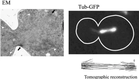
Three-dimensional ultrastructural analysis of yeast mitotic spindle microtubules has been accomplished by reconstruc-tion from serial thin secreconstruc-tions of cells (Wineyet al.1995) and by electron tomography of thick sections of cells (O’Toole et al. 1999) (Figure 1). The work confirmed that kineto-chores are attached by a single microtubule as suggested in early high-voltage electron microscopy (EM) studies of isolated yeast spindles (Peterson and Ris 1976). Reconstruc-tion of yeast mitotic spindle microtubules from serial thin sections or electron tomograms revealed that the spindle is a highly stereotypic microtubule array in these cells. In metaphase, it is 1.4–1.5mm long, and haploid cells contain 20 microtubules from each SPB. These microtubules can be divided into shorter microtubules that do not interact with antiparallel microtubules from the other SPB, suggest-ing that they are kinetochore microtubules, particularly since there are very nearly 16 per haploid SPB (Winey et al. 1995). Once formed, the 1.5- 2.0-mm spindle length
is constant for15–20 min. At this stage the spindle appears fairly rigid and can rotate up to 90obliquely to the mother/ bud axis. Anaphase onset is characterized by rapid linear elongation of the spindle with a velocity of1mm/min to a length of 6 mm. These rates are consistent with earlier measurements taken from low-level DAPI-stained cells (Palmeret al. 1989). In anaphase the kinetochore microtu-bules shorten from their plus-end (anaphase A). As spindle elongation ensues (anaphase B), the number of interpolar microtubules decreases to one or two interpolar microtubules from each pole prior to spindle disassembly.
are detectable only by electron tomography and hold the kinetochores very close to the SPB at the end of mitosis (O’Tooleet al.1999).
The remaining few nonkinetochore microtubules from each SPB interact (defined as being close to each other in 3D space) with nonkinetochore microtubules from the other SPB (O’Tooleet al.1999). These microtubules are thought to form a core bundle or the central spindle that lengthens during anaphase B to maintain spindle integrity and contrib-utes to driving the SPBs apart. In shorter spindles, these microtubules can be difficult to distinguish from the kineto-chore microtubules, but as the spindle lengthens this group of six to eight microtubules (three to four from each SPB) become very apparent as they are tightly packed (40 nm from each other) and even twist around each other (Winey et al. 1995). Crosslinking structures can be detected
be-tween these microtubules that could be various motors or microtubule-associated proteins (MAPs), but those molecu-lar identifications have not been made (Wineyet al.1995). Finally, the long microtubules of the anaphase B central spindle seem to persist into G1 after the spindle is severed during karyokinesis. The fact that the budding yeast SPB is embedded in the nuclear envelope throughout the entire life cycle of the organism is consistent with the observation that microtubules are continually present in the nucleus. The G1 nuclear microtubule array have a median length of 150 nm (O’Tooleet al.1999).
The Parts List
Spindle pole bodies
The S. cerevisiaeSPB wasfirst observed in the electron mi-croscope by Robinow and Marak (1966) (Figure 2). As the sole microtubule-organizing center (MTOC) in these cells that have a closed mitosis (the nuclear envelope stays intact throughout the cell cycle), the SPB must have access to the nucleoplasm to form the microtubules of the mitotic spindle and to the cytoplasm to form the astral microtubules that will position the nucleus. This is accomplished by position-ing the SPB in the nuclear envelope where it faces formposition-ing microtubules in each cellular compartment, the nucleus, and the cytoplasm. When observed under the electron micro-scope, a longitudinal section of SPB reveals a layered struc-ture made up of a series of plaques (flat disks,80 nm in diameter in a haploid and 160 nm in a diploid) apparently stacked on each other150 nm tall (Byers and Goetsch 1974, 1975). By definition, the inner plaque of the SPB is in the nucleus, the central plaque is in the plane of the nuclear envelope, and the outer plaque is the cytoplasm. In addition to the plaques of the SPB, there is an associated region of the nuclear envelope called the “half-bridge”that is modified with the addition of membrane proteins and a layer of proteins on the cytoplasmic face of this small re-gion of the envelope.
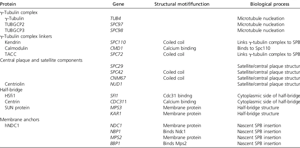
Importantly, it is likely that genetic analysis, proteomic screens, and genome-wide GFP tagging of yeast ORFs has led to the identification of all of the SPB components (Huh et al.2003; Kecket al.2011). There are 18 core SPB com-ponents, those being components found in the structure throughout the mitotic cell cycle (Table 1). Many additional
Figure 2 The mitotic spindle in yeast (A, left) is formed from spindle pole bodies (A, right) that are composed offive subcomplexes (B). (A, left) Immunofluoresence of a large-bud-ded mitotic yeast cell showing SPBs marked by Spc42-GFP (green), microtubules (red), and DNA (blue) and electron micrograph (A, right) showing trilaminar ultrastructure. Bar, 100 nm. (Eileen O’Toole, University of Colorado, Boulder). (B) Schematic of thefive major func-tional centrosome subcomplexes. Thisfigure is from Kecket al.(2011) and is reprinted with permission.
proteins are transiently associated with the SPB during the cell cycle in its functions as a signaling platform or as a MTOC (Caydasiet al.2010a,b). As befits an essential cel-lular structure, 16 of the 18 core SPB components are encoded by essential genes. The two nonessential genes,
SPC72 and CNM67, are essential in some genetic
back-grounds, and cells containing the null alleles of these genes are slow growing (Brachat et al.1998; Soues and Adams 1998; Hoepfner et al. 2000, 2002; Schaerer et al. 2001). Conditional mutations in any of the 16 essential genes encoding core SPB components cause SPB structural defects in mutant cells incubated at the restrictive condition. These defects range from the loss of microtubules to failed assem-bly of new SPBs to disintegration of parts of the SPB. Despite the clear structural differences between SPBs and the cen-trosome of vertebrate cells with its centrioles, 11 of the 18 core SPB components have vertebrate orthologs, most of which are centrosomal proteins (Table 1).
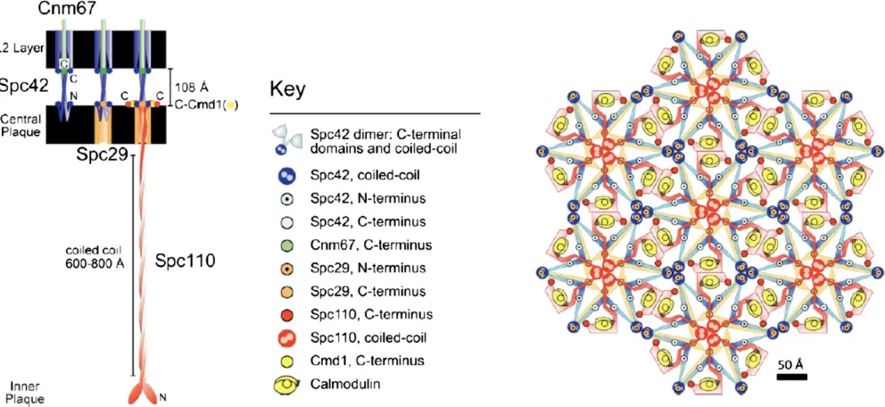
In addition to our knowledge of its composition, the SPB has been subjected to extensive structural analyses including electron tomography (O’Tooleet al.1999), cryo-EM (Bullitt et al. 1997), and mapping fluorescence resonance energy transfer (FRET) interactions in vivo (Muller et al. 2005) (Figure 3). A stunning finding from this work is that the central plaque is organized around a paracrystalline array of the Spc42 protein (Bullitt et al. 1997). This protein is nearly all coiled coil for which there is a crystal structure (Zizlspergeret al.2008), and it packs in a hexagonal array as seen in cryo-EM of isolated SPBs (Bullittet al.1997). This structural feature of the SPB was also observedin situusing electron tomography (Geiser et al. 1993; Kilmartin et al. 1993; O’Tooleet al. 1999).Spc42 interacts with the other major central plaque proteins,Spc29and the C terminus of
Spc110bound to calmodulin (Cmd1).Spc42spans the gap from the central plaque to part of the outer plaque where it interacts withCnm67. These interactions have been verified in vivoby FRET analysis, and a model of their organization has been developed (Mulleret al.2005) (Figure 3).
The central plaque is attached to the inner (nuclear side) and outer (cytoplasmic side) plaques, respectively, by“strut” proteins that span the gap between these layers of the SPB. The inner and outer plaques are critically important because they are the sites from which microtubules are nucleated. The sole“strut”protein connecting the central plaque to the inner plaque is Spc110(Geiseret al.1993; Kilmartinet al. 1993). The protein has a central coiled-coil domain that forms homodimers, and the C-terminal end binds calmodu-lin (Cmd1) and is where it interacts withSpc42in the cen-tral plaque. Interestingly, Spc110 is an essential target of calmodulin in budding yeast, and this function of calmodu-lin does not require calcium binding (Geiser et al. 1991, 1993; Stirlinget al.1994). In an elegant set of experiments altering the coiled-coil domain withinSpc110, John Kilmar-tin showed that Spc110 is the sole determinant of central plaque to inner-plaque distance (Kilmartinet al.1993). At the inner plaque, the N terminus ofSpc110binds theg-tubulin complex and may be integral to its function in nucleating microtubules (Knop and Schiebel 1997; Nguyenet al.1998; Vinhet al.2002; Kollmanet al.2010).
Outer plaque construction is more complex, involving
Cnm67,Nud1, andSpc72.Cnm67spans the gap from cen-tral to outer plaque. It is a dimeric, coiled-coil protein with globular N- and C-terminal domains. The C-terminal domain ofCnm67bindsSpc42and is sufficient for SPB localization, and an X-ray crystallographic structure was recently reported (Klenchin et al.2011). The structure reveals a novel dimeric, Table 1 SPB components
Protein Gene Structural motif/function Biological process
g-Tubulin complex
g-Tubulin TUB4 Microtubule nucleation
TUBGCP2 SPC97 Microtubule nucleation
TUBGCP3 SPC98 Microtubule nucleation
g-Tubulin complex linkers
Kendrin SPC110 Coiled coil Linksg-tubulin complex to SPB
Calmodulin CMD1 Calcium binding Binds to Spc110
TACC SPC72 Coiled coil Linksg-tubulin complex to SPB
Central plaque and satellite components
SPC29 Satellite/central plaque structure
SPC42 Coiled coil Satellite/central plaque structure CNM67 Coiled coil Satellite/central plaque structure
Centriolin NUD1 Satellite/central plaque structure
Half-bridge
HSfi1 SFI1 Cdc31 bindng Cytoplasmic side of half-bridge
Centrin CDC311 Calcium binding Cytoplasmic side of half-bridge
SUN protein MPS3 Membrane protein Half-bridge structure
KAR1 Membrane protein Half-bridge structure
Membrane anchors
hNDC1 NDC1 Membrane protein Nascent SPB insertion
NBP1 Binds Ndc1 Nascent SPB insertion
MPS2 Membrane protein Nascent SPB insertion
interdigitated, all a-helical fold with the very C-terminal eight residues being disordered. Nonetheless, these residues are crit-ical for protein folding and structural stability. The N-terminal region ofCnm67bindsSpc72, which is the protein that directly contacts theg-tubulin complex (Knop and Schiebel 1998). In-terestingly, the N-terminalg-tubulin-complex-binding domain of
Spc72can replace the N terminus of Spc110, suggesting that this domain of these two proteins serves largely the same pur-pose (Knop and Schiebel 1998).Spc72is also found on the half-bridge attached toKar1during a period in SPB duplication in which cytoplasmic microtubules originate from the half-bridge (Pereiraet al.1999), andSpc72interacts with the microtubule dynamicity factor Stu2 (Chen et al. 1998; Gruneberg et al. 2000). Finally, Nud1is also found at the outer plaque, where it connectsCnm67toSpc72(Gruneberget al.2000).Nud1is also the SPB component that binds several signaling molecules of the mitotic exit network (MEN) (e.g., Gruneberget al.2000; Yoshidaet al.2002).
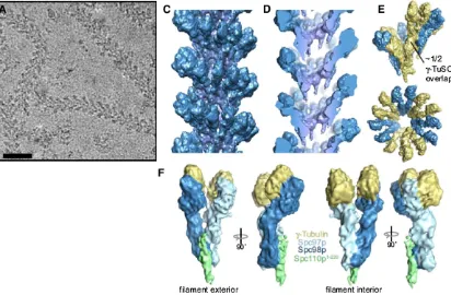
In the end, the function of the SPB is to nucleate microtubule formation. This SPB function is carried out by the g-tubulin complex that is composed of the conserved components Tub4(g-tubulin), Spc97, andSpc98 (Schiebel 2000; Vinh et al. 2002; Kollman et al. 2010) (Figure 4). Together, these proteins form the g-tubulin small complex with a stoichiometry of two g-tubulin molecules for each
Spc97andSpc98molecule (Kollmanet al.2008, 2010). This complex is assembled in the cytoplasm and can bind the cytoplasmic face of the SPB. The complex is transported into the nucleus by virtue of a nuclear localization signal (NLS) onSpc98(Pereiraet al.1998). The complex does not appear to nucleate microtubules when not at the SPB and that may be the result of microtubule nucleation requiring a higher-order structure (Kollmanet al.2010).
At the SPB, the g-tubulin complex likely forms the cap seen on the minus ends of the microtubules that werefirst detected in isolated SPBs (Byers et al.1978) and also were observed in electron tomograms of SPBs (O’Toole et al. 1999). It is not really known if microtubule formation is constitutive, meaning that the addition of g-tubulin com-plexes to the SPB immediately leads to microtubule forma-tion. Alternatively, there may beg-tubulin complexes at the SPB that are not active in nucleation and lack an attached microtubule. A domain ofSpc110, in addition to the three-proteing-tubulin complex, is necessaryin vitroto enable the formation of cap-like structures that can nucleate microtu-bule formation (Vinhet al.2002; Kollmanet al.2010). How-ever, thisin vitro nucleation is inefficient, raising questions about whether all of the proteins and their appropriately modified forms have been incorporated in the assay. The N-terminal domain of Spc110 interacts primarily with
Spc98, contributing to the formation of the higher-order complex. This domain of the protein is known to be phos-phorylated by bothCdc28(CDK) andMps1, suggesting that this modification may control assembly or nucleation activ-ity (Friedmanet al.2001; Huismanet al.2007).
SPBs and nuclear pore complexes (NPCs) (Chialet al.1998) and is required to insert both structures into the envelope during assembly (Lau et al.2004; Onischenko et al.2009).
Mps2 is also found at the SPB/nuclear envelope interface (Munoz-Centeno et al.1999). Both of these proteins are in complex with a second protein—Ndc1withNbp1(Arakiet al. 2006) andMps2 withBbp1(Schrammet al. 2000)—which are thought to serve as linkers from their membrane protein partner to the core SPB structure.
The remaining two SPB membrane proteins, Kar1 and
Mps3, are found in the special modified region of the nu-clear envelope adjacent to the SPB, the half-bridge (Vallen et al.1992; Spanget al.1995; Jaspersenet al.2002). The half-bridge is critically important to SPB assembly yet is poorly understood. It is seen in the electron microscope as an 90-nm electron-dense region of the nuclear enve-lope on one side of the SPB (oddly, it does not surround the SPB) (Byers and Goetsch 1975). The membrane likely stains dark because of the presence of the membrane pro-teins. Electron microscopic studies revealed a structure lay-ered over the cytoplasmic face of the half-bridge (O’Toole et al. 1999). This layer is likely composed of Sfi1and its bound centrin (Cdc31) molecules (Kilmartin 2003; Liet al. 2006). Interestingly, both Mps3 and Kar1 have been reported to bind Cdc31 (Biggins and Rose 1994; Spang et al. 1995; Jaspersen et al. 2002). Perhaps this binding can occur when Cdc31 is in complex with Sfi1, allowing
for the retention of the Sfi1/centrin complex at the half-bridge and for the overall organization of the half-half-bridge. Indeed, mutations in any of these four genes lead to losing the half-bridge structure (Byers 1981; Rose and Fink 1987; Jaspersenet al.2002).
polymer as microtubules encounter changes in force from chromosome attachment and bi-orientation in mitosis. In diploid cells, each spindle pole body nucleates at least 36 microtubules (32 kinetochore microtubules plus 4 interpolar microtubules). The spindle pole is operating at, or close to, its microtubule-carrying capacity. Nonetheless, major ques-tions remain as to what limits microtubule nucleation. In addition, it is not known how close to the edge of the SPB microtubules can be nucleated. There may be a region at the periphery of the SPBs that cannot form microtubules. There could be a dynamic interplay between kinetochore microtu-bule capture and SPB size/microtumicrotu-bule nucleation capacity. This idea stems from the observation that SPBs grow and microtubule nucleation increases in cdc20 mutants held at their restrictive growth temperature (O’Toole et al. 1997). These spindles contain approximately fourfold the number of microtubules (81 vs. 23) relative to a wild-type haploid spindle pole. This observation raises the notion that check-point activation is able to drive SPB assembly and more microtubule formation, giving the cell a better chance of capturing the unattached kinetochores, and raises the ques-tion of whether there are other limiting factors for the num-ber of microtubules nucleated from each pole (Yoderet al. 2003). In addition, MTs exhibit different angles of exit from the spindle pole body, and their minus ends can be slightly displaced from the pole. The area occupied by theg-tubulin complexes is larger than the central plaque and provides a greater nucleation capacity than strictly defined by the SPB.
Microtubules
Microtubules are large polymeric filaments composed ofa -and b-tubulin heterodimers. The polymeric form is com-posed of 13 protofilaments arranged cylindrically to form the 25-nm diameter microtubule. There are3·105
mol-ecules/cell (Abruzzi et al. 2002) and 3· 104 free tubulin molecules (Sprague et al. 2003), or 30–35 mM tubulin is polymerized into microtubules, and 3 mM tubulin is free in solution (see Tables 2 and 3). TUB1 and TUB3 encode a-tubulin and TUB2 encodes b-tubulin. TUB1 accounts for 90% ofa-tubulin protein in the cell, withTUB3 contribut-ing the remaincontribut-ing 10%. These genes are constitutively expressed, as microtubules are present throughout the cell cycle. Nuclear and cytoplasmic (or astral) microtubules are evident throughout the cell cycle. Cytoplasmic astral micro-tubules are needed for the orientation and positioning of the nucleus during the mitotic cell cycle of budding yeast. This subset of microtubules is thought to help in orienting the mitotic spindle by a combination of pushing and pull-ing forces exerted on the cell cortex, coupled with micro-tubule assembly and disassembly. Upon SPB duplication and the onset of mitosis, the new SPB must gain compe-tence to nucleate nuclear microtubules that, together with nuclear microtubules from the old SPB and microtubule-based motors, develop into the mitotic spindle.
Microtubules are inherently polar polymers with a“plus” and“minus”end that have distinct structures and dynamic properties. The minus end is nucleated at the SPB and the plus end extends away from the spindle pole. The plus end is considerably more dynamic and undergoes frequent changes from growth to shortening. Switching from growth to shortening (catastrophe) or from shortening to growth (rescue) is stochastic. This behavior is known as dynamic instability and is believed to be responsible for kinetochore attachment and chromosome segregation. Fluorescence re-covery after photobleaching (FRAP) andfluorescent speckle microscopy shows that minus ends contribute little or noth-ing to the assembly dynamics of cytoplasmic or nuclear microtubules (Maddox et al. 2000b). Unlike microtubule dynamics in several other systems, polymer flux through Table 2 Spindle parts list
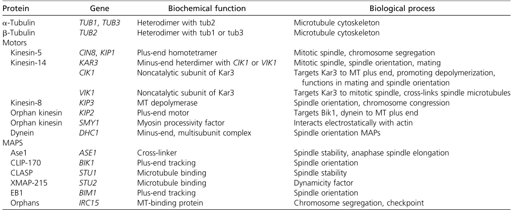
Protein Gene Biochemical function Biological process
a-Tubulin TUB1,TUB3 Heterodimer with tub2 Microtubule cytoskeleton
b-Tubulin TUB2 Heterodimer with tub1 or tub3 Microtubule cytoskeleton
Motors
Kinesin-5 CIN8,KIP1 Plus-end homotetramer Mitotic spindle, chromosome segregation Kinesin-14 KAR3 Minus-end heterdimer withCIK1orVIK1 Mitotic spindle, spindle orientation, mating
CIK1 Noncatalytic subunit of Kar3 Targets Kar3 to MT plus end, promoting depolymerization, functions in mating and spindle orientation
VIK1 Noncatalytic subunit of Kar3 Targets Kar3 to mitotic spindle, cross-links spindle microtubules
Kinesin-8 KIP3 MT depolymerase Spindle orientation, chromosome congression
Orphan kinesin KIP2 Plus-end motor Targets Bik1, dynein to MT plus end
Orphan kinesin SMY1 Myosin processivity factor Interacts electrostatically with actin
Dynein DHC1 Minus-end, multisubunit complex Spindle orientation MAPs
MAPS
Ase1 ASE1 Cross-linker Spindle stability, anaphase spindle elongation
CLIP-170 BIK1 Plus-end tracking Spindle orientation
CLASP STU1 Microtubule binding Spindle stability
XMAP-215 STU2 Microtubule binding Dynamicity factor
EB1 BIM1 Plus-end tracking Spindle orientation
the minus end is negligible or nonexistent in budding yeast (Table 3).
More recently, microtubule assembly dynamics have been examined at the nanoscale (Kerssemakerset al.2006; Schek et al. 2007). These studies reveal extensive variability in growth rate. The plus end of the microtubule undergoes frequent excursions of shortening (up tofive layers of tubu-lin) throughout net growth. Thus net changes of state at the macro scale (observable in the light microscope) hide the mechanics of tubulin subunit addition and subtraction oc-curring at the nano level. These hidden fluctuations are likely to be critical for the sensitivity of the switch between growth and shortening and regulation of the switch through tension or compression (Howard and Hyman 2009).
Microtubules are stiff (Young’s modulus of 1.2 GPa) (Gitteset al.1993), meaning that they are structurally rigid like plastics such as polycarbonate centrifuge tubes. One of the challenges in understanding the behavior of polymers such as microtubules in living cells is the realization that, at the size scale of the molecules in question, there is essen-tially no inertia. Instead, thermal fluctuations and viscous forces dominate reactions, and the force required to drive a given reaction may be only slightly greater than that of thermal motion. We must turn to physical definitions that define a material’s properties. Young’s modulus is a measure of the stiffness of a material and is the ratio of stress (pres-sure) to strain (change in length). Persistence length describes afilament’s resistance to thermal force and is the distance over which the correlation of the direction of the two ends of a polymer is lost. The persistence length of a microtubule is6 mm. This is three orders of magnitude longer than a typical yeast cell; thus, microtubule bending observed in live cells reflects active chemical processes. Spindle microtubules self-assemble from a pool of tubulin subunits in vivo. The process is endothermic and driven by the loss of ordered water surrounding the tubulin dimer
(Inoue et al. 1975; Salmon 1975). The free energy differ-ence between dimer and polymeric states is small; therefore, the dimervs.polymer concentrations are comparable (since neither state is energetically favored over the other). In fact, in interphase mammalian cells, the experimental measure-ments bear this out (Schliwaet al. 1979; Morris and Lasek 1984).
Microtubule motor proteins
Microtubule motor proteins convert chemical energy to mechanical energy to generate forces that can slide antipar-allel microtubules apart, couple cargo (e.g.chromosomes) to growing or shortening microtubule plus ends, regulate chro-mosome position in metaphase, and cross-link microtubules into bundles with specific polarity patterns to fortify spindle stability. In budding yeast, the repertoire of molecular motors is restricted to six kinesin-like proteins and one dy-nein (Table 2). Through pioneering experiments from Saun-ders and Hoyt to dissect kinesin function, it was found that
Cin8andKip1provide a pushing force for spindle elongation that is antagonized byKar3(Roofet al.1992; Saunders and Hoyt 1992). In addition, yeast can survive with only two motors: Cin8 and either Kar3 or Kip3 (Cottingham et al. 1999) The Cin8 motor is able to support cell viability in the presence of small quantities of benomyl to dampen mi-crotubule dynamics. The major spindle motors are thus the class 5 bipolar kinesins,Cin8andKip1. Their dominant func-tion is to slide antiparallel microtubules apart (Roof et al. 1991, 1992). These kinesins are regulated directly through phosphorylation (Chee and Haase 2010) and contribute to spindle formation, stability, and anaphase spindle elonga-tion. In addition, kinesin-5 motors have been found to be responsible for chromosome congression via spatial gra-dients in microtubule catastrophe rates (Gardner et al. 2008a).
Kar3, originally isolated in a screen for karyogamy mutants (hence Kar) (Meluh and Rose 1990), is antagonistic to the plus-end-directedCin8andKip1(Saunders and Hoyt 1992; Saunderset al.1997b).Kar3is a minus-end-directed kinesin-14 family member (Endowet al.1994). It was pro-posed thatKar3provides an inward-directed force, opposing the outward-directed pushing force of the kinesin-5 family members (Saunders and Hoyt 1992). This antagonistic mo-tor model for mitotic spindle function was revolutionary for thefield in the late 1990s and provided an important frame-work for thinking about motor function and dynamic micro-tubules in spindle function (Saunders et al. 1997b). Kar3
also provides structural support to the spindle by cross-linking antiparallel microtubules in anaphase (Gardneret al.2008b). The inward force opposing Cin8and Kip1could be due to minus-end-directed walking of Kar3 on antiparallel micro-tubules or to its ability to cross-link antiparallel microtu-bules. Defects in Kar3 function will effectively antagonize
Cin8andKip1via two distinct mechanisms. One mechanism counteracts directional force. Forces are exerted in opposite directions—an outward extension due toCin8andKip1vs. Table 3 Rates and numbers
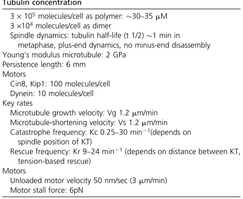
Tubulin concentration
3·105molecules/cell as polymer:30–35mM
3·104molecules/cell as dimer
Spindle dynamics: tubulin half-life (t 1/2)1 min in metaphase, plus-end dynamics, no minus-end disassembly Young’s modulus microtubule: 2 GPa
Persistence length: 6 mm Motors
Cin8, Kip1: 100 molecules/cell Dynein: 10 molecules/cell Key rates
Microtubule growth velocity: Vg 1.2mm/min Microtubule-shortening velocity: Vs 1.2mm/min Catastrophe frequency: Kc 0.25–30 min21(depends on
spindle position of KT)
Rescue frequency: Kr 9–24 min21(depends on distance between KT,
tension-based rescue) Motors
an inner contraction fromKar3. A second, not mutually ex-clusive, mechanism is the loss of structural stability of the spindle. The loss ofKar3cross-linking antiparallel microtu-bules leads to reduced spindle extension since the effective-ness of the outward motors is reduced. The latter model is an indirect mechanism but one that satisfies the genetic findings of shorter spindles inkar3D mutants (Saunders et al.1997b).
Analysis of Kar3 function during mating revealed that
Kar3 binds to microtubule plus ends and also functions as a microtubule depolymerase, thereby bringing two nuclei together in the mating projection for efficient fusion (Molk and Bloom 2006). The ability to visualize microtubules dur-ing matdur-ing provided an alternative hypothesis to the original sliding model of Meluh and Rose (1990).Kar3is complexed with either of two noncatalytic subunits,Cik1orVik1(Page et al. 1994; Manninget al.1999).Kar3is complexed with
Cik1in mating cells and along the spindle in vegetative cells. At spindle poles,Kar3is complexed withVik1in vegetative cells.Kar3’s function can be tuned depending on the identity
of the associated proteins Vik1 or Cik1 (Manning et al. 1999). In elegant biophysical experiments, it has been shown how the nucleotide-insensitive subunit (Vik1) coor-dinates the interaction of theKar3motor with microtubules (Allinghamet al.2007).
Kip3is a kinesin-8 family member and exhibitsbonafide microtubule depolymerase activity (Guptaet al.2006; Varga et al.2006). In the cytoplasm,Kip3regulates astral cytoplas-mic cytoplas-microtubule length and, together with cytoplas-microtubule plus-end-binding proteins Bim1, Kar9, Bni1, and Bud6, plays a critical role in directing the mitotic spindle to the bud (reviewed in Pearson and Bloom 2004). In the nucleus,
Kip3 contributes to kinetochore clustering in metaphase (Wargackiet al.2010). It has been found that longer micro-tubules accumulate higher concentration of MAPs and that, remarkably, Kip3 depolymerizes longer microtubules faster than shorter ones (Vargaet al.2006). How does a motor know how long the microtubule is? If the rate of motor movement is faster than microtubule dynamics, then motors accumulate at MT plus ends. Once there, various motors, such asKip3, might enhance the catastrophe frequency.
Kip2is another predominantly cytoplasmic kinesin and is antagonistic toKip3. Astral microtubules are extremely short in kip2 mutants, indicative of potential microtubule poly-merization activity of Kip2. Kip2 targets proteins such as
Bik1 and cytoplasmic dynein to microtubule plus ends (Carvalho et al.2003, 2004), where they interact with cy-toplasmic cues (Kar9,Num1) to orient the spindle toward the bud and ensure chromosome and nuclear segregation into mother and daughter cells.
The sixth kinesin protein in yeast, Smy1, was found in a genetic screen as synthetic lethal with myosin (Lillie and Brown 1992, 1994). It has since been found to be a proces-sivity factor in vitro (Aliet al. 2008; Hodges et al. 2009).
Smy1 enhances myosin processivity and intracellular trans-port by electrostatic interactions with actin. This passive
electrostatic tether is likely to be an important new mecha-nism that contributes to the effective processivity of individ-ual motor proteins for a given track.
Dynein is a minus-end microtubule motor whose exclu-sive function is in spindle orientation (Eshel et al.1993; Li et al.1993). The heavy chain itself (Dyn1orDhc1) is a proc-essive motorin vitro(Reck-Petersonet al.2006). The heavy chain is complexed with several intermediate and light chains (IC/Pac11, LIC/Dyn3, LC8/Dyn2) (Geiser et al. 1997) as well as with the dynactin complex (p150glued/
Nip100, p24/YLL049w, dynamitin/Jnm1, Arp1, Arp11/
Arp10), Bik1 (a CLIP-170 ortholog), and Kip2 (Carvalho et al. 2004). Bik1 is carried out to microtubule plus ends via Kip2 where they target dynein to its site of function. Dynein exerts its function by force generation at the micro-tubule-binding site on the cortex (Lee et al. 2005; Moore et al.2009b).
The motor field has exploded from the early days when antagonistic motor function was the reigning paradigm. It is now clear that, in addition to plus-end- and minus-end-directed motility, motors can function as microtubule poly-merases or depolypoly-merases by biasing fluctuations in dimer addition or subtraction at the plus end and by cross-linking of antiparallel or parallel microtubules, electrostatic couplers to enhance motor processivity and in organelle transport (e.g., mitochondria). While some of these functions are seg-regated to one class of kinesin (Kin-5 sliding motors, Kip3
depolymerase), in other kinesin classes the functions may be dictated by interacting proteins (Kar3-Vik1cross-linkingvs.
Kar3-Cik1depolymerase) (Sproulet al. 2005) or yet addi-tional mechanisms that remain for future investigations (e.g.,
Cin8function as a depolymerase at plus ends) (Gardneret al. 2008a).
Microtubule-associated proteins
MAPs regulate motor protein function, kinetochore micro-tubule dynamics, spindle orientation, and stability in meta-phase and anameta-phase. Historically, MAPs have been defined as any microtubule-binding protein. As such, the list is large and diverse. Recent biophysical experiments reconstituting microtubule plus-end-tracking activity and the structural determination of microtubule plus-end-tracking proteins have revealed mechanistic insights into MAP function (Figure 5) (Slep 2010). The major plus-end-tracking non-motor proteins in yeast are Bim1 (EB1), Bik1 (CLIP-170),
of condensin and cohesin and are proposed to promote pro-tein–protein interactions [HEAT:Huntingtin, elongation fac-tor 3 (EF3), protein phosphatase 2A (PP2A), and the yeast PI3-kinaseTOR1] (Neuwald and Hirano 2000). These repeats undergo force-induced conformational changes and may be critical in linking mechanical responses with microtubule-based force generation (Grinthalet al.2010).
Bim1 requires a GTP hydrolysis-competent cap at the microtubule plus end and recognizes unique features of the plus end. Bim1promotes lateral a/b-tubulin contacts.
Bim1 in turn recruits CAP-GLY domains. The Tog domains such as found inStu2 fold into intra-HEAT loops that form a facade delineating the tubulin-binding site and catalyze
the incorporation of a tubulin dimer (Widlundet al. 2011). The biological function of the major MAPs in yeast bears out the diversity gleaned from the structural information. Bim1
tracks microtubule plus ends in yeast and is involved in spin-dle orientation (Milleret al., 2000; Hwanget al.2003). In the nucleus it is found on kinetochore microtubule plus ends. Its spindle function is not a critical determinant in mitosis. In contrast, it is critical at astral microtubule plus ends to orient relative to asymmetric protein cues in the bud (Hwanget al. 2003). Bik1 (Clip-170, CAP-GLY domains) is transported to plus ends viaKip2(Carvalhoet al.2004), where it has been proposed to stimulate dynein function. More recently, Bik1
has been shown to interact with Bim1.Bim1andBik1form Figure 5 Domain structure and hierarchy of microtubule plus-end-tracking proteins Bim1 (EB1), Bik1 (CLIP-170), Nip100 (p150glued), Stu1 (CLASP), and
Stu2 (XMAP215). (Top left) Bim1 (EB1 family) is delineated by an N-terminal calponin homology domain (CH), an EB1 domain that confers dimerization and a C-terminal EEY motif that binds CAP-Gly domains. Bik1, of the CLIP-170 family, contains an N-terminal CAP-Gly domain, a central coiled-coil (CC) dimerization domain, a C-terminal Zn2+knuckle domain that can inhibit the ability of CAP-Gly domains to bind tubulin, and a C-terminal ETF motif akin
to the EEY motif found in EB1. Nip100 (of the p150gluedfamily) contains a CAP-Gly domain. In the CLASP ortholog, Stu1 contains a microtubule-binding
domain (MBD) as well as a kinetochore-binding domain (KtBD). Stu2 contains two N-terminal TOG domains followed by a C-terminal CC dimerization domain. (Top right) Bim1 and Stu2 bind to and regulate plus-end microtubule dynamics (green arrows). SKIP-motif proteins and the CAP-Gly-containing proteins Nip100p150and Bik1 show a Bim1-dependent microtubule plus-end-tracking activity (blue arrows). Whether Stu1 can plus-end-track
auton-omously or is Bim1-dependent remains to be elucidated. Regulatory elements are depicted in red. Adapted with permission from K. Slep (2010). (Bottom left) EB1 structure and microtubule binding. Structure of the EB1 N-terminal calponin homology domain (left, orange) reveals seven peripheral helices packed around the central conserveda3 helix. The structures of human EB1 (orange), EB3 (dark gray), andS. cerevisiaeBim1p (light gray) superimposed (bottom) reveal a high degree of structural identity across the EB1 family. The conserved C-terminal EB1 dimerization domain is formed through a coiled coil that folds back at its C-terminal region to form a four-helix bundle. The interface between the coiled coil and the four-helix bundle contains a signature FYF motif involved in SKIP-motif binding. (Bottom middle) Structure and mechanism of the TOG domain-containing Stu2. A cartoon representation of theDrosophilaXMAP215 TOG2 domain, delineating the six HEAT Repeats (HR), A–F, forms the elongated domain. Each HEAT repeat is formed by two antiparallel helices bridged by an intra-HEAT loop, positioned here at the top of the domain. (Bottom right) The G59S mutation of human p150gluedconstructed in yeast. Predicted structures of residues 25–83 for wild-type Nip100 and the Nip100-G45S mutant. The sequence of
Nip100 was threaded onto the structure of p150glued. Mooreet al.(2009a) have demonstrated that the CAP-Gly domain has a critical role in the
homodimers that interact to create a tetramer that in turn binds microtubule plus ends (Blake-Hodek et al. 2010). Combinatorial interactions among the different MAPS pro-vide a mechanism to increase the number of distinct activ-ities from a limited number of proteins. Different complexes may generate novel functions, provide modes of inhibition or activation (Blake-Hodek et al.2010), or modulate the strength of microtubule-binding interactions at distinct sites. The homolog ofStu2, XMAP215, is a microtubule poly-merase, which accelerates the growth of microtubules 10-fold after fertilization in frog eggs. In yeast,Stu2seems to be a dynamicity factor.In vivo,Stu2acts to increase the num-bers of cytoplasmic microtubules, consistent with a stabiliz-ing activity. At kinetochores, Stu2 appears to promote turnover of attached microtubule ends, suggesting a destabi-lizing activity (Pearson et al. 2003). Pure Stu2 has been reported to destabilize microtubules in vitro as well, but aXenopushomolog, XMAP215, has been found to promote both assembly and disassembly, depending on conditions (Shirasu-Hiza et al. 2003). Given this variety of reported activities,Stu2seems to act as a dynamicity factor, stabiliz-ing or destabilizstabiliz-ing microtubule dynamics, dependstabiliz-ing on the local environment and protein interaction (Pearson et al. 2003; Brittle and Ohkura 2005). Most recently, the TOG domains and a basic region have been engineered to reca-pitulate almost full microtubule polymerase activity (Widlund et al.2011).Stu2is therefore a significant catalyst of tubulin polymerization at microtubule plus ends.
The CLASP (CLIP-associated protein) homolog is Stu1
(Akhmanova and Hoogenraad 2005; Ortiz et al. 2009). CLASPs are plus-end-tracking proteins that stabilize MT plus ends by facilitating the incorporation of tubulin subunits (Maiatoet al.2005). In addition, by localizing at specialized MT regions distinct from plus ends, at least one CLASP member contributes to the stability of MT bundles in in-terphase (Bratman and Chang 2007). Metazoan CLASPs bind to KTs, where they regulate the dynamic behavior of KT MTs (kMTs) and, consequently, chromosome congres-sion (Maiato et al. 2002, 2003; Cheeseman et al. 2005; Mimori-Kiyosue et al. 2006). Stu1is required for spindle formation (Yin et al. 2002) and is found on the spindle. More recently, it has been shown that Stu1 binds unat-tached kinetochores in budding yeast (Ortiz et al.2009). Unattached kinetochores sequesterStu1to prevent prema-ture spindle stabilization, thereby keeping the two spindle poles together to facilitate sister kinetochore bi-orientation. The spindle midzone is the region of overlapping antiparallel microtubules, which, on average, constitutes about eight microtubles (three to four from each spindle pole) (Wineyet al.1995). The interpolar microtubules vary in length and rarely span the length of the spindle. The region of overlap is3/4mm in length. As the spindle elon-gates in anaphase, polar microtubules increase in length and decrease in number, generating an elongated late-anaphase spindle with a pair of overlapping microtubules. This struc-ture provides physical support until spindle disassembly.
Proteins binding to the region of overlap (known as the midzone) provide a critical function in spindle stability. One of the major midzone proteins isAse1(anaphases pin-dleelongation) (Pellmanet al.1995).Ase1is a member of a conserved family of cross-linking proteins (PRC1 in humans, fascetto and sofe in Drosophila, MAP65 in plants).
Ase1forms a dimer and has a microtubule-binding domain. It diffuses randomly on single microtubules and plays a key role in cross-linking antiparallel microtubules. In mammals, a kinesin 4 moves theAse1homolog PRC1 to the plus ends of antiparallel microtubules. Ase1phosphorylation plays an important role in regulating spindle assembly as well (Kotwaliwale et al. 2007). Thus, while these MAPs have primary functions in specific spatial or temporal domains of spindle morphogenesis, such as the spindle midzone, their functions are not restricted to these domains. More subtle phenotypes and contribution to a variety of spindle features will undoubtedly be discovered as we continue to tease apart the molecular nuts and bolts of the spindle.
With the advent of large-scale genomic screens applied to spindle morphogenesis (Hwang et al. 2003; Vizeacoumar et al. 2010) and spindle disassembly (Woodruff et al. 2010), it is certain that MAPs will continue to be identified. Of note isIrc15(Keyes and Burke 2009), which is found to bind microtubules in vitroand localize to spindles in vivo. Cells mutant inIrc15have a prolonged duration of mitosis. DNA, cohesion, and condensin springs in the spindle
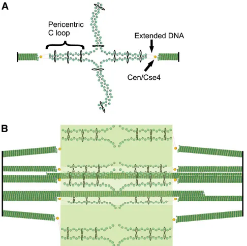
The 16 kinetochore microtubules (on average 0.35 mm in length in the half-spindle) and 4 interpolar microtubules (1 mm in length from each spindle pole) yield20 mm of microtubule polymer in the spindle. At 8 nm per tubulin dimer, 13 protofilaments per microtubule, and 110 kDa per dimer, 3.25 · 106 kDa of tubulin is polymerized in the spindle (Figure 1). The number of Cin8andKip1 microtu-bule motor proteins (100 kDa) in the spindle is on the order of just a few hundred (A. P. Joglekar and K. Bloom, unpublished results), and their mass is dwarfed by that of tubulin. Likewise, each kinetochore (see below) is5·103 kDa (De Wulfet al.2003), and the 32 per spindle constitute 0.16·106kDa, or,5% the mass of tubulin. The mass of chromatin in a cell is on the order of 1.4 · 107bp · 660 Da = 10·106kDa. Thus the spindle is just 1/3 the mass of the DNA (and1/6 the mass of the chromatin) that needs to be faithfully segregated.
chromatin constitutes roughly one-fifth the mass of the seg-regation apparatus (0.8 ·106kDa/0.8 + 3.25·106kDa), a significant fraction relative to spindle microtubules. To-gether with microtubules, the pericentric chromatin pro-vides a structural component that contributes to mitotic spindle function. The pericentric chromatin is organized into a molecular spring that opposes the outward-directed micro-tubule-based pushing forces. The spring is bound to the spindle at the kinetochore/microtubule interface and linked to its sister chromatid via cohesin. Thus, motors bound to antiparallel microtubules provide struts in the spindle, while DNA springs are elastic elements in the spindle. These loops provide an inward force to counterbalance outward-pulling forces generated by kinetochore and spindle microtubules. The loops are compliant and contribute to mechanical strain in response to tension generated in mitosis.
It is unlikely that the elastic spring is dictated by the properties of DNA itself. In addition to histones, the major constituents of chromatin in the pericentric chromosomal region include topoisomerase 2, cohesion, and condensin. Cohesin and condensin are members of the structural maintenance of chromosome (SMC) proteins that assemble into complexes that adopt a ring-like conformation. The backbone of the ring is formed by the SMC proteins themselves (MukB in bacteria,Smc2andSmc4inS. cerevi-siae condensin, and Smc1and Smc3in S. cerevisiae cohe-sion) (reviewed in Melby et al. 1998). In eukaryotes, the SMC monomer is folded in an antiparallel coiled coil. At one end, the two monomers associate to form a hinge, and at the other end is an ATP-binding head domain. Clo-sure of the ring at the head domain is carried out by proteins known as kleisins, includingScc1(also known asMcd1) and
Brn1. Each dimer is associated with additional proteins [e.g.,
Ycs4,Ycg1,Scc3(also known asIrr1),Rad61, andPds5] at the head domain to form a functional complex in vivo. In bacteria, the SMC coiled coils are bound by ScpA and ScpB. The primary biochemical function of cohesin is to hold to-gether sister-chromatid strands while condensin functions in chromatin compaction (Hirano 2006; Nasmyth and Haering 2009). Cohesin and condensin are enriched threefold in the pericentromeric chromatin where they contribute to the spring -like elastic properties of this region of the chromosome (Gerlichet al.2006; Bachellier-Bassiet al.2008; D’Ambrosio et al. 2008; Ribeiro et al. 2009; Samoshkin et al. 2009). Cohesin and condensin exhibit distinct patterns of localiza-tion within the spindle (Stephens et al. 2011). Cohesin (Smc3) exhibits a bi-lobed structure when viewed from the side and a cylindrical array when viewed on end (Yehet al. 2008) (Figure 6). In contrast, condensin (Smc4) is enriched along the spindle axis (Bachellier-Bassiet al.2008). Pericen-tric cohesin is radially displaced from the spindle microtu-bules, while condensin lies proximal to the spindle axis where it is primarily responsible for axial compaction of pericentric chromatin. Together with the intramolecular centromere loop, these SMC complexes constitute a molecular spring that balances spindle microtubule forces in metaphase (Stephens et al. 2011). The segregation apparatus is therefore a com-posite material of microtubules and chromatin (Bouck and Bloom 2007; Boucket al.2008).
The kinetochore
The kinetochore is a large multiprotein complex that links the sister chromatids to the mitotic spindle during chromo-some segregation. The physical organization of the kineto-chore into a trilaminar structure (Brinkley and Stubblefield 1966; Jokelainen 1967) is visible in electron microscopy images of vertebrate kinetochores, and the molecular composition in yeast follows this three-layer organization (Joglekaret al.2006, 2008, 2009; Cheeseman and Desai 2008; Andersonet al.2009; Santaguida and Musacchio 2009) despite the inability to see yeast kinetochores in the electron microscope (Peterson and Ris 1976). The kinetochore forms on the microtubule plus end as Figure 6 DNA springs in the spindle: model of the organization of cohesin
a“basket”of elongated molecules (namelyNdc80) that re-cruit the outer-kinetochore components of the KMN network (which comprises the Knl1 complex, the Mis12 complex, and theNdc80complex). These outer-kinetochore proteins dangle from the expanded basket surface generated by
Ndc80 and can interact with the chromatin and proteins of the constitutively centromere-associated network. The purpose of the basket is to allow the outer-kinetochore com-ponents to move over a greater distance and increase the likelihood of encountering an unattached centromere. In addition, this basket-like structure is likely to be part of the solution that contributes the mechanism in which a dy-namic microtubule remains attached to the kinetochore. Namely, tubulin subunits must be free to diffuse into and away from the microtubule plus end while it is embedded in the kinetochore. The geometry of the pericentric chromatin loops predisposes the centromere DNA (CEN) to be at the surface of the chromosome, where CEN is able to recruit a centromere histone H3 variant (CENPA) as well as the centromere DNA-binding proteins (CBF3). The outer and inner halves of the kinetochore can then interact and form a stable attachment, connecting the chromosome to the mi-crotubule. The kinetochore serves several important roles during chromosome segregation: it links chromosome move-ment to microtubule dynamics, monitors chromosome bi-orientation, and serves as a site of catalysis for synchroniz-ing chromosome segregation with cell cycle events.
We propose that the whole budding yeast mitotic spindle might serve as a model for a single regional centromere with multiple microtubule attachments per chromosome (Zinkowski et al. 1991) and that the cruciform structure found at bud-ding yeast centromeres is analogous to the looping model for more complex centromeres (Figure 6). The cruciform structure of the pericentromere places the centromeres at the apex of the intramolecular loop loaded with cohesin, maximizing the distance between sister centromeres and thus reconciling increased cohesin and maximal spot sepa-ration during mitosis (Yeh et al.2008). Further work has revealed that the formation of this structure is promoted by the DNA-binding components of the kinetochore (Anderson et al. 2009). This function is likely to be inherent in the structure of the proteins. For example, theS.cerevisiae pro-tein Ndc10 (also known as Cbf3a) is needed to form the looping cruciform structure; it is thought to bind as a dimer, and it is possible that these dimers serve to bring two regions of chromatin together to form a loop. Given the high level of conservation in composition between yeast and higher eukaryotic kinetochores (Joglekaret al.2006, 2008, 2009), one view is that multiple binding-site kinetochores of regional centromeres are repeats of the basic kinetochore of budding yeast, as proposed by the repeated subunit hypoth-esis (Zinkowski et al.1991). However, electron microscopy work has suggested that the mammalian kinetochore is dis-organized and lacks the recurring subunits proposed by the repeated subunit hypothesis (Donget al.2007; McEwen and Dong 2010). A view that reconciles these perspectives is that
the inner-kinetochore–centromere interface resembles a wo-ven fabric, rather than two separate fixed structures.
Building the Spindle
Spindle pole duplication and separation
SPB duplication is the cell cycle process by which the two SPBs responsible for mitotic spindle assembly are formed. The first morphologically recognizable steps of the process begin early in the cell cycle during G1. However, the first regulatory events may occur at the end of the previous cell cycle in the form of a licensing event conceptually similar to the licensing of chromosomal DNA replication (Hasse et al. 2001). The classic work of Breck Byers and Loretta Goetsch revealed the morphological pathway of SPB duplication (Byers and Goetsch 1975). They also demonstrated that SPB duplication is coordinated with the cell cycle, such that the numerous cdc2 mutants identified in the neighboring laboratory of Lee Hartwell not only gave uniform arrests by cell morphology and DNA content, but also arrested with uniform SPB and spindle morphologies (Byers and Goetsch 1974). This profound and fundamental observation is now taken for granted. Also fundamentally important in this early work was the discovery of a single cdc2 mutant that violated the coordinated arrest finding. Strains mutant in
CDC31failed to duplicate their SPBs, yet went onto a mitotic
arrest that should have included a bipolar spindle (Byers 1981). CDC31 was the first gene identified in yeast, or in any organism, to have a specific function in spindle pole duplication. It is not surprising that this cellular process in yeast, like most others, has been dissected genetically in great detail.
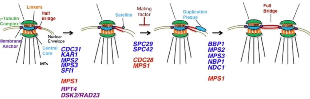
Thefirst recognizable intermediate in SPB duplication is the satellite-bearing stage (Figure 7). This stage is most easily observed in mating factor-arrested cells or in cells at
the cdc28 arrest (budding yeast CDK) (Byers and Goetsch
1974). This stage is also observed in cycling cells, indicating that it is a normal intermediate in SPB duplication. At this early point in duplication, the half-bridge has lengthened from 90 nm to nearly twice its length, and a tuft of dark staining material (called the “satellite”) is observed at the SPB-distal end of the bridge on the cytoplasmic side. It is interesting that both the lengthening of the half-bridge and the deposition of the satellite occur in G1, but how these events are regulated is not completely known.
Presumably, lengthening of the half-bridge occurs prior to the addition of the satellite. John Kilmartin’s discovery of
head-to-tail arrangement that is not significantly altered by cal-cium binding (Kilmartin 2003; Li et al. 2006). Kilmartin estimated the length of Sfi1 bound to centrin as90 nm, or the length of the half-bridge throughout most of the cell cycle. Using epitope-tagged alleles ofSfi1, Kilmartin showed that the N terminus ofSfi1was proximal to the SPB and that the C terminus was distal from the SPB. This result suggests thatSfi1determines the length of the half-bridge and that its orientation in the bridge offers a molecular basis for asymmetry in the bridge that is likely important in SPB du-plication (Liet al.2006). Importantly, the elongated bridge seen in mating factor-arrested cells or seen between two SPBs (called the full bridge) has two apparent end-to-end populations of Sfi1 along its length. In these cases, the N termini are at the ends of the long half-bridge and the C-terminal ends are in the middle, potentially interacting with each other (Liet al.2006; Andersonet al.2007). It has been suggested that the N terminus ofSfi1not associated with the existing SPB defines the site of SPB assembly, potentially by recruiting satellite components (Liet al.2006).
Adams and Kilmartin (1999) showed that the satellite is composed of the core SPB componentsSpc42,Spc29,Nud1, and Cnm67. After the satellite is formed, it will expand while still on the cytoplasmic face of the nuclear envelope, forming what the authors termed a “duplication plaque” that appears similar to the central plaque of the SPB (Adams and Kilmartin 1999) (Figure 7). Expansion of the duplica-tion plaque is concurrent with its inserduplica-tion into the nuclear envelope. This is accomplished by the creation of a fenestra at the distal end of the half-bridge into which the growing duplication plaque is inserted, giving it access to the nucle-oplasm. How the membrane fenestration is created is un-known, but it appears to require the functions of the SPB membrane proteins Ndc1 and Mps2 and their respective binding partners Nbp1 and Bbp1 (Winey et al. 1991,
1993; Schramm et al. 2000; Araki et al.2006). Mps2 also interacts with the half-bridge membrane and SUN-domain protein Mps3, but this interaction is involved with bridge assembly or maintenance (Jaspersenet al.2006). Nonethe-less, Mps3 is also required for insertion of the duplication plaque into the nuclear envelope (Sue Jaspersen, personal communication).
The still mysterious relationship between SPBs and NPCs was first hinted at by the discovery thatNdc1 is found at both structures (Chial et al.1998). Ndc1was known to be required for SPB assembly (Wineyet al.1993) and has been subsequently shown to be required for the assembly of NPCs into the intact nuclear envelope of yeast (Lau et al.2004; Onischenko et al. 2009). However, the relationship goes beyond a shared component. EM studies have shown that NPCs are found clustered near SPBs in mitotic cells, possibly to ensure the segregation of a sufficient number of NPCs to the daughter cells with each SPB (Wineyet al. 1997). Fur-thermore, Adams and Kilmartin (1999) reported observing an NPC-like structure at the site of insertion of the SPB into the nuclear envelope, raising a potential role for NPCs in SPB duplication. Various genetic interactions have been detected between alleles of genes encoding SPB components and NPC components. For example, the deletion of genes coding for the nonessential membrane nucleoporins
Pom152 or Pom34 suppressed the SPB insertion defects caused by certain mutant alleles of NDC1, MPS2, or BBP1 (Chialet al.1998; Sezenet al.2009). This includes the in-triguing finding that the null allele of the essential MPS2 gene can be rescued by deletion of Pom152 or Pom34
SPB size (Greenland et al. 2010). These interactions be-tween SPBs and NPCs are complex and poorly understood, but involve shared components (Chialet al. 1998), shared environment in the nuclear envelope (Witkin et al. 2010), and coordinated translational control of component synthe-sis (Sezenet al.2009).
Upon insertion into the nuclear envelope, the duplication plaque matures into a SPB by its acquisition of microtubule nucleation capacity. The resulting structure is called the duplicated side-by-side SPB. As noted above, this activity is dependent on g-tubulin complexes, the outermost compo-nents of the outer and inner plaques. Outer plaque develop-ment is complicated because of MT nucleation from the bridge of duplicated side-by-side SPBs on the basis of the interaction ofSpc72with the bridge componentKar1(Pereira et al.1999). The fact that the duplicated SPBs at this stage are different from each other with respect to age leads to the older “mother” SPB being able to nucleate microtubules, whereas the younger“daughter” SPB lags in the cell cycle in gaining this activity, which is controlled by Cdc28/Clb5
(Segal et al. 2000; Pereira et al. 2001). Furthermore, the phosphorylation ofg-tubulin (Tub4) also influences the be-havior of the cytoplasmic microtubules (Vogel et al.2001). Inner-plaque formation involves the localization of both the g-tubulin complex and its inner-plaque linker, Spc110, into the nucleus. This is accomplished by NLS on Spc110
(Adams and Kilmartin 1999) and onSpc98, which is suffi -cient to transport the entireg-tubulin complex into the nu-cleus (Pereira et al.1998). Mutation of the NLS in Spc110
relocalizes it to the cytoplasm, and overexpression of this mutant form of the protein will interfere with SPB duplica-tion (Adams and Kilmartin 1999). Furthermore, proper interactions between Spc110 and calmodulin (Cmd1) are required for normal assembly (Sundberget al.1996). Sim-ilar to Spc110, mutation of an NLS in Spc98blocks locali-zation of theg-tubulin complex to the nucleus and is lethal (Pereira et al. 1998). This mutant allele enabled Schiebel and co-workers to show that anMps1-phosphorylated form ofSpc98is found only in the nucleus, but the function of this phosphorylation event is not known (Pereiraet al.1998).
Conditional mutations in the various genes encoding SPB components block every step of the duplication pathway. The generic phenotype of these types of mutants is mitotically arrested cells (large-budded cells with replicated chromosomes) displaying aberrant microtubule organiza-tion arising from the single SPB instead of two SPBs forming a bipolar spindle. The single SPB is the “mother”SPB that the cells possessed prior to attempting duplication, and it forms a monopolar spindle (e.g., Wineyet al.1991). These monopolar spindles arising from SPB duplication defects cause a mitotic arrest by triggering the spindle assembly checkpoint (Weiss and Winey 1996). Examination of the defective SPBs by electron microscopy has proven instruc-tive about the nature of the failed assembly process. As mentioned above, mutations in the genes encoding compo-nents of the half-bridge lose this structure at the restrictive
temperature and block duplication by eliminating the site of satellite assembly (Byers 1981; Rose and Fink 1987; Jaspersen et al.2002; Kilmartin 2003).
Failure to insert the duplication plaque into the nuclear envelope is observed in cells mutated in the membrane anchor proteinsNdc1andMps2or in their respective bind-ing partners, Nbp1 and Bbp1 (Winey et al. 1991, 1993; Schramm et al.2000; Arakiet al.2006). Specific alleles of the SUN-domain membrane protein Mps3 also cause this phenotype (Sue Jaspersen, personal communication). Mu-tant cells include not only the monopolar spindle formed from the mother SPB, but also a second defective SPB de-rived from the duplication plaque. This defective SPB is at-tached to the cytoplasmic face of the nuclear envelope and is assembled to the point where it has cytoplasmic microtu-bules (Winey et al. 1991, 1993). Oddly, these cytoplasmic microtubules can exert force on the defective SPB, presum-ably via cortical attachments, and pull it away from the mother SPB and into the bud on a thin extension of the nuclear envelope that lacks chromatin.
Finally, inner-plaque defects can be observed inSpc110,
Cmd1, and some alleles of the genes encoding components of theg-tubulin complex. In one such allele (tub4-34), the inner plaque appears to be missing although the SPB has been inserted into the envelope (Marschall et al. 1996). CMD1alleles exhibit pleiotropic defects because of the nu-merous functions of calmodulin, but SPB defects are evident (e.g., Sun et al. 1992). More exotic are alleles of Spc110
defective in calmodulin binding that form aberrant struc-tures in the nucleus that act as MTOCs (Sundberg et al. 1996). In addition, the spc110-226 allele causes a loss of integrity of SPB structure at the restrictive temperature (Yoderet al.2005). In these mutants, the inner-plaqueg -tu-bulin complexes along with the mutantSpc110can “delam-inate”from the SPB and form foci in the nucleoplasm that are associated with the minus end of microtubules. This observation of SPB failure in mitosis suggests that significant forces are applied to the structures at this point in the cell cycle when the chromosomes are achieving biorientation.
the kinesin-like motor proteins, Cin8andKip1 (Roofet al. 1992; Saunders and Hoyt 1992). Mutations in the C-terminal domain ofSfi1block separation (Andersonet al.2007). This is the domain of the protein that is at the center of the bridge that may be exposed once the bridge is severed, pro-ducing the site for bridge elongation during the next round of duplication. How the C-terminal ends of theSfi1function to create a dissolvable junction in the middle of the bridge, whether it be via direct interactions or via interacting with an another bridge component, is not known. Separation of the SPBs does require force to be applied to the SPBs such that they move in the nuclear envelope. The kinesin-like motor proteinsCin8andKip1are required for SPB separa-tion and presumably work by cross-linking the microtubules from each of the SPBs. Upon moving toward the plus ends of the microtubules during spindle assembly, the motors par-ticipate in driving the SPBs apart. Cytoplasmic microtubules can exert forces on SPBs (Tolic-Norrelykke 2010), but SPB separation will occur without these microtubules (Huffaker et al. 1988; Jacobset al. 1988; Liet al. 1993) or without dynein function (Li et al.1993). Finally, the bridge is not severed in cyclin B (CLB1-4)-depleted cells (Fitch et al. 1992) or in cells overexpressing Swe1, which inhibits CDK activity (Limet al. 1996). These results reveal that the in-creasing CDK/CyclinB activity in mitotic cells is required to separate SPBs.
Regulation of SPB duplication
Most SPB components are phosphorylated (Wigge et al. 1998), and this modification is required for the proper reg-ulation and assembly of several components (e.g., Jaspersen et al.2004). Recent phospho-proteomics studies of overex-pressed g-tubulin complexes (Tub4, Spc97, Spc98) (Lin et al. 2011) and of entire intact SPBs (Keck et al. 2011) revealed extensive phosphorylation of SPB components. For example, Kecket al.(2011) report 297 phosphorylation sites over the 17 of 18 core SPB components; only Mps3
lacked mapped sites, likely due to poor coverage of this membrane protein. Furthermore, these authors showed that there are substantial changes in phosphorylation-site usage at different points in the cell cycle. Both groups showed that phosphorylation of g-tubulin (Tub4) is functionally impor-tant, as previously shown for Y445 (Vogelet al.2001; posi-tion Y445 verified in Keck et al. 2011). Linet al. (2011) found that phospho-mimetic mutations in TUB4 at S74, S100, and S360 are lethal. Kecket al.(2011) showed that the highly conservedTub4-S360 was phosphorylatedin vivo and is a CDK site in vitro as predicted by its sequence. In these authors’ research, the tub4-S360D allele displayed a temperature-sensitive lethal phenotype (Kecket al.2011). At the restrictive temperature, the cells displayed significant defects in the mitotic spindle similar to what was reported by Lin et al. (2011). At the permissive temperature, the tub4 -S360Dstrains exhibit abberations in spindle elongation dur-ing anaphase, revealdur-ing an intrigudur-ing link betweeng-tubulin complex phosphorylation and spindle dynamics.
A few protein kinases have been implicated in SPB assembly and function, but unique among these is Mps1
protein kinase (Winey and Huneycutt 2002). This is the only yeast protein kinase-encoding gene for which SPB duplica-tion defective alleles have been identified. At the restrictive condition, the original allele, mps1-1, produced monopolar spindles formed by SPBs with a long half-bridge. This result indicated that Mps1 is required for satellite formation, but not for bridge elongation (Wineyet al.1991). Further anal-ysis of an allelic series and an analog-sensitive allele revealed that Mps1is involved in bridge elongation, in sat-ellite formation and utilization, and in membrane insertion of the duplication plaque (Pereira et al. 1998; Schutz and Winey 1998; Friedman et al. 2001; Castillo et al. 2002; Jones et al.2005; Holinger et al.2009; Arakiet al.2010). This may not be surprising in retrospect now that Cdc31,
Spc29, Spc42, Spc98, and Spc110 are known substrates (Pereira et al. 1998; Friedman et al. 2001; Castillo et al. 2002; Holingeret al.2009; Arakiet al.2010). The relevant substrate(s) for SPB nuclear envelope insertion is not known. In addition to the multiple SPB phenotypes observed in mps12 mutant strains, it was also observed that these cells failed to arrest in mitosis similar to other SPB duplica-tion-defective mutant strains (Winey et al.1991). This was found to result from failure of the spindle assembly check-point in which Mps1 also functions (Weiss and Winey 1996). Like other mitotic protein kinases,Mps1has multiple functions during mitosis, including important functions at the kinetochore (e.g., Maureet al.2007; Dobraet al.2011). As with chromosomal DNA replication, the precise duplica-tion of the SPBs to produce a single new SPB per cell cycle is ultimately under the control ofCdc28, the CDK that regulates the budding yeast cell cycle. As mentioned above,Cdc28 activ-ity in mitotic cells is required to separate duplicated side-by-side SPBs from the mitotic spindle. Similar to the chromosome cycle, CDK activity must be inactivated during anaphase to allow for SPB duplication (Hasseet al.2001). Thisfinding hints at a li-censing event, but molecular basis of that event is unknown. Finally, CDK activity in late G1 is required to proceed past the satellite-bearing stage. Cdc28 is known or thought to phosphorylate several SPB components, including Spc29,
Spc42,Spc110,Nbp1,Bbp1, andNud1(Friedmanet al.2001; Ubersax et al. 2003; Jaspersen et al. 2004; Park et al. 2004, 2008; Holinger et al. 2009). The function of most of these phosphorylation events is unknown, butCdc28phosphorylation of Spc42 is required for proper assembly (Jaspersen and Winey 2004; Kecket al.2011). Other potential regulators of the SPB duplication pathway are the yeast polo kinaseCdc5
The step-wise assembly and separation of SPBs tells only part of the story of SPB assembly (Figure 8). The exchange of transiently expressed GFP-tagged SPB components into SPBs (Jaspersen and Winey 2004), FRAP of GFP-tagged SPB components (Yoder et al. 2003), and the continued growth of SPBs in mitotically arrested cells (O’Toole et al. 1997) all reveal dynamic behavior of SPB components. Not known are the mechanism of the exchange, the extent to which any given SPB component can be exchanged, and the biological significance of the exchange. It possible that the exchange of components allows for the repair of damaged SPBs because there is no known instance of de novo SPB formation. Hence, cells must maintain their SPBs because they cannot be replaced. Some studies have begun to ad-dress this exchange of components. The exchange of tran-siently expressedSpc42 into SPBs fails in cells mutated for its binding partnerSpc29and fails in cells lackingCdc28or
Mps1 activities, suggesting that the exchange can share requirements with assembly during SPB duplication (Jas-persen et al. 2004). A system to specifically damage SPBs was engineered by inserting TEV protease sites into the
Spc110protein (Greenlandet al.2010). Cleavage ofSpc110
leads to a spindle checkpoint-mediated cell cycle delay. This system was used for a screen for genes required for SPB remodeling and identified mutations in genes with various functions in the nuclear envelope and in protein turnover (Greenlandet al.2010). Maintenance of MTOC structures is an emerging and important area of study (Pearsonet al.2009) that is accessible to genetic analysis in budding yeast.
Spindle Dynamics
Regulation of microtubule dynamics, kinetochore and interpolar microtubules
The half-life of a tubulin dimer in the microtubule lattice is 50 sec (Maddox et al.2000a). The microtubules do not shorten completely prior to rescue. Rather, there is a gradient in dynamics, with the most rapid turnover exhibited at the plus ends (near the spindle midzone) and the least turnover at the minus ends (proximal to the spindle pole) (Figure 9). Thus kinetochore microtubules make many short excursions into catastrophe followed by frequent rescue. It has been proposed that these robust short excursions contribute to mechanisms responsible for thefidelity of chromosome seg-regation (Pearsonet al.2006).