Evaluation of molecular tests for
prenatal diagnosis of chromosome
abnormalities
GM Grimshaw
A Szczepura
M Hultén
F MacDonald
NC Nevin
F Sutton
S Dhanjal
HTA
Health Technology Assessment
How to obtain copies of this and other HTA Programme reports.
An electronic version of this publication, in Adobe Acrobat format, is available for downloading free of charge for personal use from the HTA website (http://www.hta.ac.uk). A fully searchable CD-ROM is also available (see below).
Printed copies of HTA monographs cost £20 each (post and packing free in the UK) to both public and private sector purchasers from our Despatch Agents.
Non-UK purchasers will have to pay a small fee for post and packing. For European countries the cost is £2 per monograph and for the rest of the world £3 per monograph.
You can order HTA monographs from our Despatch Agents: – fax (with credit card or official purchase order)
– post (with credit card or official purchase order or cheque) – phone during office hours (credit cardonly).
Additionally the HTA website allows you either to pay securely by credit card or to print out your order and then post or fax it.
Contact details are as follows:
HTA Despatch Email: [email protected]
c/o Direct Mail Works Ltd Tel: 02392 492 000 4 Oakwood Business Centre Fax: 02392 478 555
Downley, HAVANT PO9 2NP, UK Fax from outside the UK: +44 2392 478 555 NHS libraries can subscribe free of charge. Public libraries can subscribe at a very reduced cost of £100 for each volume (normally comprising 30–40 titles). The commercial subscription rate is £300 per volume. Please see our website for details. Subscriptions can only be purchased for the current or forthcoming volume.
Payment methods
Paying by cheque
If you pay by cheque, the cheque must be in pounds sterling, made payable to Direct Mail Works Ltd and drawn on a bank with a UK address.
Paying by credit card
The following cards are accepted by phone, fax, post or via the website ordering pages: Delta, Eurocard, Mastercard, Solo, Switch and Visa. We advise against sending credit card details in a plain email.
Paying by official purchase order
You can post or fax these, but they must be from public bodies (i.e. NHS or universities) within the UK. We cannot at present accept purchase orders from commercial companies or from outside the UK.
How do I get a copy of HTA on CD?
Please use the form on the HTA website (www.hta.ac.uk/htacd.htm). Or contact Direct Mail Works (see contact details above) by email, post, fax or phone. HTA on CDis currently free of charge worldwide.
The website also provides information about the HTA Programme and lists the membership of the various committees.
abnormalities
GM Grimshaw
1*NC Nevin
4A Szczepura
1F Sutton
5M Hultén
2S Dhanjal
2F MacDonald
31
Centre for Health Services Studies, University of Warwick, Coventry, UK
2
Biological Sciences, University of Warwick, Coventry, UK
3
West Midlands Regional Genetic Laboratory and Consultancy Services,
Birmingham Womens Health Care Trust, Birmingham, UK
4
University of Belfast, Belfast, Northern Ireland
5
Statistician, Learning and Skills Council, Coventry, UK
*
Corresponding author
Declared competing interests of the authors: none
Published May 2003
This report should be referenced as follows:
Grimshaw GM, Szczepura A, Hultén M, MacDonald F, Nevin NC, Sutton F, et al. Evaluation of molecular tests for prenatal diagnosis of chromosome abnormalities. Health Technol Assess 2003;7(10).
NHS R&D HTA Programme
T
he NHS R&D Health Technology Assessment (HTA) Programme was set up in 1993 to ensure that high-quality research information on the costs, effectiveness and broader impact of health technologies is produced in the most efficient way for those who use, manage and provide care in the NHS.Initially, six HTA panels (pharmaceuticals, acute sector, primary and community care, diagnostics and imaging, population screening, methodology) helped to set the research priorities for the HTA Programme. However, during the past few years there have been a number of changes in and around NHS R&D, such as the establishment of the National Institute for Clinical Excellence (NICE) and the creation of three new research programmes: Service Delivery and Organisation (SDO); New and Emerging Applications of Technology (NEAT); and the Methodology Programme.
This has meant that the HTA panels can now focus more explicitly on health technologies (‘health technologies’ are broadly defined to include all interventions used to promote health, prevent and treat disease, and improve rehabilitation and long-term care) rather than settings of care. Therefore the panel structure has been redefined and replaced by three new panels: Pharmaceuticals; Therapeutic Procedures (including devices and operations); and Diagnostic Technologies and Screening.
The HTA Programme continues to commission both primary and secondary research. The HTA Commissioning Board, supported by the National Coordinating Centre for Health Technology Assessment (NCCHTA), will consider and advise the Programme Director on the best research projects to pursue in order to address the research priorities identified by the three HTA panels. The research reported in this monograph was funded as project number 94/43/04.
The views expressed in this publication are those of the authors and not necessarily those of the HTA Programme or the Department of Health. The editors wish to emphasise that funding and publication of this research by the NHS should not be taken as implicit support for any
recommendations made by the authors.
Criteria for inclusion in the HTA monograph series
Reports are published in the HTA monograph series if (1) they have resulted from work
commissioned for the HTA Programme, and (2) they are of a sufficiently high scientific quality as assessed by the referees and editors.
Reviews in Health Technology Assessment are termed ‘systematic’ when the account of the search, appraisal and synthesis methods (to minimise biases and random errors) would, in theory, permit the replication of the review by others.
HTA Programme Director: Professor Kent Woods
Series Editors: Professor Andrew Stevens, Dr Ken Stein, Professor John Gabbay, Dr Ruairidh Milne and Dr Chris Hyde
Managing Editors: Sally Bailey and Sarah Llewellyn Lloyd
The editors and publisher have tried to ensure the accuracy of this report but do not accept liability for damages or losses arising from material published in this report. They would like to thank the referees for their constructive comments on the draft document.
ISSN 1366-5278
© Queen’s Printer and Controller of HMSO 2003
This monograph may be freely reproduced for the purposes of private research and study and may be included in professional journals provided that suitable acknowledgement is made and the reproduction is not associated with any form of advertising.
Applications for commercial reproduction should be addressed to The National Coordinating Centre for Health Technology Assessment, Mailpoint 728, Boldrewood, University of Southampton, Southampton, SO16 7PX, UK.
Published by Core Research, Alton, on behalf of the NCCHTA.
List of abbreviations ... i
Executive summary ... iii
1 Introduction and background... 1
Introduction ... 1
Chromosome abnormalities in the fetus... 1
Prenatal risk assessment ... 3
Prenatal diagnostic testing ... 4
Developments in prenatal diagnostic testing ... 4
Background to the current research ... 5
Aims of the research ... 6
Study design ... 6
2 Development and technical evaluation of molecular tests... 9
Introduction ... 9
Background to the FISH test ... 9
Background to the Q-PCR test... 9
Development of the Q-PCR test... 10
Technical evaluation of molecular tests ... 11
Fineberg level 1: technical capacity of molecular tests ... 12
Fineberg level 2: diagnostic accuracy of molecular tests ... 16
Sensitivity and specificity of molecular tests ... 18
Conclusions ... 23
3 Current laboratory testing practices ... 25
Introduction ... 25
UK survey of NHS genetics laboratories ... 25
Existing and planned use of molecular tests in NHS cytogenetics laboratories ... 25
Staffing and equipment for molecular tests in NHS cytogenetics laboratories ... 27
Prenatal services and activity in NHS cytogenetics laboratories ... 28
Study sites: structuring of prenatal testing services ... 30
Conclusions ... 32
4 Costs of karyotyping and molecular tests... 33
Introduction ... 33
Capital costs... 34
Labour costs ... 35
Consumable costs ... 36
Cost per sample tested ... 37
Test costs under different laboratory conditions ... 40
Discussion ... 42
5 Methods used to evaluate benefits to parents and other test users... 45
Patient parameters ... 45
Clinician surveys ... 47
Health commissioners surveys ... 49
Summary ... 49
6 Patient and physician choices... 51
Introduction ... 51
Results of regional and national survey of obstetricians ... 51
Obstetricians’ test choice for individual patients ... 55
Midwives’ test preferences ... 57
Stakeholders’ test preferences ... 57
Discussion ... 58
7 Estimating the cost-effectiveness of molecular tests... 61
Introduction ... 61
Cost-effectiveness of molecular tests as a replacement for karyotyping ... 62
Cost-effectiveness of molecular tests as a routine add-on to karyotyping ... 64
Cost-effectiveness of testing regimes with varying levels of karyotyping replacement .. 65
Costs of alternative testing regimes ... 66
Incremental cost-effectiveness ratios ... 71
Discussion ... 72
8 Valuing benefits for parents and other stakeholders... 75
Introduction ... 75
Parental anxiety ... 75
Parental health status ... 77
Cost–utility analysis ... 80
Extending the cost-effectiveness analysis .... 81
Valuing preferences: the cost–benefit approach ... 86
Overview of economic evaluation techniques ... 91
9 Summary and conclusions ... 97
Service delivery ... 97
Evaluation framework... 99
Conclusions ... 104
Recommendations ... 105
Contents
Acknowledgements ... 107
References... 109
Appendix 1 Development and
preliminary assessment of single Q-PCR
test for Down syndrome ... 113
Appendix 2 The number and type of
abnormalities undetected if using 5-probe FISH or multiplex Q-PCR ... 117
Appendix 3 First questionnaire
to women ... 119
Appendix 4 Obstetricians’ criteria
for undertaking amniocentesis for
specific patients... 123
Appendix 5 Influence of throughput on
test costs... 125
Appendix 6 Calculation of weighted
cost-effectiveness analysis ... 127
Appendix 7 General public questionnaire.... 129
Appendix 8 Willingness to pay: contingent
valuation and conjoint analysis ... 133
Appendix 9 Possible test strategy ... 135
Appendix 10 Aspects of assessing
diagnostic technologies ... 137
Appendix 11 National Screening
Committee’s criteria for appraising the viability, effectiveness and appropriateness of a screening programme ... 145
Health Technology Assessment reports
published to date ... 147
Health Technology Assessment
i
List of abbreviations
All abbreviations that have been used in this report are listed here unless the abbreviation is well known (e.g. NHS), or it has been used only once, or it is a non-standard abbreviation used only in figures/tables/appendices in which case the abbreviation is defined in the figure legend or at the end of the table.
CVS chorionic villus sampling
FISH fluorescence in situ hybridisation
HTA Health Technology Assessment
MLA Medical Laboratory Assistant
MTO Medical Technical Officer
NS not significant
QALY quality-adjusted life-year
QoL quality of life
Q-PCR quantitative polymerase chain reaction
ROC receiver operating characteristic
STR small tandem repeat
WTP willingness to pay
iii
Background
Many women undergo prenatal tests for chromo-some abnormalities in their baby, usually following identification of an increased risk of the baby having Down syndrome. One test that can show up abnormalities likely to lead to mental or physical handicap is done by sampling the amniotic fluid that surrounds the baby, usually around 14 weeks of pregnancy. There is a very small risk of mis-carriage with this procedure and parents are warned of this. If the test shows there is chromo-some abnormality, parents may want to discuss whether to continue with the pregnancy.
Until now parents have had to wait for up to 3 weeks for the results of this test (karyotyping), which is based on culturing the cells sampled from the amniotic fluid. Karyotyping allows examination of all the baby’s chromosomes. New DNA tests have been developed that can give results in 2–3 days. These new molecular tests, using fluorescence in situ hybridisation (FISH) or the quantitative polymerase chain reaction (Q-PCR), search for abnormalities in specific chromosomes. Errors in chromosomes other than in those tested will not be disclosed. The abnormalities not tested for are much more rare. For example, only 4 in every 1000 babies tested will have one of these rarer abnormalities and some of these may be identified during other examinations, for example during routine ultrasound examinations.
Many parents will welcome the quicker result from a more focused test but some may be prepared to wait for the result of a test that examines all the chromosomes.
Objectives
The objectives of this study were to:
• measure the technical performance of FISH and Q-PCR tests versus karyotyping • estimate the relative costs of molecular tests
under various conditions
• establish the value to women, clinicians and others of more rapid molecular test results
• assess the cost-effectiveness of molecular tests, and consider possible changes in current testing protocols.
Design
Two-stage trial; technical performance assessed through a blinded comparison of molecular tests against the accepted gold standard (karyotyping) in a laboratory setting in the first stage; effective-ness and cost-effectiveeffective-ness measured in a service setting in the second stage. Measurement of anxiety and health status of women; willingness to pay (WTP) for four stakeholder groups; and survey of UK obstetricians and midwives.
Setting
Two study sites – the catchment areas for the West Midlands Regional Genetic Laboratory and the Northern Ireland Regional Genetics Centre, Belfast.
Participants
Blinded samples: 2376 Down only molecular tests; 1576 multiplex/5-probe tests; 3952 karyotyping.
Trial: 194 women (141 intervention group; 53 control group).
WTP: 1000 general public; 141 women;
84 partners; 105 health commissioners.
Interventions (diagnostic tests)
• Molecular tests for the five most common chromosome abnormalities.
• Molecular tests for Down syndrome only. • Karyotyping.
Main outcome measures
Technical capacity, diagnostic accuracy, diagnostic impact, patient outcome and cost-effectiveness.
Executive summary
iv
Results
Technical capacity – does the test perform reliably and deliver accurate (i.e. precise) information?
FISH and Q-PCR test results are as reliable and precise as karyotyping for the five most common chromosome abnormalities.
Diagnostic accuracy – does the test contribute to an accurate diagnosis (of chromosome abnormalities)?
The ability to detect the five most common chromosome abnormalities, absolute sensitivity and specificity, are 1.00 and 1.00 for FISH and 0.9565 and 0.9997 for Q-PCR, respectively.
The ability to detect all clinically significant chromosome abnormalities, relative sensitivity and relative specificity, are 0.8605 and 0.9999 for FISH and 0.8234 and 0.9996 for Q-PCR, respectively.
Diagnostic impact – will the test replace other diagnostic tests or procedures?
Preferences of clinicians, women and other stakeholders will influence diagnostic impact.
Fifty-seven per cent of obstetricians expressed a preference for molecular tests for most patients and karyotyping for a minority; only 15% would choose both tests. The views of midwives were similar.
Most women (67%) and 54% of partners expressed a pre-test preference for molecular tests. Health commissioners were undecided. The general public expressed a preference for karyotyping (60%).
Patient outcome – does the test contribute to improved health/reduced anxiety for the patient?
Quality of life measure (EuroQol EQ-5D)
demonstrated significantly increased health status linked to more rapid test results. Anxiety measure (Speilberger) exhibited similar impact.
Cost-effectiveness – does the test use improve cost-effectiveness compared to alternative interventions?
Molecular tests are less expensive than karyo-typing. As a replacement within larger laboratories (> 1100 specimens per annum), Q-PCR is pre-ferred; for smaller laboratories (< 450), FISH is
preferred. Five testing regimes were assessed in terms of cost-effectiveness:
1. Molecular test and karyotyping for all women. 2. Molecular test as a replacement for karyotyping 3. Molecular test for all plus karyotyping for
high-risk women.
4. Karyotyping for all plus molecular test for high-risk women.
5. Parental choice plus karyotyping for high-risk women.
Simple cost-effectiveness analysis based on the cost per case detected (all cases) demonstrates that regimes 2, 3 and 5 are more cost-effective than karyotyping and 1 and 4 are not. This pattern does not change if cost-effectiveness analysis is limited to clinically significant cases only.
Cost–utility analysis estimates a cost per quality-adjusted life-year gained of £23,542–£41,939 for regime 1; it was not possible to assess regimes 2–5 using this technique.
Regimes 2, 3 and 5 will not detect some rare chromosome abnormalities (approximately 2–4, 1–2 and 1 per 1000 women tested, respectively).
Introduction of regime 1 could increase annual UK test costs by up to £2.8 million. Regimes 2 and 3 should result in savings of up to £1.76 million per annum, and regime 5 approximately two-thirds of these savings. Regime 4 would be largely cost neutral.
Conclusions
Implications for healthcare
In the current climate, the use of prenatal testing is determined by individual clinicians, laboratories and hospitals. There is evidence of a lack of equity of provision, and of regional and local variations with regard to primary risk assessment. This may well be replicated with regard to final diagnosis if molecular tests are introduced without discussion of appropriate implementation protocols based on this report. Debate and consensus will be necessary to develop clinical protocols for introduction of molecular tests and prevent continuation of inequities and variations. Important ethical issues must not be overlooked and crucial to this debate will be the needs and wishes of parents as well as the views of other stakeholders such as scientists,
v
Recommendations for future research
It was not possible to assess the impact on quality of life and anxiety of replacing karyotyping with molecular tests for all women or selected groups of women within this study. This could be addressed ethically as tests are introduced into service and should form part of the implementation.Introduction
In the UK, a diagnostic test for fetal chromosome abnormalities is available during pregnancy to all women who are considered to have higher than average risk factors or, less commonly, to be anxious about their pregnancy. Unlike most diagnostic tests, the purpose of this prenatal test is not to identify and treat a predefined disorder. Rather, the aim of the current diagnostic test (karyotyping) is to provide information on the presence and nature of any chromosome abnor-malities in the fetus before birth, so that the parents can make informed choices about the course of the pregnancy. Termination is one option, but should the parents decide that the pregnancy will continue, birth and neonatal treatment planning can be used to optimise the outcome for the child. The information provided may also enable parents to plan for future children, whether the present pregnancy is to be continued or not.
Although karyotyping can detect a range of abnormalities, it is primarily used for detection
of Down syndrome.*The test is based on an
examination of fetal cells cultured either from a sample of amniotic fluid or from a sample of cells taken from the chorionic villi. Amniotic fluid is the liquid that surrounds the fetus in the uterus, and chorionic villi are tissue samples obtained from the placenta. Both amniocentesis and chorionic villus sampling (CVS) are not without some risk to the fetus. Both procedures are invasive and involve the use of a fine needle inserted through the abdominal wall under ultrasonic guidance to remove a sample of fluid or placenta cells. Amniocentesis is usually carried out in the second trimester of pregnancy (around 16 weeks); CVS is performed in the first trimester (11–14 weeks). Around 0.5–1% of amniocenteses and 1–3% of chorionic villus
samples result in miscarriage.1CVS is the less
common of the two procedures in the UK and is normally only available at centres of fetal medicine. Throughout this report, where
amniocentesis samples are referred to, this term also applies to samples acquired through CVS, unless stated otherwise.
Chromosome abnormalities
in the fetus
The chromosome abnormalities that karyotyping might detect in the fetus are of two types,
numerical or structural. Numerical abnormalities involve the gain or loss of one or more chromo-somes. This form of abnormality is termed
aneuploidy. The presence of a single extra chromosome is referred to as trisomy and the presence of two or more sets of chromosomes
is referred to as polyploidy.2The other main type
of abnormality detected consists of structural chromosome rearrangements, and these result from chromosome breakage with subsequent reunion in a different configuration. These abnormalities include translocations, which involve the exchange of genetic material between chromosomes.
The most common abnormalities diagnosed prenatally are aneuploidies such as trisomy 21 (Down syndrome), trisomy 18 (Edward syndrome), trisomy 13 (Patau syndrome), and X and Y sex
chromosome aneuploidies.3Together these
account for up to 95% of live-born chromosomal
abnormalities.4Statistics on the occurrence of the
various chromosome abnormalities are compli-cated by the fact that certain abnormalities have such serious consequences that the chances of survival for the fetus are seriously compromised. Available data on occurrence of different abnor-malities are therefore sometimes presented in relation to the number of births (with or without stillbirths), at other times in terms of pregnancies, or sometimes in relation to numbers of amniotic samples tested.
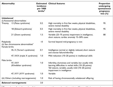
Table 1 illustrates the types of chromosome
abnormalities commonly detected and their frequency in relation to birth numbers. This
1
Chapter 1
Introduction and background
* During 1999 the recognised term for the syndrome trisomy 21 became, by published convention, ‘Down syndrome’,
Introduction and background
2
shows that the overall incidence of all chromosome abnormalities is approximately 9.1 per 1000 in newborns. Of these, the table shows that a majority of cases (6.1 per 1000 newborns) have some extra or missing chromosome material. Such chromo-some abnormalities are termed ‘unbalanced’ and are linked to varying levels of physical and mental disability. Included in this group are trisomies 21, 18 and 13, and the numerical sex
chromosome abnormalities.
The remainder of abnormalities (found in approximately 3 per 1000 newborns) are termed ‘balanced’ chromosome abnormalities. Individuals with these abnormalities do not usually have any symptoms. However, as carriers they are at in-creased risk of having chromosomally unbalanced offspring. In turn, these children have increased risk of miscarriage and/or the birth of children with learning difficulties and physical disabilities.
The most common chromosome abnormality is trisomy 21, commonly termed Down syndrome,
where there is an extra chromosome 21. Trisomy 21 occurs in approximately 1.5 per
1000 newborns.6Children with this condition
may suffer from learning difficulties and/or certain physical defects. For example, it is estimated that Down syndrome accounts for about 25% of all cases of moderate and severe learning difficulties in children of school age; it may also be associated with congenital heart malformations occurring in 15–50% of
children with the syndrome.5,7
Currently in the UK, the prenatal testing process for detection of Down syndrome and other chromosome abnormalities is conducted in two steps:
• Step 1: risk assessment and case selection for step 2
• Step 2: prenatal diagnostic testing.
Effective prenatal diagnosis of chromosome abnormalities is thus dependent on both accurate
TABLE 1 Chromosome abnormalities commonly detected, their frequency and consequences
Abnormality Estimated Clinical features Proportion
incidence undergoing
per 1000 spontaneous
births5 pregnancy
loss (%)
Unbalanced
(i) Autosomal abnormalities
Trisomy 13 (Patau syndrome) 0.2 High mortality in first few weeks, physical disabilities, 95 severe mental disability
18 (Edward syndrome) 0.3 High mortality in first few weeks, physical disabilities, 95 severe mental disability
21(Down syndrome) 1.5 Variable (25–75 points) impairment in intelligence, 80 short stature, cardiac anomaly 15–50% cases
Polyploidy ~0 Survival beyond mid-pregnancy is rare (ii) Sex chromosome abnormalitiesa
Female births
45, X (Turner’s syndrome) 0.1 Intelligence normal or slightly reduced, short stature and ovarian failure/infertility
47, XXX (triple X syndrome) 1.0 Mild reduction (10–20 points) in intellectual skills Male births
47, XXY 1.0 Infertility, clumsiness and variable, but usually mild, (Klinefelter syndrome) learning difficulties in verbal skills (10–20 points)
Tall stature, variable, usually mild (10–20 points) impairment in intelligence
47, XYY (XYY syndrome) 1.0 Variable
(iii) Others (including rearrangements) 1.0 Risk of having chromosomally unbalanced offspring
Balanced rearrangements 3.0
a
[image:14.595.72.538.111.473.2]3
case selection and effective diagnostic testing, and steps 1 and 2 are therefore closely linked.
Prenatal risk assessment
Step 1, which consists of prenatal risk assessment, is based on a combination of epidemiological risk factors (e.g. age, family history) and test results (e.g. serum screening). Risk is normally expressed in terms of the probability of a fetal chromosome abnormality being present. Thresholds are sub-sequently used to select those cases that should be offered prenatal diagnostic testing. For example, if the risk is assessed to be 1 in 250 or greater,
diagnostic testing may be advised.†Careful
assessment is necessary because of the invasive nature of the diagnostic test procedure (i.e. amniocentesis). The likelihood of a severe fetal abnormality must be set against the possibility of loss of a ‘normal’ fetus.
Women with a family history of chromosome abnormalities or previous births with chromosome abnormalities are usually judged to be at risk and offered diagnostic testing. For other women, single or combined selection criteria are used to identify those who should be offered amniocentesis. These include:
• maternal age, thresholds typically set by individual clinicians in the range 35–37 years of age
• abnormal levels of serum marker(s) detected during routine screening of maternal blood • structural fetal defects or malformations,
and/or abnormal thickening of the nuchal fold detected during ultrasound scanning • those couples where either parent is a carrier
of a structural chromosome abnormality • parental anxiety.
It is important to note that the vast majority of women undergo amniocentesis based on an assessment of risk for Down syndrome, although the diagnostic test used will in fact detect a large number of other chromosome abnormalities,
as illustrated in Table 1.‡
Maternal age
Historically, an age of 35 years or greater was generally accepted to be an important risk factor for Down syndrome. However, if maternal age is used as the only criterion for selection for diagnostic testing, it has been found that only approximately 30% of cases of Down syndrome
are identified prenatally.7,9This is because the
majority of Down syndrome babies are born to women below the selected cut-off age. Although the absolute risk of chromosome abnormality is lower for younger women, birth rates are higher. The shortcomings of a single age-based selection criterion for diagnostic testing is now widely
recognised.10–13
Maternal serum screening tests
A more accurate estimation of risk of Down syndrome can be achieved by combining maternal age with the result of a maternal serum screening test using various biochemical markers. Although practice varies between Trusts, routine maternal serum screening of all pregnant women (using varying combinations of biochemical markers)is now well established in the NHS.14
Serum screening not only enables improved prenatal diagnosis by identifying younger women who are at high risk, thereby increasing detection rates, but it may also exclude older women who are found to be at lower risk, thereby reducing the risks to the normal pregnancy associated with amniocentesis. A recent report for the Health Technology Assessment (HTA) Programme sug-gests that Down detection rates, using a combined maternal age and serum screening approach, and accepting a false-positive rate of 5%, range from 59% for two serum markers to 76% for
four serum markers.8In general, selection of
women for amniocentesis based on serum screening and maternal age is also reported to be more cost-effective than the use of
maternal age alone.7–9,15–18
Nevertheless, the role of routine serum screening continues to be reassessed as the sensitivity of serum screening tests in general use is constantly
re-examined.14,19–22
† Cut-off risk thresholds for diagnostic testing vary from hospital to hospital but are normally in the range 1:250 to
1:300. In many cases, the exact figure is decided regionally in conjunction with specialist genetics services.
‡ There is research evidence to show that the quality of information given and the understanding of patients about
Introduction and background
4
Ultrasound scans
Most women in the UK receive an ultrasound scan between the 18th and 22nd weeks of pregnancy (second trimester scan). This scan is used both to check the accuracy of estimates of gestational
age and to examine the fetus.19
It has recently been argued that a mid-trimester ultrasound examination, together with maternal age, may provide a valid option for risk assessment for abnormalities such as Down syndrome, without
the use of serum screening.19Several studies
have looked specifically at the use of ultrasound scanning to identify high-risk pregnancies for Down syndrome. Earlier anomalies scanning at 11–12 weeks (first trimester) and measurement of the thickness of the translucent nuchal fold (skin at the back of the neck) may prove useful in this
respect.10It has been demonstrated that increased
nuchal translucency can be indicative of Down
syndrome.8However, trials in a routine setting
have to date given varied results, suggesting that technical issues related to the test need further work before transfer from specialist centres is feasible. Nevertheless, a diagnosis of nuchal translucency, or absolute values for the thickness of the nuchal fold, combined with age, have become an additional criterion for selection
for amniocentesis in some UK centres.19,23
Parental anxiety
Psychological indicators are not formally part of hospital protocols for selection of women for amniocentesis. In specific instances, however, parental anxiety is known to be used as a clinical indication for amniocentesis. It appears that use of such an indicator is varied and dependent on
individual obstetric consultants.24
Prenatal diagnostic testing
Once prenatal risk assessment has identified a pregnancy as ‘at risk’, the second stage in the testing process is to offer the woman a diagnostic test to identify whether a fetal chromosomal abnormality is present or not. If present, the test will identify the nature of any abnormality.
The current gold standard test used for diagnosis,
karyotyping, is a test based on culture and exam-ination of fetal cells obtained through amnio-centesis or CVS. In the second trimester, collection of fetal cells is normally by amniocentesis and in the first trimester by CVS (and occasionally amniocentesis). Not all attempts to obtain fetal cells are successful; around 2% of all attempts
to obtain amniotic fluid are unsuccessful, and
between 1.3 and 5% of CVS attempts fail.8,25,26
Definitive diagnosis is then accomplished using conventional cytogenetic analysis of the fetal cells cultured from the amniotic fluid (or chorionic villus sample). Failure to culture the fetal cells obtained occurs in a small number of cases; the average rate reported in the UK in 1999 was 0.3%
of cases.27In addition, there is a slight possibility
of misleading results because of overgrowth of maternal cells, estimated to occur in around 0.2%
of cultures.28Chorionic villus samples can also
undergo direct analysis (without culture).
Karyotyping is a reliable and accurate test for trisomy 21 (Down syndrome). The karyotyping detection rate for Down syndrome for those samples that have been successfully cultured is
99.7%.29This test also allows the detection of
all chromosome abnormalities.30,31However, the
test is non-automated and involves a lengthy procedure requiring specialist scientific expertise; it is therefore slow and expensive.
From the patient’s point of view, the main
disadvantage of the karyotyping test is that samples must be cultured for an extended period of time. The mean time to diagnosis is currently 13.4 days (weighted mean related to number of samples
processed in laboratory, range 7.3–20.2 days).27
Large variations in the rate at which cells grow, for individual specimens, leads to a wide range of reporting times. Obstetricians usually therefore advise patients that the wait for results following amniocentesis could be between 3 and 4 weeks. This waiting for diagnostic test results can place a significant emotional burden on women and their partners.
Developments in prenatal
diagnostic testing
Ideally, a prenatal diagnostic test should provide speedy as well as accurate information, and a number of methods have been proposed over the years to reduce the time required for cytogenetic diagnosis using karyotyping. These have included the harvesting of amniotic fluid cells in situ rather
than from tissue culture flasks,32the introduction
of specialised culture media,33,34and the use of
lower percentages of oxygen in culture.35Some
laboratories now record an average time to
report-ing of results of 7 days.19,27However, all these
5
could only reduce analysis time to 6 days in
individual cases.36
Three important trends predicted for future prenatal testing for Down syndrome are: (i) improved targeting of screening for identification of high-risk pregnancies; (ii) a shift from second to first trimester screening; and (iii) the introduction
of molecular tests to provide more rapid results.10
A recent report from the HTA Programme has examined the first of these, the possibility of improving current criteria for selection for
pre-natal chromosome analysis.8The second trend
is currently the subject of considerable research because moves towards earlier diagnosis of fetal abnormality (i.e. sampling of fetal cells in the first rather than the second trimester) would have obvious advantages. One approach being suggested is the wider use of CVS. However, this technique has a higher miscarriage rate than second trimester amniocentesis. Furthermore, there is a risk of inducing limb defects before 10 weeks. No formal, large-scale evaluation exists regarding the safety and efficacy of CVS relative to second trimester
amniocentesis.1,25,26An alternative approach may
be first trimester amniocentesis. This is technically possible, although the volume of amniotic fluid currently required for cell culture and karyotyping is considered to be larger than would be feasible
without further increasing the risk to the fetus.37
First trimester amniocentesis therefore remains to be fully evaluated in terms of its safety and efficacy relative to CVS and second trimester amniocentesis.
An alternative approach to earlier diagnosis of fetal abnormalities would be to reduce the time between sampling and diagnosis. Recent advances in genetic techniques mean it is now possible for laboratories to introduce new molecular methods to reduce the time between sampling and chromo-some diagnosis to 24–48 hours. This would have the effect of potentially providing diagnosis up to 2 weeks earlier. These techniques also require smaller amounts of cellular material than karyo-typing. Thus, the volume of amniotic fluid required for molecular testing is much smaller, and there may in future be an increased
possibility of first trimester amniocentesis using
molecular technology.§
Two main types of molecular method are currently available for prenatal diagnosis of chromosome abnormality. These are fluorescence
in situ hybridisation (FISH) and the quantitative
polymerase chain reaction (Q-PCR). These tests, which are described in greater detail in chapter 2 (see page 9), can be performed without the need to culture fetal cells.
The FISH test involves the use of chromosome-specific probes, which have fluorescent labels attached. The probes are applied to cellular preparations made without a lengthy process of fetal cell culture. Detection systems involve the use of fluorescence microscopy equipment or other similar image analysis systems. FISH is amenable to a certain amount of automation, and there are currently a few automated FISH systems on the market, but they have not yet been clinically evaluated. Some are also under
development in universities.38
The Q-PCR test, the alternative to FISH, is based on a versatile and widely used amplification tech-nique through which even the smallest amount of a defined DNA target can be amplified to provide quantities that are detectable and identifiable. Amplification utilises the action of an enzyme (DNA polymerase), which catalyses the rapid synthesis of new strands of DNA from an original strand using a primer. Detection requires a DNA analyser, an expensive item of equipment.
Background to the
current research
Pressure from medical champions, the media, special interest groups and industry to introduce new tests before they have been fully evaluated, in terms of both their technical performance and their cost-effectiveness, has always been a challenge for those commissioning services. Currently, the NHS faces a number of difficult challenges posed
by new genetic tests.10,13,39,40Although much of the
current debate centres on the introduction of
§ Much effort is also currently being devoted to finding new diagnostic techniques that avoid the invasive procedure
Introduction and background
6
entirely new diagnostic testing, made possible by genetic advances, in some instances the same technological developments can also be applied to existing diagnostic procedures. Prenatal diagnosis is one such area, with the possibility of introducing new molecular methods to provide more rapid diagnosis of fetal chromosome abnormalities.
With over 650,000 pregnancies per annum in England and Wales, piecemeal introduction of a new prenatal test technology could lead to a substantial increase in costs, especially if the new molecular tests were to be used as an ‘add-on’ to existing karyotyping. Furthermore, widespread roll-out of these molecular tests could also have substantial implications for the organisation of genetics services, including counselling services, and for training of laboratory staff in these new techniques. As was highlighted in a recent HTA publication, the current pattern of service in the UK already demonstrates “inequality of access to service and ... current multiple stepwise
unco-ordinated screening of Down’s syndrome”.8
It is important, therefore, that any widespread introduction of new molecular methods in the antenatal setting should include careful con-sideration of the costs and benefits of different testing strategies and their implementation. For this to be possible, research evidence is required to support decision-making.
Aims of the research
The present project was commissioned by the NHS HTA Programme in 1997. The purpose of the study was to evaluate molecular prenatal diagnosis for Down syndrome by comparing two molecular alternatives (FISH and Q-PCR) with traditional laboratory karyotyping.
The main aim of the research project was to provide reliable evidence for the introduction of molecular methods into NHS laboratories for prenatal diagnosis of chromosome abnormalities (principally Down syndrome). Within this overall aim, the objectives of the research were:
• to establish the reliability, sensitivity/specificity and turnaround time of FISH and Q-PCR
diagnosis (respectively on uncultured
amniocytes and DNA extracted from amniotic fluid samples) compared with the existing culture-based test (karyotyping)
• to estimate the cost of these new techniques relative to the existing test under various conditions (e.g. different throughputs, staffing structures, diagnostic testing strategies, etc.) • to assess the value to women and clinicians of
more rapid diagnosis
• to estimate the cost-effectiveness, cost–utility and cost–benefit of these molecular tests, and to model the impact of possible changes in current testing protocols.
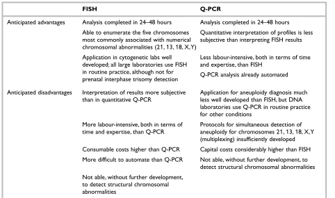
Table 2 provides an overview of the characteristics of
the two molecular tests at the outset of the study.
Study design
The study design involved a phased introduction of the technology, with the objective of deter-mining the extent to which routine use of molecular tests on a large scale would be reliable, effective and cost-effective compared to existing cytogenetic methods.
The design chosen was a two-stage trial with technical performance assessed in a laboratory setting in the first stage, and effectiveness and cost-effectiveness measured in a service setting in the second stage. The evaluation was conducted at two study sites (the West Midlands Regional
Genetic Laboratory¶and Northern Ireland
Regional Genetics Centre, Belfast) and the Trusts served by these sites.
The evaluation framework used in the study was based on the hierarchy formulated by Fineberg
in 1977 for diagnostic technologies in general.41
An adapted form was developed for prenatal diagnosis with the following five levels:
• Technical capacity – does the test perform reliably and deliver accurate (i.e. precise) information?
• Diagnostic accuracy – does it contribute to an accurate diagnosis of chromosome abnormalities?
¶ Six months after commencement of the study, the West Midlands Regional Genetic Laboratory (based at the
7
• Diagnostic impact – does the test replace other diagnostic tests or procedures?
• Patient outcome – does the test contribute to improved health or reduced anxiety for the patient?
• Cost-effectiveness – does use of the
molecular test improve the cost-effectiveness of healthcare compared to alternative interventions?
Stage
1
At the outset of the study, the developmental state of the two tests was slightly different. One test (FISH) was already developed and available as a commercial kit to test for trisomies 21, 13 and 18, and X and Y aneuploidy. The other molecular test (Q-PCR) still required further development research in order to produce a multiplex test that could detect trisomies 21, 13 and 18, and X and Y aneuploidies simultaneously. This essential development work was therefore conducted as part of stage 1 of the study.
During the first part of the study, the technical performance of the new molecular methods was assessed through an independent masked comparison of each molecular test against the
accepted gold standard, karyotyping.41–43For
Q-PCR, this included assessment of both a Down only Q-PCR test and a later multiplex Q-PCR test
capable of detecting trisomies 21, 13 and 18, and X and Y sex chromosome abnormalities. For FISH, a 5-probe test, able to identify the same abnormalities as the multiplex Q-PCR test, was assessed. All amniotic samples, where sufficient residual fluid was available after allocation for karyotyping, underwent a molecular test in addi-tion to karyotyping. For each sample, one of the molecular tests (Q-PCR or FISH) was performed because there was generally insufficient fluid for both molecular tests and karyotyping to be carried out on a single sample. The scientists performing the molecular tests were blind to the karyotyping results, and vice versa. Molecular test results were not available to clinicians during this technical evaluation stage.
[image:19.595.55.537.112.397.2]A detailed costing study of the two molecular tests and of karyotyping was performed. In addition, a survey of all UK cytogenetics laboratories was carried out to identify the molecular test preferences of laboratory staff, and to gather information on training require-ments, laboratory skill-mix, karyotyping test prices, access to DNA sequencers, and any planned introduction of molecular tests. In addition, a questionnaire survey of obstetricians in the two study regions was carried out to identify clinical perceptions of the benefits of faster molecular test results.
TABLE 2 Relative advantages and disadvantages of FISH and Q-PCR at outset of study
FISH Q-PCR
Anticipated advantages Analysis completed in 24–48 hours Analysis completed in 24–48 hours
Able to enumerate the five chromosomes Quantitative interpretation of profiles is less most commonly associated with numerical subjective than interpreting FISH results chromosomal abnormalities (21,13,18, X,Y)
Application in cytogenetic labs well Less labour-intensive, both in terms of time developed; all large laboratories use FISH and expertise, than FISH
in routine practice, although not for Q-PCR analysis already automated prenatal interphase trisomy detection
Anticipated disadvantages Interpretation of results more subjective Application for aneuploidy diagnosis much than in quantitative Q-PCR less well developed than FISH, but DNA
laboratories use Q-PCR in routine practice for other conditions
More labour-intensive, both in terms of Protocols for simultaneous detection of time and expertise, than Q-PCR aneuploidy for chromosomes 21,13,18, X,Y
(multiplexing) insufficiently developed Consumable costs higher than Q-PCR Capital costs considerably higher than FISH More difficult to automate than Q-PCR Not able, without further development, to
detect structural chromosomal abnormalities Not able, without further development,
Introduction and background
8
All patients with abnormal karyotyping test results, identified by both genetics services during stage 1, were followed up with a questionnaire (anony-mised to the research team) to the individual’s obstetrician. This questionnaire asked about the selection criteria used in each specific case, and recorded the obstetrician’s retrospective test preference, supposing a molecular test had been available for this patient. A similar-sized sample of normal results was followed up in the same way.
At the end of stage 1, a preliminary evaluation of the two molecular tests was performed, taking into account costs and technical performance, in order to select one or both tests for further assessment in a routine service setting during stage 2 of the project.
Stage 2
During this second stage of the study, the benefits to women and their partners of a more rapid result were explored in a routine service setting. Although there was some experience of using FISH in a service setting in UK laboratories, including the study sites, there was no similar experience of using multiplex Q-PCR. The effective transfer of the Q-PCR test to the service setting was therefore expected to require more time and, possibly, some adaptation. Once a molecular test was established in the service setting, its technical performance could be assessed under routine service conditions during stage 2.
Although abnormal test results are important when considering the clinical benefits of a more rapid diagnostic test, these represent only a very small minority of prenatal test results (3% of cases). An important benefit to the whole group of women being tested, therefore, was expected to be associated with normal test results (97% of
cases), linked to the reduced period of anxiety while waiting for test results.
During stage 2 of the study, molecular test results were made available to clinicians and patients in hospitals served by the two centres, with confirm-atory karyotyping results being provided at a later date. The benefits of a molecular test result were assessed in a number of ways during this stage of the project.
The impact of molecular tests on levels of anxiety and health status of a sequential sample of women who received a molecular test was compared with a control group of women receiving only the traditional karyotyping test. Women in each group were asked to record their health status/quality of life (QoL), together with anxiety at specific points during the test wait period until 7 weeks after amniocentesis. The last measurement was there-fore approximately 4 weeks after the karyotyping result. This latter measurement was made as there is evidence that anxiety may persist beyond receipt of test results.44
9
Introduction
Of the two molecular tests assessed in this project, FISH was the one with a longer history in prenatal diagnosis and at the start of the study, FISH was already playing an important role in diagnostic cytogenetics, although not for routine prenatal testing. In contrast, Q-PCR was less well developed in terms of its application for detection of fetal chromosome abnormalities, but was widely used in genetics for the diagnosis of other genetic disease. Although they differed in their history, both molecular tests represented a major technical advance, which could potentially prove to be very valuable in prenatal diagnosis.
Background to the FISH test
FISH was first introduced in clinical cytogenetics
in the USA in 1988,45following which there was a
rapid development in the technology.46The test
was first introduced into the UK in 1991.47The
technique uses chromosome-specific probes with fluorescent labels attached; these probes are now available in commercial kit form. Detection requires microscope systems, which may range from simple fluorescence microscopes to more expensive image analysis systems such as micro-scope and cooled charged-coupled device cameras. FISH is a less labour-intensive test than the current karyotyping technique. Also, although FISH is relatively expensive in terms of consumables, the capital (equipment) costs are relatively low compared with Q-PCR.
History of the FISH test in
prenatal diagnosis
Early FISH studies on uncultured amniocytes made use of single chromosome-specific probes (e.g. for chromosome 21). However, these first probes (centromeric repetitive or alphoid) were found to show cross-hybridisation between certain
chromosomes (for example, the two probes inter-acted for chromosomes 13 and 21). This discovery led to the development of different types of probe (chromosome cosmid contig probes and YACs). The use of the first of these to detect Down syn-drome (trisomy 21) was successfully demonstrated in a prospective study of 500 uncultured amniotic
fluid samples as early as 1994.48Around 2 years
later, the YAC probes started to be used in the UK to provide rapid preliminary reports on samples (Lowther G, Duncan Guthrie Institute of Medical Genetics, Glasgow: personal communication, 1996). At the same time, it was demonstrated that a combination of five FISH probes could be used to examine all five chromosomes most commonly associated with chromosomal abnormalities
(21, 18, 13, X, Y) in a single*multicolour FISH
hybridisation experiment.3Subsequently, 5-probe
FISH test kits were developed and marketed by commercial manufacturers.
Background to the Q-PCR test
Q-PCR, the alternative to FISH, was at the outset of the study a less well established test for prenatal diagnosis. The technique amplifies DNA by the polymerase chain reaction method using selected primers, and it is theoretically possible to amplify DNA from a single copy of the target sequence. The cost of reagents for Q-PCR is lower than for FISH, although a royalty payment has to be made for service use of Q-PCR, negotiated by the NHS with the licence holders. Quantitative interpret-ation of Q-PCR chromosome profiles is also less subjective than using the human eye to interpret
FISH test results.49In addition, Q-PCR is less
labour-intensive than FISH and is more easily automated; Q-PCR products can be automatically analysed using a DNA sequencer and associated software. The capital cost of this equipment is high [e.g. Applied Biosystems, approx. £93,000 (all prices in this report are based on 1998
Chapter 2
Development and technical evaluation
of molecular tests
* The hybridisation is carried out with the 13 and 21 probes applied to one area of the microscopy slide and the 18,
Development and technical evaluation of molecular tests
10
prices)], but many genetic services already have sequencers, or have negotiated access to sequencers run by others.
History of the Q-PCR test in
prenatal diagnosis
Successful Q-PCR amplification was first described
in 1991 to detect X chromosome differences,50
at about the same time as the FISH test was being introduced into service in the NHS. The technique was then modified by Mansfield in order to be able to detect trisomies 21, 18 and the triple-X syndrome using small tandem repeat (STR)
markers combined with the Q-PCR reaction.51
A small trial (134 cases) investigating the accuracy of this technique (using the STR marker D21S11) in detecting trisomy 21 in varied samples (amniotic fluid, chorionic villus biopsy and fetal blood
samples) was reported 3 years later, in 1994.52
Subsequently, a number of small trials reported, where this technology was applied to other selected aneuploidies. Eventually, in 1995, it was shown that it is possible to use a combi-nation of Q-PCR markers (rather than a single marker) to simultaneously diagnose trisomies 21 and 18, and X and Y chromosome
abnor-malities.53,54However, by 1997 (the start of the
present study), none of these multiplex assays had been tested for prenatal diagnosis, either in large patient populations or under routine conditions in NHS laboratories.
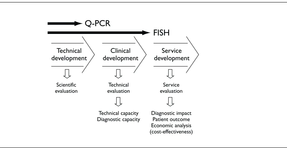
At the outset of the study, therefore, Q-PCR had not yet reached the same level of development as FISH for prenatal testing, as illustrated in Figure 1.
Development of the Q-PCR test
As has been reported above, at the start of the study, although a 5-probe FISH test was available to identify trisomies 21, 13 and 18 and X, Y-related abnormalities in a single process, a similar five chromosome Q-PCR (multiplex) test was not yet available as a single test. Translational research was therefore required at the outset of this HTA project to produce a Q-PCR test that was com-parable to 5-probe FISH. This research was carried out in two stages. Firstly, Q-PCR was refined and underwent scientific and preliminary technical evaluation for Down syndrome (trisomy 21) detection only, while simultaneously, a multiplex test was developed. Then, the multiplex test similarly underwent scientific and preliminary technical evaluation in the laboratory for the five most common chromosome abnormalities. By the end of this process, both FISH and Q-PCR were at a stage to undergo comprehensive technical and service evaluation (Figure 1).
Preliminary assessment of single
Q-PCR test (for Down syndrome
detection)
Before the start of the main study, Q-PCR was first assessed for the detection of trisomy 21 (Down syndrome) only at Heartlands Hospital, Birmingham (supported by the West Midlands Locally Organised Research Scheme). The material used (1430 samples) was surplus to that required for amniotic fluid cell culture. Testing of these samples using single Q-PCR demonstrated that the use of only two markers was informative
Technical development
Clinical development
Service development
Scientific evaluation
Technical evaluation
Service evaluation
Technical capacity Diagnostic capacity
Diagnostic impact Patient outcome Economic analysis (cost-effectiveness)
Q-PCR
[image:22.595.73.537.532.772.2]FISH
11
in 97.6% of samples. In the remaining 2.4% of cases (34/1430), an additional marker would have been required for trisomy 21 diagnosis to be achieved. In the 1430 samples tested, only 1 cytogenetically verified trisomy 21 case fell into the group requiring an additional marker, while another 20 trisomy 21 cases were readily identified by the single Q-PCR. There were no (0/1430) false-positive or false-negative diagnostic errors per se. The only complication encountered was maternal blood contamination, which occurred in less than 1.5% of amniotic fluid samples.
At the start of this study a further 737 samples were analysed. There was no significant change in the earlier findings with these additional samples included in the analysis. Overall a total of 2139 samples were analysed, as a total 28 out of 2167 of samples (1.3%) were bloodstained and excluded. Details of the full assessment of 2167 samples, including additional samples processed as part of this study, are provided in appendix 1.
Development and preliminary
assessment of the multiplex Q-PCR test
It proved more complicated than anticipated to combine markers for the detection of aneuploidies other than trisomy 21. In particular, problems were encountered in identifying which specific markers could be successfully combined and used together (multiplexed). The research team therefore had to undertake a more elaborate and staged technical development process than was initially anticipated. First, three markers were combined and used to-gether for the detection of trisomy 21 (D21S11, IFNAR and D21S1270). Once this element of the test was optimised, other markers were added one by one. Eventually, two separate combinations of multi-plexing were devised, each containing 5–6 markers. The Q-PCR test finally selected for this study used five markers for chromosome 21 (D21S11, IFNAR, D21S1270, D21S1412, D21S1411); three markers for chromosome 18 (MBP-A, MBP-B, D18S535); two markers for chromosome 13 (D13S631, D13S634); one marker (AMXY) for sexing; and one marker for the X chromosome (XHPRT).Technical evaluation of
molecular tests
Following test development and preliminary scientific evaluation, the evaluation of a diagnostic
technology then focused on comprehensive tech-nical and service evaluation of the test (Figure 1). In the most widely accepted hierarchy (Fineberg)
for evaluating diagnostic tests,41,55the first two
stages of such an evaluation are:
• Technical capacity – does the test perform reliably and deliver accurate (i.e. precise) information?
• Diagnostic accuracy – does the test contribute to an accurate diagnosis (of chromosome abnormalities)?
Both molecular methods (FISH and Q-PCR) were assessed in terms of these two aspects of test performance relative to karyotyping. During stage 1 of the study, the samples tested (anony-mised to the research team) were drawn from amniotic fluids received at three centres: Birming-ham Heartlands Hospital Trust, BirmingBirming-ham Women’s Hospital and the Northern Ireland Regional Genetics Centre (Belfast City Hospital Trust). The following approach was then adopted:
• a karyotyping test was carried out on each sample
• any amniotic fluid in excess of that required for cell culture and karyotyping was used to perform molecular tests (approximately
1–2 ml†was necessary for each test).
In the original research protocol, the plan was to perform both molecular tests, Q-PCR and FISH, in addition to karyotyping, for each sample. How-ever, in most cases there proved to be insufficient fluid to perform both molecular tests. As a result, the research protocol was adapted to include allocation of samples to only one of the molecular tests, FISH or Q-PCR, as well as karyotyping.
Data recorded for all samples processed as part of the study included indications for referral; estimated gestational age; amount of fluid; pres-ence of visible blood contamination; turnaround time for laboratory processing (molecular tests and karyotyping); any technical difficulties encountered in performing the test; and karyo-typing and molecular test results. Molecular test results were not released to clinicians or women during the first, technical evaluation stage of the study. The only use made of molecular test results was within the laboratory, and this was left to local decision-makers. In the service laboratories, an
† In practice, it was found that the volume required was dependent on gestation. More fluid was needed for earlier
Development and technical evaluation of molecular tests
12
abnormal molecular test result was likely to lead to earliest possible harvesting of cell cultures and therefore a more rapid karyotyping result.
Test repeatability (the variance in a test result that occurs when the test is repeated on the same specimen) could not be assessed because insufficient amniotic fluid was available after fluid had been allocated for karyotyping and the selected molecular test.
Fineberg level
1
: technical
capacity of molecular tests
Technical capacity – whether a tests performs reliably and delivers accurate (i.e. precise) infor-mation – was assessed in the study laboratories for three molecular test variants: the 5-probe commercial FISH kit (AneuVysion, Vysis Ltd) and two forms of in-house Q-PCR test, a Down syndrome test and the multiplex Q-PCR test. The 5-probe FISH and multiplex Q-PCR both tested for trisomies 21, 18 and 13 and numerical sex chromosome abnormalities.
Technical difficulties were systematically recorded throughout the study. There were no reported incidences of major equipment malfunction in either stage 1 or stage 2 of the research project (described earlier, see pages 7 and 8). However, some very early failures of Q-PCR were observed in stage 2 of the study as the test entered into the routine laboratory setting, moving from the
University of Warwick to the Regional Genetic Laboratory in Birmingham. These were a result of attempts to speed up reporting time by using an alternative test protocol, leaving the sample for DNA precipitation in the freezer at –70°C for 2 hours rather than at –20°C overnight. No Q-PCR amplifi-cations were obtained for any of these samples.
The main study findings relating to the technical capacity of molecular tests are summarised in
Tables 3 and 4 for FISH and Q-PCR tests
respec-tively. The top section of each table (labelled i) first lists the total number of samples allocated to testing using 5-probe FISH or both types of Q-PCR test (Down syndrome only and multiplex Q-PCR). Using 5-probe commercial FISH kits, a total of 859 samples were processed (400 samples at the University of Warwick and 459 in Northern Ireland). Using the Down only Q-PCR test, 2367 samples were processed (2167 samples at the Uni-versity of Warwick and 200 in Northern Ireland). Following final development of the multiplex test for the other trisomies (13, 18) and numerical sex chromosome abnormalities, a further 717 samples were processed at the University of Warwick using the multiplex Q-PCR test.
When a prenatal test is carried out on a sample, there are three main types of test outcome that may be classified as a ‘failure’. Firstly, the test process may itself fail. In the present instance, this might be because of a failure to culture cells from the amniocytes (in the case of karyotyping) or a failure to amplify (Q-PCR) or hybridise
TABLE 3 Results of technical evaluation of FISH molecular tests in stage 1
FISH
Northern West Total
Ireland Midlands (%)
i Total samples allocated to test 459 400 859 (100) Karyotyping tests where no result obtained 1 1 2 (0.2) Molecular tests where no result obtained 54 4 58 (6.8) ii No. of abnormalities disclosed by karyotyping 23 15 38 (4.4) No. of abnormalities disclosed by molecular test 23 13 36 (4.2) No. of abnormalities not disclosed by molecular test 0 2a 2a(0.2) iii Specimens excluded from molecular tests because of visible macroscopic 26 UnknownbUnknownb
bloodstaining
Evidence during test of maternal cell contamination in original specimenc 26 10 36 (4.2)
a
(a) 45XY, der(13:14) (p10:p10) pat and (b) 46XY del(15)(q26.1), neither expected to be detectable by FISH using the Aneuvision protocol. In both these cases clinical risk of abnormality was assessed to be high because of (i) familial history and (ii) abnormal scan result, which would imply that karyotyping would be performed
b
All specimens with visible bloodstaining excluded before referral to laboratory in West Midlands
c
13
(FISH) DNA. In such a case, no test result would be obtained.
A second outcome may be that although the test process does not fail, the result obtained is inconclusive or uninformative. Such an outcome may occur in the case of Q-PCR, for example, when a second sequencer run is required to secure a reportable result but there is insufficient DNA available. In such a case also, no test result would be obtained. Finally, a test may be completed and a result produced, but there may be an incorrect result or test error.
It has long been recognised that evaluation of diagnostic tests tends to concentrate on the last of these and that the first two types of outcome (test failure and inconclusive/uninformative
results) are rarely reported in the literature.56
The present study recorded all cases where no test result was obtained, including the first two types of outcome.
The top sections of Tables 3 and 4 (rows labelled i) indicate the number of karyotyping and molecular tests where no result was obtained. For FISH,
Table 3 shows that 58/859 molecular tests fell into
this category and therefore only 801 molecular tests produced a result. For Q-PCR, Table 4 shows that there were 47/2367 Down only tests in which
no result was obtained (2320 produced a result). Similarly for multiplex Q-PCR, 68/717 tests pro-duced no result and in the remaining 649 cases a result was obtained. Both FISH and Q-PCR therefore exhibited a relatively high incidence of test failure or inconclusive/uninformative results during the introduction period: 9.5% for multiplex Q-PCR, 6.8% for 5-probe FISH, and 2.0% for Down only Q-PCR. The comparable level for karyotyping was less than 0.2%.
Factors influencing the level of test failure or inconclusive/uninformative results varied. In the case of multiplex Q-PCR, during the development stage insufficient amniotic fluid sometimes made it impossible to repeat the test three times when the first two markers were uninformative (because the individual was not polymorphic for either of these first two markers). This accounted for many of the multiplex Q-PCR tests where no result was obtained. Another possible factor influencing Q-PCR tests, maternal cell contamination, is discussed on page 15 (‘Molecular test results and maternal cell contamination’). Factors influencing the level of failed tests for FISH are discussed in more detail on page 14 (‘Molecular test results and gestational age’).
In the middle section of Tables 3 and 4 (section labelled ii) information is presented, for all the
TABLE 4 Results of technical evaluation of Q-PCR molecular tests in stage 1
Q-PCR
Northern West Midlands Total (%)
Ireland
Down Down Multiplex Down Multiplex
only only only (%) (%)
i Total samples allocated to test 200 2167 717 2367 (100) 717 (100) Karyotyping tests where no result obtained 4a 0 0 4a(0.2) 0 (0.0)
Molecular tests where no result obtained 32 15b 68c 47 (2.0) 68c(9.5)
ii No. of abnormalities disclosed by karyotyping 11 37d 12 48 (2.0) 12 (1.7) No. of abnormalities disclosed by molecular test 8 32 10 40 (1.7) 10 (1.4) No. of abnormalities not disclosed by molecular test 3e 5e 2f 8e(0.3) 2f(0.3) iii Estimate of total cases of maternal cell contamination 22 28 Unknowng 50 (2.1) Unknowng
a
These specimens were part of the batch affected by an outbreak of bacterial contamination within the cell culture
b
No more DNA left for reanalysing the cases homozygous, and therefore non-informative, for both D21S11and IFNAR
c
Includes technical problems in development pilot phase, e.g. when trying to speed up report time (see page 12) or if insufficient sample size for initial multiplexing of Q-PCR
d
Q-PCR testing only for Down syndrome in this series. Five abnormalities were detected at karyotyping, none was Down syndrome
e
Molecular test not capable of detecting these chromosome abnormalities, none was Down syndrome
f
Both samples 47XXY (Klinefelter syndrome), the single polymorphic marker for X chromosome was uninformative
g