DIGITAL EVALUATION OF FUNCTIONAL OCCLUSION PARAMETERS
TEMPOROMANDIBULAR DISORDERS
1
Dr. Saraswathi Gopal, K., *
,2Dr.
1Professor & Head, 2Post Graduate Student Meenakshi A
4Chief Dental Implant surgeon, Best LASER Dental clinic, Valasaravakkam
ARTICLE INFO ABSTRACT
Background:
main role in determining the teeth health, chewing, muscle and TMJ function. Dysfunctional occlusal contacts can lead to disorders in dento
symptoms for the patient.
Aim and Objective:
analyses occlusal contact forces, occlusion time, and the effect of reducing disclusion time on symptoms of temporomandibular joint
Materials and Method
criteria for temporomandibular disorders (TMD), including the clinical protocol outlined by the International RDC/TMD consortium network (version: January 20, 2014) to assess the effi reduced disclusion time in lateral excursions to resolve the myofascial pain symptoms. As per the inclusion and exclusion criteria, static and dynamic interocclusal record was scanned and analysed using image software
The questionnaire was used for symptom assessment at every recall visit.
Results: Among the occlusal parameters, TMD patients had prolonged occlusion and disclusion time, variations in right and left qua
compared to post therapeutic evaluation in same individuals. Changes in the intensity of symptoms from reducing the disclusion time to <0.4 seconds were statistically significant with p 0.001from treatment day 1, and onward through the 3 months period of observation.
Conclusion:
musculoskeletal symptoms of myofascial pain, such that this meth therapeutic success.
Copyright©2017, Saraswathi Gopal et al. This is an open access article distributed under the Creative Commons Att use, distribution, and reproduction in any medium, provided the original work is properly cited.
INTRODUCTION
Temporomandibular Disorders (TMD) is a collective term which includes a group of clinical conditions affecting the stomatognathic system, in particular the muscles of mastication and the Temporomandibular joints (TMJ), which is characterized by a group of commonly reported
fairly localized pain, limited or asymmetric mandibular movements and TMJ noises (crepitations or clickings) (Okeson, 1997; Alberto Baldini et al., 2014). Because of the different methods used to assess TMD symptoms, which are often subjective, literature reports that the world prevalence of TMD varies between 7% and 84% of the population (Luther, 2007).
*Corresponding author: Dr. Padma, M.
Post Graduate Student, Department of Oral Medicine and Radiology,
ISSN: 0975-833X
International Journal of Current Research
Vol.
Article History:
Received 03rd
November, 2016 Received in revised form 14th
December, 2016
Accepted 08th
January, 2017
Published online 28th
February,2017
Citation: Dr. Saraswathi Gopal, K., Dr. Padma, M., Dr. Mahesh Kumar, P. and Dr. Murugavel, C.
parameters using t scan in temporomandibular disorders”,
Key words: Temporomandibular disorders, Occlusal contacts, Disclusion time, Occlusion time, Occlusal forces, Myofascial pain, T-scan.
RESEARCH ARTICLE
DIGITAL EVALUATION OF FUNCTIONAL OCCLUSION PARAMETERS USING T SCAN IN
TEMPOROMANDIBULAR DISORDERS
Dr. Padma, M.,
3Dr. Mahesh Kumar, P. and
4Dr. Murugavel
Student, 3Senior Lecturer, Department of Oral Medicine and Radiology, Meenakshi Ammal Dental College, Chennai
Chief Dental Implant surgeon, Best LASER Dental clinic, Valasaravakkam, Chennai
ABSTRACT
Background: Temporomandibular disorder (TMD) is a multifactorial disease and occlusal force has main role in determining the teeth health, chewing, muscle and TMJ function. Dysfunctional occlusal contacts can lead to disorders in dento-maxillary system, which may be accompanied by painful symptoms for the patient.
Aim and Objective: To evaluate the digital occlusal parameters using
T-analyses occlusal contact forces, occlusion time, and the effect of reducing disclusion time on symptoms of temporomandibular joint- muscular disorders.
Materials and Method: Myofascial pain symptomatic patients was recruited as per the diagnostic criteria for temporomandibular disorders (TMD), including the clinical protocol outlined by the International RDC/TMD consortium network (version: January 20, 2014) to assess the effi reduced disclusion time in lateral excursions to resolve the myofascial pain symptoms. As per the inclusion and exclusion criteria, static and dynamic interocclusal record was scanned and analysed using image software. 25 cases were treated in four visits. Recall occlusal parameters were recorded. The questionnaire was used for symptom assessment at every recall visit.
Among the occlusal parameters, TMD patients had prolonged occlusion and disclusion time, variations in right and left quadrant occlusal forces, more incidence of occlusal premature contacts compared to post therapeutic evaluation in same individuals. Changes in the intensity of symptoms from reducing the disclusion time to <0.4 seconds were statistically significant with p 0.001from treatment day 1, and onward through the 3 months period of observation.
Conclusion: The results indicate that T scan evaluation and then treating the dentition relieves the musculoskeletal symptoms of myofascial pain, such that this meth
therapeutic success.
is an open access article distributed under the Creative Commons Attribution License, which use, distribution, and reproduction in any medium, provided the original work is properly cited.
Disorders (TMD) is a collective term which includes a group of clinical conditions affecting the stomatognathic system, in particular the muscles of mastication and the Temporomandibular joints (TMJ), which is characterized by a group of commonly reported symptoms: fairly localized pain, limited or asymmetric mandibular movements and TMJ noises (crepitations or clickings) 2014). Because of the different methods used to assess TMD symptoms, which are literature reports that the world-wide n 7% and 84% of the
Department of Oral Medicine and Radiology,
Mostly it seems to afflict 10% of the population over 18 years of age, where chronic pain is experienced in the Temporomandibular region (LeResche, 1997). In fact, because of the variability of the symptoms of this cluster disorder, potentially confirm a diagnosis of TMD, it has been quite problematic to isolate a primary etiology that predisposes the patients to suffer symptoms, leading researchers to advocate that TMD is of multifactorial etiology. Possible causes for TMD are Bruxism, mandibular muscle activity, facial growth, and also other systemic, postural, metabolic, structural, traumatic, psychological, social and behavioral influences, which have been identified as possible predisposing, initiating, and maintaining factors for TMD (Greenberg
Baldini et al., 2013; De Boever and Carlsson, 1994). For many years Dental Medicine had considered an unfavorable dental occlusion as a principal cause of the occurrence of TMD, because TMD appeared to be more frequently
International Journal of Current Research
Vol. 9, Issue, 02, pp.46362-46369, February, 2017
INTERNATIONAL
OF CURRENT RESEARCH
Dr. Saraswathi Gopal, K., Dr. Padma, M., Dr. Mahesh Kumar, P. and Dr. Murugavel, C. 2017. “Digital evaluation of functional occlusion
”, International Journal of Current Research, 9, (02), 46362-46369.
USING T SCAN IN
Dr. Murugavel, C.
Department of Oral Medicine and Radiology,
, Chennai
Temporomandibular disorder (TMD) is a multifactorial disease and occlusal force has main role in determining the teeth health, chewing, muscle and TMJ function. Dysfunctional occlusal maxillary system, which may be accompanied by painful
-Scan device that senses and analyses occlusal contact forces, occlusion time, and the effect of reducing disclusion time on
Myofascial pain symptomatic patients was recruited as per the diagnostic criteria for temporomandibular disorders (TMD), including the clinical protocol outlined by the International RDC/TMD consortium network (version: January 20, 2014) to assess the efficacy of reduced disclusion time in lateral excursions to resolve the myofascial pain symptoms. As per the inclusion and exclusion criteria, static and dynamic interocclusal record was scanned and analysed visits. Recall occlusal parameters were recorded.
Among the occlusal parameters, TMD patients had prolonged occlusion and disclusion time, drant occlusal forces, more incidence of occlusal premature contacts compared to post therapeutic evaluation in same individuals. Changes in the intensity of symptoms from reducing the disclusion time to <0.4 seconds were statistically significant with p value of 0.001from treatment day 1, and onward through the 3 months period of observation.
The results indicate that T scan evaluation and then treating the dentition relieves the musculoskeletal symptoms of myofascial pain, such that this methodology increases clinical
ribution License, which permits unrestricted
Mostly it seems to afflict 10% of the population over 18 years of age, where chronic pain is experienced in the Temporomandibular region (LeResche, 1997). In fact, because of the variability of the symptoms of this cluster disorder, that potentially confirm a diagnosis of TMD, it has been quite problematic to isolate a primary etiology that predisposes the patients to suffer symptoms, leading researchers to advocate that TMD is of multifactorial etiology. Possible causes for ruxism, mandibular muscle activity, facial growth, and also other systemic, postural, metabolic, structural, traumatic, psychological, social and behavioral influences, which have been identified as possible predisposing, initiating, s for TMD (Greenberg et al., 2012; 2013; De Boever and Carlsson, 1994). For many years Dental Medicine had considered an unfavorable dental occlusion as a principal cause of the occurrence of TMD, because TMD appeared to be more frequently observed INTERNATIONAL JOURNAL
OF CURRENT RESEARCH
in patients with compromised or excessive vertical dimensions (De Boever et al., 2000). However, later studies suggested TMD had a multifactorial origin such that dental occlusion was considered to be only one factor that may affect the adaptive capacity of the Stomatognathic system, which could lead to the development of TMD (McNamara, 1997). The introduction of the Research Diagnostic Criteria for TMD (RDC/TMD) Axis – I provided standardized criteria for the diagnosis and classification of the different forms of TMD’s, categorizing them in three groups: Group I muscle disorders, Group II disc displacements and Group III other common joint disorders. The RDC facilitated creating statistically better intra- and interexaminer reliability when clinicians were observing and reporting patient signs and symptoms and provided a common model for any examiner (Manfredini et al., 2006; Baldini et al.,
2012).
In 1987 Maness et al., (1987) reported the development of the first computerized system designed for occlusal analysis (T-Scan I, Tekscan, Inc. Boston, MA, USA). The T-(T-Scan VIII system allows for the recording the Occlusion Time (OT) which is a dynamic parameter defined as the elapsed time from initial tooth contact until maximum intercuspation is reached in centric occlusion (Kerstein and Grundset, 2001), Disclusion time (DT) is the elapsed time in seconds measured from the beginning of a jaw movement made in one direction, when all teeth are relieved in complete contact, through until only canines or incisors are in contact. Disclusion Time can be measured in 3 different jaw movements; the Right excursion, the Left excursion, and the Protrusive excursion. With lengthy disclusion time present in an occlusal scheme, excessive muscle contraction occurs over time and leads to the buildup of toxic muscle contraction by- products (namely lactic acid) within the muscle fibers (Robert Kerstein, 2015). In a given day, a patient’s level of toxins will exceed the muscle’s ability to metabolize these toxic substances. This would then initiate an ischemic state followed by the clinical appearance of MPDS symptoms. Continued daily, weekly, monthly, and yearly, lengthy occlusal compressions of the posterior teeth & their periodontal ligament, as a result of lengthy disclusion time, would perpetuate the high levels of muscle contractions and establish an ongoing state of chronic ischemia and muscle dysfunction. Thus a study was conducted to evaluate & compare digital occlusal parameters in before and after dental treatmentoftemporomandibulardisorder(myogenous) patients using T scan and objective is to correlate the chewing pattern and occlusal force in TMD patients, to evaluate the occlusal parameters and comparison in both genders and to evaluate and compare the occlusal force, occlusion and disclusion time, and symptomatic relief of pain in before and after treatment in TMD patients.
MATERIALS AND METHODS
Twenty five Myofascial pain symptomatic patients, who met the below inclusion criteria were recruited based on the diagnostic criteria for temporomandibular disorders (TMDs), outlined by the International RDC/TMD consortium network (version: January 20, 2014) (Dworkin and LeResche, 1992; Satheesh and Haralur, 2013). Patients with symptoms of headache, spastic muscles, dentinal hypersensitivity were checked for digital occlusal parameters using T scan and recorderd both before and after dental treatment procedures.
Inclusion Criteria
Age- between 20-40 years
Patients diagnosed with signs and symptoms of TMD- RDC- ( MYALGIC SYMPTOMS )
sex matched individuals ( both male & female)
Exclusion Criteria
Patients with history of trauma, congenital
abnormalities to temporomandibular joint
Undergoing orthodontic treatment, absence of
orthognathic surgery.
patients with any medical illness, rheumatoid arthritis, patients under steroid therapy
Equipment and procedure
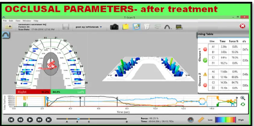
The T-Scan VIII is a sophisticated computerized occlusal analysis system, that illustrates both static and dynamic occlusion. It can be used to analyze tooth contact patterns that occur within every mandibular movement (centric relation, centric occlusion, lateral excursions and protrusion), which can all be recorded in various mandibular postures, and body postures, as well. The T-Scan VIII device (Tek-Scan Inc., Boston, MA, USA) is approved as a ‘‘medical index center – scanning sensor system. It has multiple levels of recording sensitivity that can be adjusted at will, to match the recording level within the recording sensor, to the occlusal force exerted by the patient. The hardware comprised of a recording handle (which holds the recording sensor) that is connected to the computer through a USB port. Held securely within the recording handle is a ‘‘sensor support’’, which keeps the 100 micron thick recording sensor properly extended flat within the mouth, during all intraoral recording procedures as shown in Figure 1 a & b (Koos et al., 2010; Hirano et al., 2002) When a force is exerted on the sensor, occurs a voltage drop on its conduction paths that is immediately recorded by the software upon each tooth contact, and is then displayed for clinical analysis. The T-Scan VIII computerized occlusal analysis system was used to record the subjects, Occlusion Time during 8 mandibular opening–closing movements. The patient should sit upright in the dental chair with their head on the headrest and mandibular occlusal plane parallel to the horizontal Frankfurt plane. The Occlusion time (OT) and Disclusion Times (DT), Occlusal forces in both right and left quadrant were recorded by placing the sensor between the dental arches, and asking the patient to perform 8 mandibular opening–closing movements without clenching, in and out of complete intercuspation (Kerstein and Grundset, 2001) with a velocity of about 1 movement per second. Before and after treatment parameters of a patient is shown in Figure 2 & 3. Thus, an average value of the occlusal parameters was obtained for each individuals in before and after dental treatment procedures were summarized.
Symptom assessment questionnaire
At each visit, subjects were asked to answer a symptom questionnaire that used an ordinal number scale to describe the current status of their condition [Table 1]. The ordinal scale ranged from 0 to 5 to describe symptom severity (0 ‑ no symptoms, 1 ‑ very little, 2 ‑ mild, 3 ‑ moderate, 4 ‑ severe, and 5 ‑ very severe). The common musculoskeletal symptoms that were graded were morning jaw pain, jaw fatigue, facial tension, difficulty in eating or chewing, clenching difficulty, temporal headaches, and neck pain.
Fig. 1a&1b. USB cable sensor holder and sensors, Support Handle assembly
Fig. 2. Occlusal contact force in right and left quadrants, premature contacts in 27,28 region, and occlusion time of 2.31 seconds and disclusion time of 8.46 secs
Fig. 3. Occlusal contact force in right and left quadrants disclus
USB cable sensor holder and sensors, Support Handle assembly
Occlusal contact force in right and left quadrants, premature contacts in 27,28 region, and occlusion time of 2.31 seconds and disclusion time of 8.46 secs
rce in right and left quadrants, no premature contacts and occlusion time of 0.67 & 0.48 seconds and disclusion time of 0.30 & 0.80 seconds
Occlusal contact force in right and left quadrants, premature contacts in 27,28 region, and occlusion time of
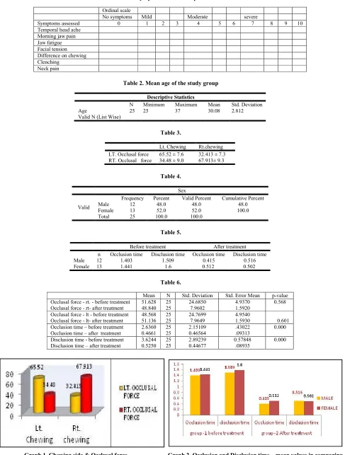
[image:3.612.94.513.221.447.2] [image:3.612.95.516.488.697.2]Table 1. Symptom assessment questionnaire
Ordinal scale
No symptoms Mild Moderate severe
Symptoms assessed 0 1 2 3 4 5 6 7 8 9 10
Temporal head ache Morning jaw pain Jaw fatigue Facial tension Difference on chewing Clenching
Neck pain
Table 2. Mean age of the study group
Descriptive Statistics
N Minimum Maximum Mean Std. Deviation
Age 25 25 37 30.08 2.812
Valid N (List Wise)
Table 3.
Lt. Chewing Rt.chewing
LT. Occlusal force 65.52 ± 7.6 32.413 ± 7.3
RT. Occlusal force 34.48 ± 9.0 67.913± 9.3
Table 4.
Sex
Valid
Frequency Percent Valid Percent Cumulative Percent
Male 12 48.0 48.0 48.0
Female 13 52.0 52.0 100.0
Total 25 100.0 100.0
Table 5.
Before treatment After treatment
n Occlusion time Disclusion time Occlusion time Disclusion time
Male 12 1.403 1.509 0.415 0.516
Female 13 1.441 1.6 0.512 0.502
Table 6.
Mean N Std. Deviation Std. Error Mean p-value
Occlusal force - rt. - before treatment 51.628 25 24.6850 4.9370 0.568
Occlusal force - rt- after treatment 48.840 25 7.9602 1.5920
Occlusal force - lt - before treatment 48.568 25 24.7699 4.9540
Occlusal force - lt- after treatment 51.136 25 7.9649 1.5930 0.601
Occlusion time – before treatment Occlusion time – after treatment
2.6360 0.4661
25 25
2.15109 0.46564
.43022 .09313
0.000
Disclusion time - before treatment Disclusion time – after treatment
3.6244 0.5250
25 25
2.89239 0.44677
0.57848 .08935
0.000
Graph 1. Chewing side & Occlusal force Graph 2. Occlusion and Disclusion time – mean values in comparing male and female patients- Before and after dental treatment
In each visits, the subjects were not allowed to review their previous answers, to avoid bias in their differing questionnaire response. The 25 treated subjects questionnaire scores were computed by mean with standard deviation. Since the mean and median of different groups of individuals were compared, a nonparametric statistic (the Wilcoxon signed‑ranks test) was used for statistical analysis (Table 7).
RESULTS
A total number of 25 patients (13 female and 12 male), with mean age 30.08(SD ± 2.812) as shown in Table 2, were included in this study.
Occlusal force was higher in the preferred chewing side (P < 0.05), maximum in right side compared to left with mean value of 67.913± 9.3 in right side and 65.52 ± 7.6 in left side as shown in Table-3 & Graph-1
Demographic data
[image:5.612.167.446.88.139.2] [image:5.612.161.451.561.611.2]According to the demographic data as shown in Table- 4 & Table- 5, Graph -2 ;female patients have increased mean value ofocclusiontimeof 1.444anddisclusion time of 1.6 compared to male patients with mean value of Occlusion time of 1.403 and disclusion time of 1.509.
Table 8. Symptom questionnaire statistical data
1. Comparison of median intensity values of ‘morning jaw pain’ from pre- treatment Day 1 to other time intervals
Jaw pain Median Mean rank Minimum Maximum Z P-value
Day 1 4 14.50 0 7 … …
Day 8 0 14.50 0 2 -4.604 <0.001
1 month 0 14.50 0 0 -4.604 <0.001
3 months 0 14.50 0 0 -4.604 <0.001
2. Comparison of median intensity values of jaw fatigue’ from pre- treatment Day 1 to other time intervals
Jaw fatigue Median Mean rank Minimum Maximum Z P-value
Day 1 4 7.50 0 7 … …
Day 8 0 7.50 0 3 -3.431 <0.001
1 month 0 7.50 0 0 -3.430 <0.001
3 months 0 7.50 0 0 -3.430 <0.001
3. Comparison of median intensity values of ‘facial tension” from Pre- treatment Day 1 to other time intervals
Facial tension Median Mean rank Minimum Maximum Z P-value
Day 1 4 8.50 0 6 … …
Day 8 0 8.50 0 0 -3.157 <0.001
1 month 0 8.50 0 0 -3.157 <0.001
3 months 0 8.50 0 0 -3.157 <0.001
4. Comparison of median intensity values of ‘‘differences in chewing/eating” from pre-treatment Day 1 to other time intervals
‘differences in chewing/eating’ Median Mean rank Minimum Maximum Z P-value
Day 1 4 6.5 0 6 … …
Day 8 0 6.5 0 0 -3.209 <0.001
1 month 0 6.5 0 0 -3.209 <0.001
3 months 0 6.5 0 0 -3.209 <0.001
5. Comparison of median intensity values of ‘clenching” from pre-treatment Day 1 to other time intervals
Clenching Median Mean rank Minimum Maximum Z P-value
Day 1 4 16.50 0 8 … …
Day 8 0 16.50 0 4 -5.27 <0.001
1 month 0 16.50 0 0 -5.297 <0.001
3 months 0 16.50 0 0 -5.297 <0.001
6. Comparison of median intensity values of ‘temporal headache” from pre- treatment Day 1 to other time intervals
Temporal headache Median Mean rank Minimum Maximum Z P-value
Day 1 4 21.0 0 6 … …
Day 8 0 21.0 0 0 -6.010 <0.001
1 month 0 21.0 0 0 -6.006 <0.001
3 months 0 21.0 0 0 -6.006 <0.001
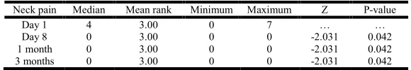
7. Comparison of median intensity values of ‘neck pain” from pre-treatment Day 1 to other time intervals
Neck pain Median Mean rank Minimum Maximum Z P-value
Day 1 4 3.00 0 7 … …
Day 8 0 3.00 0 0 -2.031 0.042
1 month 0 3.00 0 0 -2.031 0.042
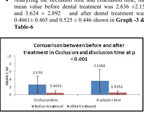
Analyzing the occlusion time and Disclusion time, the mean value before dental treatment was 2.636 ±2.15 and 3.624 ± 2.892 and after dental treatment was 0.4661± 0.465 and 0.525 ± 0.446 shown in Graph -3 & Table-6
Graph 3. Mean Value of Occlusion and Disclusion time of all Patients- before and after treatment
Using Paired sample T test (Table-6), mean value for occlusal force on the right side before dental treatment (51.528±25.6850) and after dental treatment (48.840±7.960) and the left side (48.568 ±24.76) before dental treatment and ( 51.136±7.649) after treatment was found. There was no statistically significant value of 0.568 in right side and 0.601 in left side. Mean value of Occlusion time and disclusion time before treatment was 2.636 ±2.15 and 3.624 ± 2.892 and after dental treatment was 0.4661± 0.465 and 0.525 ± 0.446 and statistically highly significant p value of 0.000 in both occlusion time and disclusion time.
Symptom pain questionnaire
[image:6.612.63.296.57.241.2]An ordinal scale questionnaire was used to assess the severity of various TMD symptoms where the mean values of the scores for the entire group were determined for statistical comparison. The changes in mean intensity and frequency of all myofascial pain symptoms studied and was found (Table -8) to be statistically significant between day 1 and day 7 (P < 0.001), day 1 and 1 month (P < 0.001) & day 1 & 3 months (P < 0.001).
Table 7. Wilcoxon signed rank test for determining the level of significance in occlusion time, disclusion time and occlusal force
Occlusal force – rt- after treatment -
Occlusal force – rt. - before treatment
Occlusal force – lt- after treatment –
occlusal force – t - before treatment
Z -.471a
-.498b
Asymp. Sig. (2-tailed) .638 .619
This test shows highly significant p value for occlusion time, disclusion time and not significant for occlusal force.
DISCUSSION
Dr. Bernard Jankelson’s study of the human dental occlusion were precursors to the neuromuscular occlusion published in 1955 lead to the recognition of the scientific methods to quantify the function of the masticatory system (Jankelson, 195555; Prafulla Thumati, 2016). Gadgets like digital analysis of occlusion using T-scan, electromyography (EMG), joint vibratography (JVA) to measure the function of the masticatory system-dental occlusion, temporomandibular joints (TMJ) and muscles were subsequently invented. These biometric diagnostic tools help in the practice and management of painful conditions related to temporomandibular disorders
(TMD) (Jankelson et al., 1975). Orofacial pain was
incorporated into the authors practice in 2010 to provide healthcare to patients who have been suffering from varied symptoms, which include headache, migraine, early morning facial stiffness, trauma to musculoskeletal tissues (muscles, ligaments, tendons), low back ache, nutritional deficiencies, nervous tension, or stress (Jankelson et al., 1975; Berger et al.,
2007). One of the predominant symptom and at the same time the main cause of the patients reporting for treatment due to TMD is a pain. Pain as an unpleasant sensory and emotional experience associated with actual or potential tissue damage. Factors of nonspecific pain within TMD are usually classified as psychosocial, occupational and personal. All these factors encourages an acute pain episode, while mental suffering due to anxiety, adjustment disorder, depressive or irritable mood, fear of somatic illness, and persistent somatizations can incite a chronic form of pain. Chronic tension is associated with an increase in parafunctional activity, which is considered to be one of the main TMD factors. Stress as well as anxiety, depression and personality traits exert an important influence on the increased frequency of parafunctional teeth contact (Rollman et al., 2000; Lakshmi et al., 2016). Kersteine in 2001 was the first person to locate centric relation prematurity with simultaneous recording of sequence of resultant tooth contacts using computerized occlusal analysis system.
According to him this method offers significant improvement in the precision of locating the first tooth contact. Correct understanding of dynamic occlusion is very critical in differentiating between the normal and pathological occlusal parameters. Digital evaluation of the occlusion by T scan will provide additional information like occlusion and disclusion time on functional occlusion. T scan analysis enables the clinician to evaluate the occlusal contacts quantitatively, also record the occlusion during continuous mandibular movement (Pyakurel et al., 2013). The patients in the present study were considered with natural complete dentition and having Angle class I occlusion exhibiting signs and symptoms of TMD. Our study showed equal gender ratio and the study age group of 20 and 40 years with mean age of 30.08 which was in accordance with Edward et al study (Edward et al., 2009). In our study the preferred chewing side was detected by declaring of the patients through questionairre, while in the literature we found that it can be detected by observing the examinees during mastication or using kinesiographic movements by using jaw tracking movement. Palinkas et al., have investigated the influence of gender and age on maximal bite force and masticatory muscles thickness. The hypothesis is the lower force applied in a non preferred chewing side is associated with a “weaker” muscles on that side therefore, with a smaller occlusal contact area And is associated with a reduced occlusal
preferred chewing side was analysed (Pyakurel et al., 2013). Occlusal force was higher in the preferred chewing side, maximum in right side compared to left with mean value of 67.913± 9.3 and 65.52 ± 7.6 in left side. The present study demonstrated the incidence of premature contacts (occlusal interferences) was higher and disclusion time was significantly longer in patients before treatment. The findings of the study are in correlation with other researchers like Cheng et al and Haralur et al in 2012 (Wang et al., 2012; Cheng et al., 2012). Premature contacts can result in condyle displacement, which may potentially cause friction and increased intra-articular pressure on the TMJ. Both situations are injurious to the TMJ and contribute to changes of the structure of TMJ. If the capacity of the subject to modify the condition is exceeded, results in the disorders of the masticatory muscles and TMD Kerstein and Radke, 2012. Analyzing the occlusion and Disclusion time, the mean value before dental treatment was 2.636 ±2.15 and 3.624 ± 2.892 and after dental treatment was 0.4661± 0.465 and 0.525 ± 0.446 and also showed statistically highly significant P value of 0.000 correlated with studies of (Kersteine 2006; Koos et al., 2010; Hirano et al., 2002). The prolonged disclusion time can lead to higher masticatory muscle activity and abnormal distribution of stresses in the disc resulting in TMD symptoms. The compression of the mechanoreceptors of molar and premolar periodontal ligaments by prolonged excursive tooth contact, activate excess muscle contractions in the masticatory muscles. The longer time of the excursive interferences results in periodontal ligaments compression and activating the masticatory muscles to contract. This cycle repeats with every posterior teeth compression during both functional and Para functional
mandibular movements and adds excessive muscle
[image:7.612.65.298.520.662.2]contractions. The additive constant and excessive masticatory muscle contractions results in lactic acid accumulation which often leads to the clinical appearance of muscular hyperactivity (Kerstein and Radke, 2012; Kerstein, 2010). Moreover, eight Occlusion and disclusion times were recorded for each subject, and a mean value was calculated in order to approach a representative OT, DT value per subject. The accuracy of the T-Scan system recording has been shown to be not altered by repeated measures, which was correlated by other studies (Koos et al., 2010; Hirano et al., 2002).
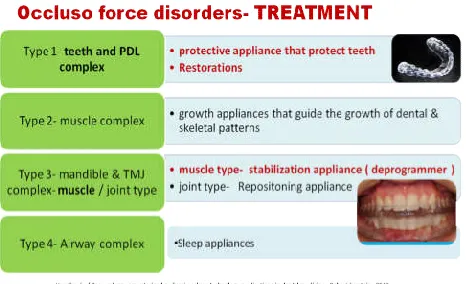
Figure 5. Treatment for Occluso force disorders
Occlusal corrections for symptomatic relief of pain in muscular TMD
Conservative and reversible self-management treatment strategies are beneficial for most patients. ie; protective
appliance to protect the teeth, restorations, stabilization appliances- deprogrammer were used as treatment of choice in our study patients as shown in Figure 5. The Tscan VIII results showed occlusal parameters and symptomatic muscular pain in the patients with TMD are significantly reduced after dental treatment. The other treatment options include vapocoolant spray and stretching of the muscles involved, injections of local anesthetic directly into the trigger point(s), massage therapy, physical therapy, exercise, elimination of stress, changing sleeping habits, the use of tricyclic antidepressants in low doses , non-steroidal anti-inflammatory drugs, muscle relaxants, counseling, etc. Despite its diverse etiology, occlusal instability has long been considered an important aetiological factor (Greenberg et al., 2012). The results of this study indicate that dental treatment will predictably reduce myofascial pain symptoms because of the physiologic muscle activity lowering effect achieved by dental procedures, which is a distinctly different occlusal adjustment approach to retruding the mandible into centric relation during traditional occlusal equilibration procedures. This study showed the multiple recall visit disclusion time mean values were statistically equivalent to the post treatment mean values of disclusion time. Further the standard deviations from recall visit to visit remained fairly constant throughout the period of observation. The mean differences also remained constant when a comparison between day 1 pretreatment and subsequent visit measurements was made. This suggests that once disclusion time is reduced <0.4 seconds, it is a lasting occlusal change. These findings are very similar to those of another disclusion time reduction study that verified that once the disclusion time was properly reduced, it remained constant, leading to the retention of proper muscle function and low symptom appearances. In this study, the population selected for treatment had their symptoms evaluated by a questionnaire at subsequent recall appointments (day 7, 1 month, 3 months), during which the subjects were not allowed to view their previous symptom responses. The results obtained from the recall date questionnaires showed there were a significant symptom resolution in the study population and was maintained during the 3 months period of observation.
CONCLUSION
A brief screening history regarding pain and activities that make the pain worse can help determine if a patient has a
potential TMD problem. A clinical examination that
incorporates an assessment of pain and joint sounds on opening and excursive movements is always recommended. TMD due to occlusal disturbances can be diagnosed using biometric T scan system provides sensitivity and specificity, higher reliability in intra-oral conditions and can be repeatedly used again in same patient. This study insists of no arbitrary grinding of the teeth and reduces the subjective interpretation of occlusal analysis data and also provides registration of dynamic occlusal information. T scan is an objective
assessment system that enhances patient education,
satisfaction, and retention and enables a more confident approach to patient care. This helps in the elimination of the cause of the disease and not just symptom relief. If the etiology is not successfully recognized and treated, the acute physical form of temporomandibular dysfunction may become a chronic pain condition.
REFERENCES
Alberto Baldini, Alessandro Nota, Paola Cozza, 2014. The association between Occlusion Time and Temporoman dibular Disorders: Journal of Electromyography and Kinesiology, 1-4
Baldini, A., Beraldi, A., Nota, A., Danelon, F., Ballanti, F., Longoni, S. 2012a. Gnathological postural treatment in a professional basketball player: a case report and an overview of the role of dental occlusion on performance. Ann Stomatol (Roma), 3(2):51–8.
Baldini, A., Nota, A., Cravino, G., Cioffi, C., Rinaldi, A., Cozza, P. 2013a. Influence of vision and dental occlusion on body posture in pilots. Aviat Space Environ Med.,
84(8):823–7.
Berger, M., J.O. Listopad, M. Marczak, J. Szymanska, 2007. Psychological aspects of temporomandibular disorders – literature review. Curr Issues Pharm Med Sci., 28(1):55-9. Cheng, H.J., Geng, Y., Zhang, F.Q. 2012. The evaluation of
intercuspal occlusion of healthy people with T-Scan II system. Shanghai Kou Qiang Yi Xue, 21:62–5.
De Boever, J.A., Carlsson, G.E., Klineberg, I.J. 2000. Need for occlusal therapy and prosthodontic treatment in the management of Temporomandibular Disorders. Part II. Tooth loss and prosthodontic treatment. J Oral Rehabil.,
27:647–59.
De Boever, J.A., Carlsson, G.E., Zarb, G.A., Sessle, B.E., Mohl, N.D. 1994. Etiology and differential diagnosis. In:.
Temporomandibular joint and masticatory muscle
disorders. 2nd ed. Copenaghen: Munksgaard – Mosby., p. 171–87.
Dworkin, S.F., LeResche, L. 1992. Research diagnostic criteria
for temporomandibular disorders: review, criteria,
examinations and specifications, critique. J Craniomandib Disord., 6: 301–355.
Edward, F., Wright, Sarah, L. 2009. Management and Treatment of Temporomandibular disorders, a clinical perspective. J of Man and Mani Therap., 17:247-54. Greenberg, M.S., Glick, M., Ship, J.A. 2012. Burket’s oral
medicine. 11th ed. New Delhi: CBS Publishers and Distributers Pvt Ltd; p.223-56.
Hirano, S., Okuma, K., Hayakawa, I. 2002. In vitro study on accuracy and repeatability of the T-Scan II system. Kokubyo Gakkai Zasshi, 69:194–201.
Jankelson, B. 1955. Physiology of human dental occlusion. J Am Dent Assoc., 50:664-80.
Jankelson, B., Swain, C.W., Crane, P.F. Radke, J.C. 1975. Kinesiometric instrumentation: A new technology. J Am Dent Assoc., 90:834-40.
Kerstein, R.B. 2010. Reducing chronic masseter and temporalis muscular hyperactivity with computer-guided
occlusal adjustments. Compend Contin Educ Dent.,
31:530-4.
Kerstein, R.B., Grundset, K. 2001. Obtaining bilateral simultaneous occlusal contacts with computer analyzed and guided occlusal adjustments. Quintessence Int., 32:7–18.
Kerstein, R.B., J. Radke, 2006. The effect of disclusion time reduction on maximal clench muscle activity levels.
Cranio., 24(3): 156-65.
Kerstein, R.B., Radke, J. 2012. Masseter and temporalis excursive hyperactivity decreased by measured anterior guidance development. Cranio., 30:243–54.
Koos, B., Godt, A., Schille, C., Göz, G. 2010. Precision of an instrumentation-based method of analyzing occlusion and its resulting distribution of forces in the dental arch. J Orofac Orthop., 71: 403–10.
Lakshmi et al., 2016. Occlusion and psychological distress in TMD: IJMDS, www.ijmds.org, July, 5(2): 1198-1207 LeResche, L. 1997. Epidemiology of Temporomandibular
Disorders: implications for the investigation of etiologic factors. Crit Rev Oral Biol Med., 8:291–305.
Luther, F. 2007. TMD and occlusion part II. Damned if we don’t? Functional occlusal problems: TMD epidemiology in a wider context. Br Dent J., 202(E3):38–9.
Maness, W.L., Benjamin, M., Podoloff, R., Bobick, A., Golden, R.F. 1987. Computerized occlusal analysis: a new technology. Quintessence Int., 18 :287–92.
Manfredini, D., Chiappe, G., Bosco, M. 2006. Research diagnostic criteria for Temporomandibular Disorders (RDC/TMD) axis I diagnoses in an Italian patient population. J Oral Rehabil., 33:551–8
McNamara, J.A. 1997. Orthodontic treatment and
Temporomandibular Disorders. Oral Surg Oral Med Oral Pathol Oral Radiol Endod., 83:107–17.
Okeson, J.P. 1997. Current terminology and diagnostic classification schemes. Oral Surg Oral Med Oral Pathol Oral Radiol Endod., 83: 61–4.
Prafulla Thumati, 2016. Diagnostic tests for
temporomandibular disorders: Journal of Advanced
Clinical & Research Insights, 3, 81–86
Prafulla Thumati: Clinical outcome of subjective symptoms in myofascial pain patients treated by immediate complete anterior guidance development technique using digital analysis of occlusion (Tek-scan) and electromyography :
ORIGINAL ARTICLE: Journal of Interdisciplinary
Dentistry, Jan-Apr 2015, Vol-5, Issue-1
Pyakurel, U., Long, H., Jian, F., Sun, J., Jha, H., Lai, W. 2013. Mechanism, accuracy and application of T-Scan system in dentistry-A review. J of Nep Dent Assoc., 3:1-5.
Robert Kerstein, 2015. Handbook of Computerized Occlusal applications in Dental Medicine
Rollman, G.B., Joanne, M., Gillespie, 2000. The Role of Psychosocial Factors in Temporomandibular Disorders.
Curr Rev of Pain., 4:71-81.
Satheesh B. Haralur, 2013. Digital Evaluation of Functional Occlusion Parameters and their Association with Temperomandibular Disorder: Journal of Clinical and Diagnostic Research, Aug, Vol-7(8): 1772-1775
Wang, C., Yin X Nanjing, 2012. Occlusal risk factors associated with temporomandibular disorders in young adults with normal occlusions. Oral Surg Oral Med Oral Pathol Oral Radiol., 114:419-23.