z
RESEARCH ARTICLE
MITOCHONDRIAL DYSFUNCTION DURING DIFFERENT SEPSIS FORMS
*,1Charalampos Martinos,
1Anastasia Kotanidou and
2Panagiotis Ηalvatsiotis
1
Department of Intensive Care Unit, Naval and Veterans Hospital of Athens, Greece
2Department of Internal Medicine Propaedeutic, Athens University Medical School, Attiko
General Hospital, Greece
ARTICLE INFO ABSTRACT
Background: Sepsis-induced multiple organ failure is the major cause of mortality and morbidity in critically ill patients. The precise mechanisms by which this dysfunction is caused remain to be elucidated. Because ATP production by mitochondrial oxidative phosphorylation accounts for more than 90% of total oxygen consumption, we postulated that mitochondrial dysfunction results in organ failure, possibly due to nitric oxide, which is known to inhibit mitochondrial respiration in vitro and is produced in excess in sepsis.
Methods: We did skeletal muscle biopsies on 25 septic patients within 24 h of admission to intensive care. The biopsy samples were analyzed for myosin heavy chains (MHC), isoform gene transcript levels using a real-time PCR technique, respiratory-chain activity and ATP production (complex V) using a real-time QPCR technique for mitochondrial enzyme gene transcript levels citrate synthase (CS), cytochrome C oxidase I-III, NADH and UCPs 2-3. Results: The gene transcription of myosin fiber types I, IIa and citrate synthase (complex V) was significant higher in skeletal muscle biopsy of septic survivors, and demonstrated lower insulin sensitivity using HOMA test. Severity of septic shock ( SAPS II) found to be higher in critically ill patients.
Conclusion: In critically ill patients, we found an association between the type of myosin fiber and ATPase gene transcription and insulin resistance that relate to organ failure and eventual outcome. These data implicate bioenergetic failure as an important pathophysiological mechanism underlying multiple organ dysfunction.
Copyright©2017, Charalampos Martinos et al. This is an open access article distributed under the Creative Commons Attribution License, which permits
unrestricted use, distribution, and reproduction in any medium, provided the original work is properly cited.
INTRODUCTION
Sepsisis the systemic inflammatory response associated with an infectious insult. It is the leading cause of death in critically ill patients, and the predominant cause of multiple organ dysfunction (Deitch, 1992). However, precise mechanisms through which organ dysfunction develops remain unknown, as do reasons for its persistence long after cessation of the acute inflammatory phase. Although microvascular flow redistribution undoubtedly occurs (Ince and Sinaasppel, 1999), we and others have shown increased tissue oxygen tension in the organs of animals and patients with sepsis (Rosser et al., 1995; Boekstegers et al., 1991). This finding suggests that the predominant defect might lie in cellular oxygen use (tissue dysoxia) rather in oxygen delivery per se. Mitochondrial oxidative
*Corresponding autor: Charalampos Martinos,
Department of Intensive Care Unit, Naval and Veterans Hospital of Athens, Greece
phosphorylation is responsible for over 90% of total body oxygen consumption and ATP generation. This take place in the mitochondrial inner membrane at the respiratory chain. The respiratory chain (electron-transport chain) includes four individua enzyme complexes (I-IV). These enzyme complexes, notably NADH-ubiquinone oxidoreductase (complex I) and cytochrome C oxidase (complex IV), can be inhibited by reactive oxygen and nitrogen species such as nitric oxide (Clementi et al., 1998; Bolanos et al., 1996; Brown and Cooper, 1994). These reactive species are produced in substantial excess during sepsis and are also generated by the mitochondria (Taylor et al., 1995). Complex I inhibition by nitric oxide is facilitated, in vitro, by depletion of the intracellular reduced glutathione. Concentrations of this antioxidant are also known to decrease in septic states (Minamiyama et al., 1996; Corbucci et al., 1985). We also know that there are three major muscle types, skeletal, cardiac and smooth muscle. Skeletal muscle is a form of muscle tissue which is under the control of the nervous system. It is made up of individual components known as
ISSN: 0975-833X
International Journal of Current Research
Vol. 9, Issue, 09, pp.57373-57379, September, 2017
INTERNATIONAL JOURNAL OF CURRENT RESEARCH
Article History: Received 27thJune, 2017
Received in revised form 24thJuly, 2017
Accepted 19thAugust, 2017
Published online 29thSeptember, 2017
Citation:Charalampos Martinos, Anastasia Kotanidou and Panagiotis Ηalvatsiotis,2017.“Mitochondrial dysfunction during different sepsis forms”, International Journal of Current Research, 9, (09), 57373-57379.
Available online at http://www.journalcra.com
Key words: Critically ill, Mitochondrial dysfunction, Gene transcription, Bioenergetic failure. z
RESEARCH ARTICLE
MITOCHONDRIAL DYSFUNCTION DURING DIFFERENT SEPSIS FORMS
*,1Charalampos Martinos,
1Anastasia Kotanidou and
2Panagiotis Ηalvatsiotis
1
Department of Intensive Care Unit, Naval and Veterans Hospital of Athens, Greece
2Department of Internal Medicine Propaedeutic, Athens University Medical School, Attiko
General Hospital, Greece
ARTICLE INFO ABSTRACT
Background: Sepsis-induced multiple organ failure is the major cause of mortality and morbidity in critically ill patients. The precise mechanisms by which this dysfunction is caused remain to be elucidated. Because ATP production by mitochondrial oxidative phosphorylation accounts for more than 90% of total oxygen consumption, we postulated that mitochondrial dysfunction results in organ failure, possibly due to nitric oxide, which is known to inhibit mitochondrial respiration in vitro and is produced in excess in sepsis.
Methods: We did skeletal muscle biopsies on 25 septic patients within 24 h of admission to intensive care. The biopsy samples were analyzed for myosin heavy chains (MHC), isoform gene transcript levels using a real-time PCR technique, respiratory-chain activity and ATP production (complex V) using a real-time QPCR technique for mitochondrial enzyme gene transcript levels citrate synthase (CS), cytochrome C oxidase I-III, NADH and UCPs 2-3. Results: The gene transcription of myosin fiber types I, IIa and citrate synthase (complex V) was significant higher in skeletal muscle biopsy of septic survivors, and demonstrated lower insulin sensitivity using HOMA test. Severity of septic shock ( SAPS II) found to be higher in critically ill patients.
Conclusion: In critically ill patients, we found an association between the type of myosin fiber and ATPase gene transcription and insulin resistance that relate to organ failure and eventual outcome. These data implicate bioenergetic failure as an important pathophysiological mechanism underlying multiple organ dysfunction.
Copyright©2017, Charalampos Martinos et al. This is an open access article distributed under the Creative Commons Attribution License, which permits
unrestricted use, distribution, and reproduction in any medium, provided the original work is properly cited.
INTRODUCTION
Sepsisis the systemic inflammatory response associated with an infectious insult. It is the leading cause of death in critically ill patients, and the predominant cause of multiple organ dysfunction (Deitch, 1992). However, precise mechanisms through which organ dysfunction develops remain unknown, as do reasons for its persistence long after cessation of the acute inflammatory phase. Although microvascular flow redistribution undoubtedly occurs (Ince and Sinaasppel, 1999), we and others have shown increased tissue oxygen tension in the organs of animals and patients with sepsis (Rosser et al., 1995; Boekstegers et al., 1991). This finding suggests that the predominant defect might lie in cellular oxygen use (tissue dysoxia) rather in oxygen delivery per se. Mitochondrial oxidative
*Corresponding autor: Charalampos Martinos,
Department of Intensive Care Unit, Naval and Veterans Hospital of Athens, Greece
phosphorylation is responsible for over 90% of total body oxygen consumption and ATP generation. This take place in the mitochondrial inner membrane at the respiratory chain. The respiratory chain (electron-transport chain) includes four individua enzyme complexes (I-IV). These enzyme complexes, notably NADH-ubiquinone oxidoreductase (complex I) and cytochrome C oxidase (complex IV), can be inhibited by reactive oxygen and nitrogen species such as nitric oxide (Clementi et al., 1998; Bolanos et al., 1996; Brown and Cooper, 1994). These reactive species are produced in substantial excess during sepsis and are also generated by the mitochondria (Taylor et al., 1995). Complex I inhibition by nitric oxide is facilitated, in vitro, by depletion of the intracellular reduced glutathione. Concentrations of this antioxidant are also known to decrease in septic states (Minamiyama et al., 1996; Corbucci et al., 1985). We also know that there are three major muscle types, skeletal, cardiac and smooth muscle. Skeletal muscle is a form of muscle tissue which is under the control of the nervous system. It is made up of individual components known as
ISSN: 0975-833X
International Journal of Current Research
Vol. 9, Issue, 09, pp.57373-57379, September, 2017
INTERNATIONAL JOURNAL OF CURRENT RESEARCH
Article History: Received 27thJune, 2017
Received in revised form 24thJuly, 2017
Accepted 19thAugust, 2017
Published online 29thSeptember, 2017
Citation:Charalampos Martinos, Anastasia Kotanidou and Panagiotis Ηalvatsiotis,2017.“Mitochondrial dysfunction during different sepsis forms”, International Journal of Current Research, 9, (09), 57373-57379.
Available online at http://www.journalcra.com
Key words:
Critically ill,
fibers. Each fiber has an outer membrane called sarcolemma which surrounds myofibrils. Between them are mitochondria and sarcoplasm. The principle cytoplasmic proteins are actin and myosin. There are two main types of fibers, type I and type II. Type I fibers appear red due to the high levels of myoglobin. They have more mitochondria and greater local capillary density. They are more suited for endurance and are slow to fatigue because they use oxidative metabolism to generate ATP (adenosine triphosphate). They generate energy for ATP re-synthesis by means of a long term system of aerobic energy transfer. On the other hand type II fibers tend to be less oxidative, they are white and a reliance on glycolytic enzymes. Compared to glycolysis oxidative phosphorylation produces 17 to 18 times as much ATP from the same amount of glucose. It is believed there are no sex or age differences in fiber distribution. Sedentary men and women have 45% type II and 55% type I fibers. Endurance athletes show a higher level of type I fibers. On the other hand sprint athletes require large numbers of type II. It has been suggested that various types of exercise can induce changes in the fibers of skeletal muscles. Type II fibers show enhancements of the oxidative capacity after high intensity endurance training which brings them to a level at which they able to perform oxidative metabolism as effectively as type I fibers of untrained subjects. This would be brought about by an increase in mitochondrial size and number and the associated related changes not a change in fiber type (MacInntosh et al., 2006).
There are two commonly used methods for fiber typing. Histochemical staining for myosin ATPase activity and immunohistochemical staining for myosin heavy chain (MHC) type which results from determination of different MHC isoforms. These methods are closely related as the MHC type is the primary determinant of ATPase activity (Smerdu et al., 1994; Pete et al., 2000). In the inner mitochondrial membrane we can also find uncoupling protein (UCP). They are transporters that work in parallel with ATP synthase, generating heat from the energy that is not used to generate ATP (Nedergaard et al., 2005; Rousset, Sophie et al., 2004). Distal muscle weakness and loss of deep tendon reflexes are usually found. It is an axonal (Latronico et al., 1996) motor-sensory polyneuropathy called Critical Illness Polyneuropathy (CIP). Polyneuropathy may develop as early as one week after the onset of SIRS, but the incidence tends to correlate with the duration of the severe illness. Τhe pathogenesis is not known. Electrophysiological findings are those of pure axonal degeneration affecting more motor than sensory fibers. The axonal damage is due to the transport system which is energy dependent. Nerve biopsy reveals axonal degeneration without demyelination or inflammation. This results in denervation and grouped atrophy of muscle (Kress et al., 2014). To address the questions whether bioenergetic status in septic patients is associated with mitochondrial dysfunction, and whether these abnormalities are related to organ failure, we undertook a systematic study of mitochondrial dysfunction in septic patients admitted to intensive care.
MATERIALS AND METHODS
Patients
After obtaining approval of PhD dissertation’s protocol from the Medical School committeeof UoA and consentto participate the study, patients were recruited from the ICU of Evaggelismos G.H. since Apr 2003 to Jul 2006. Patients
with recent-onset severe sepsis or septic shock (as defined by standard criteria) were enrolled. Those with severe coagulopathies (PLTs<30 or INR>2), immunosuppression (endogenous or long-term chemotherapeutic), or both, were excluded. Routine physiological and biochemical variables were recorded, and scores on the simplified acute physiology score (SAPS II) and sequential organ failure assessment (SOFA) calculated.
Procedures
Within 24 h of patients admission to the intensive care unit, a quadriceps femoris muscle biopsy was done microsurgical via a small incision in the middle of the muscle. Biopsy samples (averaging 750 mg total) were frozen immediately in liquid nitrogen. MHC isoform composition was determined by SDS-PAGE (sodium dodecyl sulfate-polyacrylamide gel electrophoresis) silver staining.
Statistical analysis
Quantitative variables were expressed as mean values (SD), while qualitative variables were expressed as absolute and relative frequencies. Independent samples Student’s t-tests were used for the comparison of mean values between the two groups. For the comparison of proportions.Fisher’s exact tests were used. Pearson’s correlations coefficients were used to explore the association of two continuous variables. Correlation coefficient between 0.1 and 0.3 were considered low, between 0.31 and 0.5 moderate and those over 0.5 were considered high. All reported p values are two-tailed. Statistical significance was set at p<0.05 and analyses were conducted using SPSS statistical software (version 19.0).
RESULTS
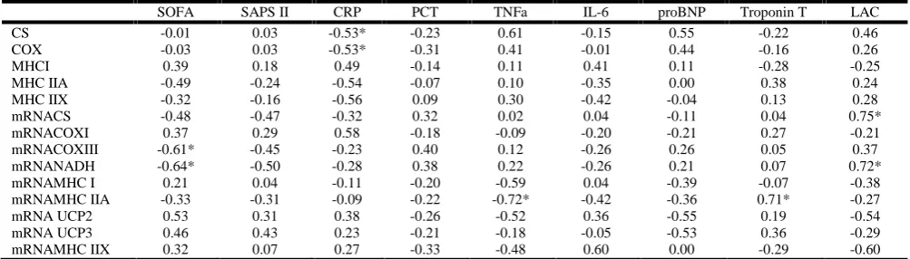
Data consists of two groups. A study group of all sepsis forms patients (33) and a control group of 10patients undergone elective orthopedic surgery. We report on the results of dividing study group into two subgroups as important. One of them with septic patients and another one with critically ill ones (severe septic and septic shock). Sample consisted of 25 patients with sepsis, 17 (68.0%) of whom were critically ill. Patients’ characteristics are presented inTable 1. Mean age for critically ill patients was 69.4 years (SD=14.0 years) and significantly higher compared to the mean age of the patients with sepsis. In both groups the majority of the patients were men with the percentages being 64.7% for the critically ill group and 62.5% for the septic group. All critically ill patients died while none of the group with sepsis died. Patients’ measurements regarding the severity of their sepsis are presented in table 2. Critically ill patients scored significantly in SAPS II, indicating greater severity of their condition, compared to the rest septic patients. SOFA index and all biochemical indexes were similar between the two groups. Owing to variation in the amount of tissue obtained, we were unable to do a full set of analyses on all patients. Information from biopsy regarding patients’ insulin tolerance is presented in Table 3. Critically ill patients had significantly lower mRNA-CS, mRNA-MHC I and mRNA-MHC IIA values compared to the rest septic patients (Figures 5-7).Τhere was no difference in biopsies between the activities of mitochondrial complexes but we found significant higher the gene transcription of septic survivors. In all septic
patients, regardless of the severity of their disease, it was found that SOFA score was significantly negatively associated with mRNA-COXIII and mRNA-NADH (Table 4). Thus, higher values in mRNA-COXIII and mRNA-NADH are associated with lower SOFA score. Similarly, higher CRP is associated
with lower CS (Figure 8) and MHCI values. Increased TNFa values or decreased troponin-T values are associated with lower mRNA-MHC IIA (Figures 9, 10). Moreover, greater lac values are associated with greater mRNA-CS (Figure 9) and mRNA-NADH values.
Demographics
F1:age F2: sex
Scores
F3: SAPS II F4: SOFA
Biopsy results
F5: mRNA CS F6: mRNA MHC I
57375 International Journal of Current Research, Vol. 9, Issue, 09, pp.57373-57379, September, 2017
patients, regardless of the severity of their disease, it was found that SOFA score was significantly negatively associated with mRNA-COXIII and mRNA-NADH (Table 4). Thus, higher values in mRNA-COXIII and mRNA-NADH are associated with lower SOFA score. Similarly, higher CRP is associated
with lower CS (Figure 8) and MHCI values. Increased TNFa values or decreased troponin-T values are associated with lower mRNA-MHC IIA (Figures 9, 10). Moreover, greater lac values are associated with greater mRNA-CS (Figure 9) and mRNA-NADH values.
Demographics
F1:age F2: sex
Scores
F3: SAPS II F4: SOFA
Biopsy results
F5: mRNA CS F6: mRNA MHC I
F7: mRNA MHC IIA
Pearson correlations
F8: CS f CRP F9:mRNA CS f lac
F10: mRNA MHC IIA f TNFa F11: MRNA MHC IIA f Troponin T
57376 Charalampos Martinos et al. Mitochondrial dysfunction during different sepsis forms
F7: mRNA MHC IIA
Pearson correlations
F8: CS f CRP F9:mRNA CS f lac
F10: mRNA MHC IIA f TNFa F11: MRNA MHC IIA f Troponin T
57376 Charalampos Martinos et al. Mitochondrial dysfunction during different sepsis forms
F7: mRNA MHC IIA
Pearson correlations
F8: CS f CRP F9:mRNA CS f lac
F10: mRNA MHC IIA f TNFa F11: MRNA MHC IIA f Troponin T
Table 1. Sample characteristics for the two study groups
Group Sepsis (SSV)
(N=8)
Critically ill (SS) (N=17)
N (%) N (%) P
Age (years), mean (SD) 54.1 (19.6) 69.4 (14.0) 0.036* Sex
Females 3 (37.5) 6 (35.3) 1.000**
Males 5 (62.5) 11 (64.7)
Death
No 8 (100.0) 0 (0.0) <0.001**
Yes 0 (0.0) 17 (100.0)
[image:5.595.156.425.250.369.2]*Student's t-test; **Fisher's exact test
Table 2. Biochemical indexes in critically ill patients and patients with sepsis
Group
Sepsis (SSV) Critically ill (SS)
Mean (SD) Mean (SD) P*
SOFA 10.4 (4.4) 12.9 (3) 0.112
SAPS II 48.1 (21.9) 68 (12.7) 0.011
CRP 16.1 (7.5) 16.4 (8) 0.925
PCT 7.6 (11.3) 8.1 (14.5) 0.949
TNFa 42.9 (26.3) 63.2 (30.5) 0.227 IL-6 898 (1576.4) 1089.3 (2062.1) 0.859 proBNP 6145.7 (4621.4) 9581 (7556.3) 0.406 Troponin T 0.32 (0.55) 0.3 (0.43) 0.962
LAC 2.24 (2.21) 3.74 (4.69) 0.505
*Student's t-test
Table 3. Biopsy results in critically ill patients and patients with sepsis
Group
Sepsis (SSV) Critically ill (SS)
N Mean (SD) N Mean (SD) P*
CS 8 6.1 (1.4) 7 9.6 (10.5) 0.373
COX 8 5.5 (2.5) 7 8.7 (7.4) 0.281
COX 6 39.2 (7.5) 4 47.3 (23.9) 0.451
MHC IIA 6 30.7 (6.6) 4 29.5 (14.2) 0.863
MHC IIX 6 27.7 (2.5) 4 24.8 (11.2) 0.545
mRNACS 6 2.6 (0.8) 5 1.3 (0.6) 0.011
mRNACOXI 6 0.70 (0.24) 5 2.01 (2.26) 0.188 mRNACOXIII 6 0.25 (0.18) 5 0.2 (0.18) 0.667 mRNANADH 6 2.53 (2.13) 5 1.67 (1.13) 0.442 mRNAMHC I 6 1.19 (0.84) 6 0.34 (0.18) 0.037 mRNAMHC IIA 6 0.58 (0.22) 6 0.29 (0.2) 0.038 mRNA UCP2 6 0.93 (0.77) 5 0.91 (0.7) 0.963 mRNA UCP3 6 1.35 (0.97) 3 3.49 (3.27) 0.161 mRNAMHC IIX 6 10.6 (10.9) 5 7.8 (7.2) 0.639 *Student's t-test
Table 4. Pearson’scorreletion between severity indexes, biochemical measurements and biopsy results
SOFA SAPS II CRP PCT TNFa IL-6 proBNP Troponin T LAC
CS -0.01 0.03 -0.53* -0.23 0.61 -0.15 0.55 -0.22 0.46
COX -0.03 0.03 -0.53* -0.31 0.41 -0.01 0.44 -0.16 0.26
MHCI 0.39 0.18 0.49 -0.14 0.11 0.41 0.11 -0.28 -0.25
MHC IIA -0.49 -0.24 -0.54 -0.07 0.10 -0.35 0.00 0.38 0.24
MHC IIX -0.32 -0.16 -0.56 0.09 0.30 -0.42 -0.04 0.13 0.28
mRNACS -0.48 -0.47 -0.32 0.32 0.02 0.04 -0.11 0.04 0.75*
mRNACOXI 0.37 0.29 0.58 -0.18 -0.09 -0.20 -0.21 0.27 -0.21
mRNACOXIII -0.61* -0.45 -0.23 0.40 0.12 -0.26 0.26 0.05 0.37
mRNANADH -0.64* -0.50 -0.28 0.38 0.22 -0.26 0.21 0.07 0.72*
mRNAMHC I 0.21 0.04 -0.11 -0.20 -0.59 0.04 -0.39 -0.07 -0.38
mRNAMHC IIA -0.33 -0.31 -0.09 -0.22 -0.72* -0.42 -0.36 0.71* -0.27
mRNA UCP2 0.53 0.31 0.38 -0.26 -0.52 0.36 -0.55 0.19 -0.54
mRNA UCP3 0.46 0.43 0.23 -0.21 -0.18 -0.05 -0.53 0.36 -0.29
mRNAMHC IIX 0.32 0.07 0.27 -0.33 -0.48 0.60 0.00 -0.29 -0.60
*p<.05; **p<.01; ***p<.001
[image:5.595.141.442.410.579.2] [image:5.595.40.542.616.759.2]DISCUSSION
We have shown, in patients with sepsis and multiple organ failure (critically ill patients), a relation between shock severity and mitochondrial dysfunction. Despite being unable to distinguish clinically between eventual survivors and non-survivors, significant differences were seen in muscle bioenergetic status taken within 24 h of intensive-care admission. Although the number of patients is relatively low and multiple comparisons were made on the data, the findings are nevertheless in keeping with cellular, animal, and the sparse human data previously reported (Brealey David et al., 2002). The respiratory chain is located in the mitochondrial inner membrane and consists of the four complexes plus specialized electron carriers. Passage of electrons down the chain creates a proton gradient across the inner membrane sufficient to drive ATP synthase (complex V) to phosphorylate ADP to ATP. Fink et al. (Fink, 2001; Fink Mitchell, 2002) has postulated that impaired cellular O2utilisation, rather than inadequate oxygen
delivery, may play an important role in sepsis development. We presume that the lengthy course of sepsis and extended exposure to reactive nitrogen species results in irreversible inhibition or permanent damage to the enzyme complex. Brealey et al. (2002) showed an association between nitric oxide overproduction, decreased ATP concentrations in skeletal muscles and mitochondrial dysfunction in critically ill patients. That’s why bioenergetic failure seems to be an important pathophysiological mechanism due to multiorgan dysfunction (Levy Richard, 2007; Vincent, 2007; Protti Alessandro and Singer Mervyn, 2006; Michael ÉvertonAndrades et al., 2011). The structure of MHC isoforms remained the same but we noticed decreased rate of transcription of genetic information from DNA to mRNA (downregulation of gene transcription) in critically ill patients. Same structure MHC isoforms is also related with decreased ATPase transcription in critically ill patients. Another important result we observed was the association of sepsis severity with the development of insulin resistance and fiber-type transitions (Halvatsiotis et al., 2015). Many studies and animal models suggest a decrease in anabolic insulin signaling within skeletal muscles of critically ill patients (Bierbrauer and Weber-Carstens, 2011; Dhar and Castillo, 2011). Insulin resistance is an adaptive mechanism that prioritizes utilization of energy for immune response in the presence of infection and a better understanding of the complex interactions between metabolism, inflammation and immunity in critically ill will lead to appropriate metabolic and immune support of these patients.Ιn conclusion TNF in the presence of H2O2 as
NF-κΒ leads to NO overproduction and decrease of ATP production. Fredriksson et al. (2006) showed mitochondrial dysfunction in leg muscles of critically ill patients because of low complex I activity. In our study, based on the former results,we proved that mitochondrial dysfunction as decreased ATP production because of low gene transcription of ATPase is correlated to the type of myosinfiber, insulin resistance and pure outcome of critically ill patients.
Acknowledgements
I would like to expressmy sincere gratitude to my advisor A. Prof. A. Kotanidou for her patience and knowledge. Besidesmy advisor, I would like to thank A. Prof. P. Halvatsiotisfor the
continuous support of my PhD study and research. At last I would like to thank my family for supporting me.
REFERENCES
Bierbrauer, J
.
and Weber-Carstens, S. 2011. Insulin resistance and protein catabolism in critically ill patients. 46(4):268-74. Anasthesiol Intensivmed Notfallmed Schmerzther. 46(4): 268-74; quiz 275. doi:10.1055/s-0031-1275784. Epub 2011 Apr 11.Boekstegers, P., Weidenhofer, S., Pilz, G., Werdan, K. 1991. Peripheral oxygen availability within skeletal muscle in sepsis and septic shock: comparison to limited infection and cardiogenic shock. Infection, 19: 317-23.
Bolanos, J.P., Heales, S.J., Peuchen, S., at al. 1996. Nitric oxide-mediated mitochondrial damage: a potential neuroprotective role of glutathione. Free Rad Biol Med., 21: 995-1001.
Brealey David, Brand Michael, Hargreaves Lain, et al. 2002. Association between mitochondrial dysfunction and severity and outcome of septic shock. Lancet, 360:219– 223.
Brown, G., Cooper, C.E. 1994. Nanomolar concentrations of nitric oxide reversibly inhibit synaptosomal respiration by competing with oxygen at cytochrome oxidase. FEBS Lett., 356: 295-98.
Clementi, E., Brown, G.C., Feelisch, M., Moncada, S. 1998. Persistent inhibition of cell respiration by nitric oxide: crucial role of S-nitrosylation of mitochondrial complex I and protective action of glutathione. Proc Natl AcadSci USA, 95: 7631-36.
Corbucci, G.G., Gasparetto, A., Gandiani, A., et al. 1985. Shock-induced damage to mitochondrial function and some cellular antioxidant mechanisms in humans. Circ Shock, 15:15-26.
Deitch, E.A. 1992. Multiple organ failure:pathophysiology and potential future therapy. Ann Surg., 216:117-34.
Dhar, A
.
, Castillo, L. 2011. Insulin resistance in critical illness. CurrOpinPediatr., 23(3):269-74. doi: 10.1097/ MOP.0b 013e3283464b3e.Fink Mitchell, P. 2002. Bench-to-bedside review: Cytopathic hypoxia. Critical Care Clinics, 6:491–499.
Fink Mitchell, P. 2002. Cytopathic hypoxia. Is oxygen use impaired in sepsis as a result of acquired intrinsic derangement in cellular respiration? Critical Care Clinics, 18:165–175.
Fink, M.P. 2001. Cytopathic hypoxia: mitochondrial dysfunction as mechanism contributing to organ dysfunction in sepsis. Crit Care Clin., 17:219–237.
Fredriksson Katarina, Hammarqvist Folke, Strigard Karin, et al. 2006. Derangements in mitochondrialmetabolism in intercostal and leg muscle of critically ill patients with sepsis-induced multiple organ failure. American Journal of Physiology-Endocrinology and Metabolism, Vol. 291, no. 5, E1044-E1050 DOI: 10.1152/ajpendo.00218.2006 Halvatsiotis, P., Stefanopoulou, S., Kotanidou, A., Orfanos, S.,
Martinos, C., Rousos, C., Ecoomopoulos, T. Raptis, S. 2015. Insulin Resistance in Sepsis and Myosin Havy Chain isoform Gene transcript levels as markersfor the outcome of a septic shock.2007-LB-4501 Diaetes Research, T 15:21:23 UTC DOI: 10.13140/RG.2.1.41
Ince, C., Sinaasppel, M. 1999. Microcirculatory oxygenation and shunting in sepsis and shock. Crit Care Med., 27: 1369-77.
Kress, John P.; Hall, Jesse B.2014. “ICU Acquired Weakness and Recovery from Critical Illness”.New England Journal of Medicine, 370(17): 1626–1635. doi:10.1056/NEJMra 1209390.
Latronico, N., Fenzi, F., Recupero, D. et al. 1996. “Critical illness myopathy and neuropathy”. Lancet, 347(9015): 1579–82. doi:10.1016/S0140-6736(96)91074-0. PMID 8667865.
Levy Richard, J. 2007. Mitochondrial dysfunction, bioenergetic impairment, and metabolic down-regulation in sepsis. Shock, 28:24–28.
MacInntosh, Brian R, Philip F, Gardiner and Alan J. MacComas. 2006. Skeletal muscle: form and function. Human kinetics, ISBN 0736045171
Michael Éverton Andrades, Arian Morina,SnežanaSpasić, and Ivan Spasojević. 2011. Bench-to-bedside: Sepsis from the redox point of view. Critical Care, 15:230.
Minamiyama, Y., Takemura, S., Koyama, K., et al. 1996. Dynamic aspects of glutathione and nitric oxide metabolism in endotoxemic rats. Y Am J Physiol., 271: G575-581.PMID 8897875
Nedergaard, J., Ricquier, D., Kozak, L.P. 2005. "Uncoupling proteins: current status and therapeutic prospects". EMBO Rep., 6 (10): 917–21. PMC 1369193 PMID 16179945. doi: 10.1038/sj.embor.7400532
Pete, Dirk, Robert S. Staron. 2000. Myosin isoforms, muscle fiber types and transitions. Microscopy research and technique 50.6:500-509.
Protti Alessandro, Singer Mervyn. 2006. Bench-to-bedside review: Potential strategies to protect or reverse mitochondrial dysfunction in sepsis-induced organ failure. Critical Care Clinics, 10:228 PMCID 1751057 doi: 10.1186/cc5014
Rosser, D.M., Stidwill, R.P., Jacobson, D., Singer, M. 1995. Oxygen tension in the bladder epithelium increases in both high and low output endotoxemic sepsis. J Appl Physiol., 79: 1878-82. PMID 8847247.
Rousset, Sophie; Alves-Guerra, Marie-Clotilde; Mozo, Julien; Miroux, Bruno; Cassard-Doulcier, Anne-Marie; Bouillaud, Frédéric; Ricquier, Daniel 2004. "The Biology of Mitochondrial Uncoupling Proteins". Diabetes. 53 (suppl 1): S130–S135. ISSN 0012 1797. PMID 14749278. doi: 10. 2337/diabetes.53.2007.S130.
Smerdu, V.I.K.A. et al. 1994. Type IIx myosin heavy chain transcripts are expressed in type IIb fibers of human skeletal muscle. American Journal of Physiology-Cell Physiology, 267.6: C1723-C1728.
Taylor, D.E., Ghio, A.J., Piantadosi, C.A.. 1995. Reactive oxygen species produced by liver mitochondria of rats in sepsis. Arch BiochemBiopsys., 316: 70-76.
Vincent, J.L. 2007. Metabolic support in sepsis and multiple organ failure: more questions than answers. Crit Care Med., 35:S436–S440.