EVALUATION OF DECALCIFICATION TECHNIQUE AND CONE BEAM COMPUTED TOMOGRAPHY IN
DETECTION OF ACCESSORY CANALS IN HUMAN PRIMARY MOLARS: AN IN VITRO STUDY
*,1
Dr. Shahid M. Shaikh,
2Dr.
Khundrakpam Eremba,
1
Department of
2
Maharana Pratap College of Dentistry and Research Center, Gwalior
3
Career Post Graduate Institute
4
MHS grade IV, Governtment
5
Saraswati
ARTICLE INFO ABSTRACT
Introduction:
worldwide. Frequent variation like apical deltas, accessory canals, and multiple orifices, have great significance in pediatric dentistry because of the close anatomic relationship b
teeth and the follicle of succedaneous permanent teeth.
Aims:
tomography (CBCT) in detection of accessory canals and to assess the prevalence, location and pattern of accessory canals in human primary molars.
Methods and Mat
molars. The presence of furcation & accessory canals were assessed by a single trained observer using a CBCT machine for radiographic technique & Microscope (Olympus SZX7) for cle
shape and position of accessory canal was tabulated under the stereomicroscope for every tooth separately.
Results:
determining the presence of access
of oval shaped and round shaped canals were non significant and their presence in furcation area also failed to reach the level of significance (p > 0.05).
Statistical analysis used: Conclusions:
comparing the different methods, decalcification was found to be significantly better than CBCT
Copyright©2017, Dr. Shahid M. Shaikh et al. This
unrestricted use, distribution, and reproduction in any medium, provided the original work is properly cited.
INTRODUCTION
The success of root canal treatment depends on complete debridement, chemico-mechanical preparation & three dimensional seal. Root canal system is a highly complex entity and rarely contains a single canal.(Kojima et al
canals, additional canals, anastomoses between canals and an apical delta all contribute to the root canal system. However these are not accessible to conventional instrumentation. (Carrotte, 2009; Carrot, 2004)The recent concept of root canal preparation has been changed from cleaning and shaping to shaping and cleaning. (Carrot, 2004)
*Corresponding author: Dr. Shahid M. Shaikh,
Department of Dentistry, Masina Hospital, Mumbai, India.
ISSN: 0975-833X
Article History:
Received 10th June, 2017
Received in revised form
20th July, 2017
Accepted 29th August, 2017
Published online 29th September, 2017
Citation: Dr. Shahid M. Shaikh, Dr. Khundrakpam Nganba, Dr. Sumedha Kushwaha, Dr. Khundrakpam Eremba, Dr. Pallavi Singh and Dr.
Shitanshu Malhotra, 2017. “Evaluation of decalcification technique and Cone Beam Computed Tomography in detection of accessory canals in h
primary molars: An in vitro study”, International Journal of Current Research
Key words:
Furcation Defects, Anatomy, Decalcification Technique, Cone Beam Computed Tomography.
RESEARCH ARTICLE
EVALUATION OF DECALCIFICATION TECHNIQUE AND CONE BEAM COMPUTED TOMOGRAPHY IN
DETECTION OF ACCESSORY CANALS IN HUMAN PRIMARY MOLARS: AN IN VITRO STUDY
Dr. Khundrakpam Nganba,
3Dr. Sumedha Kushwaha,
Khundrakpam Eremba,
5Dr. Pallavi Singh and
3Dr. Shitanshu
Department of Dentistry, Masina Hospital, Mumbai, India
Maharana Pratap College of Dentistry and Research Center, Gwalior
Post Graduate Institute of Dental sciences & Hospital, Lucknow
MHS grade IV, Governtment Dental Surgeon, Manipur, India
Saraswati Dental College & Hospital, Lucknow, India
ABSTRACT
Introduction: The complex root canal anatomy has been the reason for great research by the dentists worldwide. Frequent variation like apical deltas, accessory canals, and multiple orifices, have great significance in pediatric dentistry because of the close anatomic relationship b
teeth and the follicle of succedaneous permanent teeth.
The purpose of this study is to compare the decalcification technique and cone beam computed tomography (CBCT) in detection of accessory canals and to assess the prevalence, location and pattern of accessory canals in human primary molars.
Methods and Material: The sample comprised of 75 extracted maxillary and mandibular primary molars. The presence of furcation & accessory canals were assessed by a single trained observer using a CBCT machine for radiographic technique & Microscope (Olympus SZX7) for cle
shape and position of accessory canal was tabulated under the stereomicroscope for every tooth separately.
Results: There is a significant difference between CBCT technique and decalcification method in determining the presence of accessory canal in furcation area of primary teeth (P<0.05).The presence of oval shaped and round shaped canals were non significant and their presence in furcation area also failed to reach the level of significance (p > 0.05).
Statistical analysis used: Fisher’s Exact Test & Chi square test.
Conclusions: There is a high prevalence of accessory canal in furcation area of primary teeth. In comparing the different methods, decalcification was found to be significantly better than CBCT
This is an open access article distributed under the Creative Commons Att use, distribution, and reproduction in any medium, provided the original work is properly cited.
The success of root canal treatment depends on complete mechanical preparation & three dimensional seal. Root canal system is a highly complex entity et al., 2004) Lateral canals, additional canals, anastomoses between canals and an apical delta all contribute to the root canal system. However these are not accessible to conventional instrumentation. The recent concept of root canal reparation has been changed from cleaning and shaping to
, India.
A very close relationship exists between periodontal ligament and the pulp chamber. This communication between periodontal ligament and pulp takes place via the apical foramen, accessory canals and lateral canals in different parts of the root canal. Physiological root resorption makes the root canal treatment in primary teeth very difficult and treatment failure generally occurs because of these communications. (Poornima and Subba Reddy, 2008
formation of Hertwig’s root sheath (HE
embryonic stage and loss of odontoblastic differentiation results in lack of dentin deposition and formation of accessory canals. HERS forms the basic pathway for the diffusion of micro-organisms from the pulp to the periodontal tissues. The close proximity of permanent successor to furcation area of
International Journal of Current Research
Vol. 9, Issue, 09, pp.57172-57177, September, 2017
Dr. Shahid M. Shaikh, Dr. Khundrakpam Nganba, Dr. Sumedha Kushwaha, Dr. Khundrakpam Eremba, Dr. Pallavi Singh and Dr.
Evaluation of decalcification technique and Cone Beam Computed Tomography in detection of accessory canals in h
International Journal of Current Research, 9, (09), 57172-57177.
EVALUATION OF DECALCIFICATION TECHNIQUE AND CONE BEAM COMPUTED TOMOGRAPHY IN
DETECTION OF ACCESSORY CANALS IN HUMAN PRIMARY MOLARS: AN IN VITRO STUDY
Sumedha Kushwaha,
4Dr.
Dr. Shitanshu Malhotra
, India
Maharana Pratap College of Dentistry and Research Center, Gwalior, India
, Lucknow, India
, India
root canal anatomy has been the reason for great research by the dentists worldwide. Frequent variation like apical deltas, accessory canals, and multiple orifices, have great significance in pediatric dentistry because of the close anatomic relationship between the primary
The purpose of this study is to compare the decalcification technique and cone beam computed tomography (CBCT) in detection of accessory canals and to assess the prevalence, location and
The sample comprised of 75 extracted maxillary and mandibular primary molars. The presence of furcation & accessory canals were assessed by a single trained observer using a CBCT machine for radiographic technique & Microscope (Olympus SZX7) for cleared samples. The shape and position of accessory canal was tabulated under the stereomicroscope for every tooth
There is a significant difference between CBCT technique and decalcification method in ory canal in furcation area of primary teeth (P<0.05).The presence of oval shaped and round shaped canals were non significant and their presence in furcation area also
There is a high prevalence of accessory canal in furcation area of primary teeth. In comparing the different methods, decalcification was found to be significantly better than CBCT.
is an open access article distributed under the Creative Commons Attribution License, which permits
A very close relationship exists between periodontal ligament and the pulp chamber. This communication between periodontal ligament and pulp takes place via the apical foramen, accessory canals and lateral canals in different parts ological root resorption makes the root canal treatment in primary teeth very difficult and treatment failure generally occurs because of these communications. , 2008) The localized failure in formation of Hertwig’s root sheath (HERS) during the embryonic stage and loss of odontoblastic differentiation results in lack of dentin deposition and formation of accessory canals. HERS forms the basic pathway for the diffusion of organisms from the pulp to the periodontal tissues. The close proximity of permanent successor to furcation area of
INTERNATIONAL JOURNAL OF CURRENT RESEARCH
Dr. Shahid M. Shaikh, Dr. Khundrakpam Nganba, Dr. Sumedha Kushwaha, Dr. Khundrakpam Eremba, Dr. Pallavi Singh and Dr.
deciduous molars makes it a special point of concern. (Kumar, 2009) Hess et al in 1925 (Hess) demonstrated the accessory canals in furcation area by injecting India ink. Since then various techniques like radiographic analysis, microscopic studies, grinding of teeth, sectioning, iontophoresis, injections, metal filling of pulp cavities and corrosion, plastic embedding etc have been employed for assessing accessory canals in primary teeth. (Poornima and Subba Reddy, 2008)However, all these techniques had some serious limitations as most of the relationship of the external structure to the pulp was lost during preparation of samples. In the early 1970s, Brynolf studied the benefit of using radiographs from multiple angles to increase their diagnostic value. She found that using 3 images at different angles significantly improved the diagnosis. However, in late 1970s many authors supported the idea that reading dental radiographs was too subjective. A non destructive method to evaluate changes of root canal geometry is Cone Beam Computed Tomography (CBCT). This system has been designed for imaging of hard tissues of the maxillofacial region. CBCT is capable of providing sub-millimeter resolution in images of high diagnostic quality, with short scanning times (10-70 seconds) and radiation dosages reportedly up to 15 times lower than those of conventional scans. (Todd, 2014)The aim of this research was to compare the decalcification technique and cone beam computed tomography (CBCT) in detection of accessory canals and to assess the prevalence, location and pattern of accessory canals in human primary molars This study is one of the first in its kind to evaluate the presence and location of accessory canals on the pulpal floor of primary molars and to compare the techniques of CBCT and decalcification in North Indian population.
MATERIALS AND METHODS
Freshly extracted 76 primary molars with at least two third of their roots were collected, washed under running water and all the soft tissue was scraped from their crown and root surface. They were then stored in 10% formalin in different containers. The teeth included for the study were infected primary teeth with considerable bone loss, over retained primary teeth, primary molars with one root resorbed considerably more than the other due to the altered path of eruption of its successor and infected primary teeth with chronic recurrent infection. The exclusion criteria were teeth with more than two thirds of the root resorbed, fractured teeth, endodontically treated teeth & roots with advanced external and internal resorption. The coronal access cavity was prepared with a large round bur (Diaburs, Prime Dental Products, Mumbai, India). Roof of the pulp chamber was removed to gain access to the root canals. All the overlying dentin was removed with a tapered bur to achieve a straight line access into the root canals. All the contents of the root canals were removed using a barbed broach. Complete debridement of the root canals was done using 8 no K file under copious irrigation with 5.25% sodium hypochlorite. Two different methods were used to study the accessory canals, i.e., CBCT & Decalcification.
Cone Beam Computed Tomography (CBCT)
The specimens were first categorized according to the arch that they belong to i.e., maxillary or mandibular and according to the tooth number i.e. first or second molar. A custom made wax sheet was constructed using modeling wax (Hindustan Dental products, Hyderabad, India) with the dimension of 13 x 14 x 5 cm, which exactly matches the FOV (field of view) of CBCT machine. The palatal roots of the maxillary teeth and distal
roots of the mandibular teeth were marked with a permanent marker. The roots of each tooth were then placed on the scanning platform using the least amount of pressure and it was made sure that only 2 mm of the root apex gets embedded into the wax sheet. This was done to eliminate the superimposition of wax onto the roots during CBCT scanning. The advantage of this technique is that we can correlate the actual position of the specimen on scans conveniently. Moreover the position of each canal and tooth can be precisely monitored with this technique. Measurements were performed with CS 3D Imaging Software 3.1. (Carestream Dental, Atlanta, GA, USA), in the oblique cut, as it is the most appropriate for canal visualization. Sections were performed at 200 μm, with a screen resolution of 1366×768 pixels. After complete CBCT scan analysis all the accessory canals were identified and analyzed for their course, shape and position in all 4 planes. All the data was tabulated and the images were classified according to axial view (fig-1a, 1b) Indian ink (Lobo Chemi Pvt.Lts.Mumbai) was then injected into the canals with the help of a syringe and 26-gauge needle.
Decalcification (Gupta and Grewal, 2005; Ayasimha Raj
and Mylswamy, 2010; Parekh et al., 2011)
The teeth were placed separately in jars with a lid containing 10% nitric acid. A thread was tied around each tooth to hang it in the solution. This helped in decreasing the chances of error created in decalcification by the contact of any tooth surface with the bottom of the glass jar. The teeth were observed timely and the acid was renewed every 24 hours to maintain its efficiency in decalcifying the teeth.
Dehydration (Gupta and Grewal, 2005; Ayasimha Raj and
Mylswamy, 2010; Parekh et al., 2011)
Once the teeth became completely decalcified, they were washed under running water for eight hours, till the acid was completely washed away from the tooth surface. Teeth were then placed in ascending concentration of alcohol i.e. 70% for 16 hrs & in 80 %, 90% & absolute alcohol for 8 hrs each to achieve complete dehydration.
Method of clearing of teeth (Gupta and Grewal, 2005;
Ayasimha Raj and Mylswamy, 2010; Parekh et al., 2011)
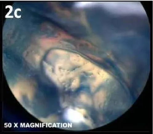
After decalcification and dehydration the samples were kept in Methyl- salicylic acid, till they become transparent and clear in appearance (nearly 2-3 hrs).(Fig-1c) The decalcified sample along with dye were analysed with stereomicroscope for anatomy, position & shape of accessory canals from the furcation area as well as from the pulpal floor .The reading of microscope were kept on two standardized point i.e. at 1.65 X and 5X so as to report the presence of accessory canal at a standardized magnification and at maximum magnification respectively .The specimens were then kept in a reverse position (furcation area toward the eyepiece {Fig-2a}). All the images were divided into oval shaped canal (Fig-2b), round shape canal (Fig-2c), the location of accessory canals were further observed i.e. exactly at the center or at the periphery.
RESULTS
Figure 1a. Presence of accessory canal in sagittal plane of CBCT
[image:3.595.48.282.229.385.2]Figure 1b. Presence of accessory canal in axial plane of CBCT showing different orifice in pulpal floor
Figure 1c. Decalcified sample after India ink injection and clearing technique
[image:3.595.310.559.288.506.2]Figure 2a. Position of specimen in stereomicroscope
Figure 2b. Presence of oval shaped accessory canal in furcation area of primary molar
Figure 2c. Presence of round shaped accessory canal in furcation area of primary molar. Present at the center and towards palatal
canal
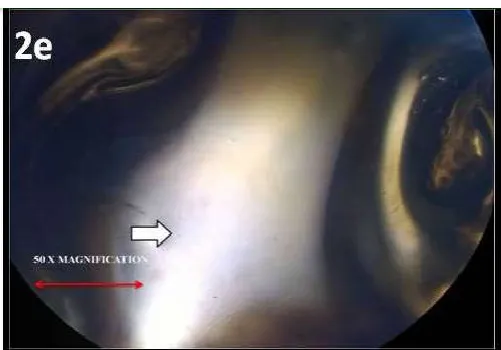
[image:3.595.49.281.426.552.2] [image:3.595.310.561.563.753.2] [image:3.595.58.271.596.778.2]Figure 2e. No accessory canal at the furcation area of primary molar
Figure 3. Shows prevalence of accessory canals in maxillary and mandibular teeth
Table 1 shows the total number of primary molars with accessory canals by two different methods. A significant difference was seen in detection between two techniques (p<0.05) when the comparison was done between maxillary and mandibular molars. Table 2 shows prevalence of oval and round shaped canals in maxillary and mandibular first and second primary molars under decalcification technique. The highest prevalence of oval and round shape canal was found in mandibular 2nd molar & least in mandibular 1st molar. Round shaped canals were mostly found in mandibular 1st molar. However their presence was statistically non significant (P>0.05) Table 3 shows the position of accessory canals in furcation area of primary molars under decalcification technique. The prevalence of accessory canals positioned in the center was maximum in maxillary 1st molar (71.42%), where as it was least near palatal root (28.57%). However the highest prevalence of accessory canals present near palatal root was 73.33% with maxillary 2nd molar, and similarly near the palatal root it was least (26.67%). Centrally positioned accessory canals were seen in the order of
Maxillary 1st molar > Mandibular 2nd molar > Mandibular 1st molar > Maxillary 2ndmolar
[image:4.595.38.292.268.433.2]Eccentrically positioned canals were either near palatal root in maxillary teeth or distal root in mandibular teeth. Table 4 describes the intergroup comparison for the location of accessory canals between maxillary and mandibular 1st and 2nd primary molars. When maxillary 1st and 2nd molars were compared greater number of accessory canals (71.42%) where seen in furcation area of maxillary 1st molar where as in 2nd molar greater number of accessory canals (73.33%) were seen along palatal canal .When mandibular 1st and 2nd molars were compared greater number of accessory canals (33.3%%) where seen in furcation area of mandibular 1st molar where as in 2nd molar greater number of accessory canals (58.33%) were seen along distal root.
Table 1. Accessory canals in two different methods
Samples Number of teeth Cbct Decalcification Difference between the methods
CBCT VS Decalcification
MAXILLARY 1ST MOLAR 19 9 (47.4%) 14 (73.7%) P*= 0.033, S
MAXILLARY 2ND MOLAR 19 11(57.9%) 15 (78.9%) P*= 0.018, S
MANDIBULAR 1ST MOLAR 19 8 (42.1%) 12 (63.2%) P*= 0.013, S
MANDIBULAR 2ND MOLAR 19 10 (52.6%) 12 (63.2%) P*= 0.001, S
Difference between different teeth regarding presence of accessory canals P**= 0.79, NS P**= 0.64, NS
*Fisher’s Exact Test, **Chi square test
Table 2.
Samples Shape of the canal
Oval & Round Round
MAXILLARY 1ST MOLAR 10 (71.42%) 4 (28.57%) P=0.196, NS
MAXILLARY 2ND MOLAR 11 (73.33%) 4 (26.67%) P=0.076, NS
MANDIBULAR 1ST MOLAR 8 (66.7) 4 (33.3%) P=0.504, NS
MANDIBULAR 2ND MOLAR 10 (83.33%) 2 (16. 67%) P=0.076, NS
*Fisher’s Exact Test
Table 3.
Samples Position of the canal
Eccentric Near palatal /distal root Center
MAXILLARY 1ST MOLAR 4 (28.57%) 10 (71.42%) P=0.196, NS
MAXILLARY 2ND MOLAR 11 (73.33%) 4 (26.67%) P=0.076, NS
MANDIBULAR 1ST MOLAR 8 (66.7) 4 (33.3%) P=0.504, NS
MANDIBULAR 2ND MOLAR 7 (58.33%) 5 (41.67%) P=0.810, NS
[image:4.595.46.546.508.579.2]Table 4.
Samples Position of the canal
Near palatal root Center
MAXILLARY 1ST MOLARS 4 (28.57%) 10 (71.42%)
MAXILLARY 2ND MOLARS 11 (73.33%) 4 (26.67%)
P VALUE 0.11, NS 0.07, NS
SAMPLES Position of the canal
Near distal root Center
MANDIBULAR 1ST MOLARS 8 (66.7) 4 (33.3%)
MANDIBULAR 2ND MOLARS 7 (58.33%) 5 (41.67%)
P VALUE 0.99, NS 0.79, NS
DISCUSSION
It is very difficult to understand the nature and position of the accessory canals in primary teeth. The furcation area of the primary molars has great significance as it encompasses the follicle of permanent teeth. Moreover the pathological changes in the periapical bone are more likely to start occurring in the interradicular region. (Kumar, 2009) Majority of the pathological changes in the region of furcation are more likely to be caused by the presence of accessory canals. (Poornima and Subba Reddy, 2008) A variety of techniques have been employed for the detection of accessory canals in primary teeth including spiral computed tomography (Vijayakumar et al., 2013), decalcification with dye penetration (Poornima and Subba Reddy, 2008; Kumar, 2009; Gupta and Grewal, 2005; Ayasimha Raj and Mylswamy, 2010; Parekh et al., 2011), SEM studies etc. (Kumar, 2009) The latest technique to construct transparent teeth without decalcifying was described by Malentacca et al in 2015 using Xylene C8H10 (Merck F.R. Germany). (Malentacca and Lajolo, 2015) Introduction of maxillofacial Cone Beam Computed Tomography in 1966 provided the clinical and practical technology which demonstrated the application of 3D imaging in Endodontics. The advantage of using CBCT over the conventional radiographic techniques has been studied since long & cannot be underestimated while making endodontic diagnosis. (14) Keeping these features in mind, we researched the presence of accessory canals and their pattern on pulpal floor of primary teeth by CBCT scanning. The presence of accessory canals was 50% and 69.73% on CBCT and decalcification technique using dye penetration and clearing respectively. This is in accordance with the study done by Lowman et al, who reported 59% patent accessory canals in molars. (Poornima and Subba Reddy, 2008)
Position of accessory canals at the furcation area is difficult to standardize as they are present abruptly all over the pulpal floor. The presence of patent and well formed accessory canals in maxillary teeth are visualized more accurately near the palatal root in maxillary primary molars. The only disadvantage of using CBCT for detection of accessory canals is that it can clearly visualize only those canals which are patent. Out of the 38 primary maxillary teeth second molars had 73.33% and 1st molar had 28.57% accessory canals near the center and at palatal region. Out of 37 primary mandibular teeth the 2nd molar had 58% accessory canals in the center and near distal root where as in mandibular 1st molar it was 66.7%. Kumar VD et al used decalcification technique and reported that most of the accessory canals were found at the center of the furcation region. (Kumar, 2009)which is in contrast to our study wherein lot of concentration of accessory canal were seen near palatal and distal roots difference might be because of many factors like method of decalcification and time of specimen analysis. The advantages of dye penetration and
clearing technique was mentioned by Okumura and Robertson et al. (Singh and Pawar, 2015) The clearing technique followed in this study resulted in clear specimens, which allowed better visualization of accessory canals in furcation area. The presence of accessory canals in Indian population was significant when compared with similar experimental studies. The observations were made by examining the specimens with a stereo - microscope at 56 X magnification under good lighting to evaluate the presence of accessory canals in the furcation area. A study done by Karl et al reported the presence of 77.5% of accessory canals in 40 primary second maxillary and mandibular molars, with prevalence of 80 % in maxillary molars and 75 % in mandibular molars. The difference in the prevalence might be attributed to the procedure applied, as in this study; decalcification was done before the injection of dye. This could have caused more opening of accessory canals and better dye penetration. (Kumar, 2009)
Conclusion
From the findings of the present study, we can conclude that there is a high prevalence of accessory canal in furcation area of primary teeth. On comparing the different methods, decalcification was found to be significantly better than CBCT.
REFERENCES
Alavi, A.M., Opasanon, A. Ng, Y.L. and Gulabivala, K. 2002. Root and canal morphology of Thai maxillary molars. Int Endod J. May; 35 (5): 478-85
Ayasimha Raj, U. and Mylswamy, S. 2010. Root canal morphology of maxillary second Premolars in an Indian population. J Conserv Dent. Jul; 13 (3): 148 51.
Carrot, P. 2004. Endodontics: Part 7. Preparing the root canal. Br Dent J. Nov 27; 197(10):603-13.
Carrotte, P.V. 2009. A Clinical guide to endodontics - update part 1. Br Dent J. Jan 24; 206(2):79-84.
Gupta, D. and Grewal, N. 2005. Root canal configuration of deciduous mandibular first molars--an in vitro study. J Indian Soc Pedod Prev Dent. Sep; 23 (3): 134-7.
Hess, W. The Anatomy of the Root Canals of the Teeth of the Permanent and Deciduous Dentition. New York: William Wood and Co. 1925.
Kiarudi, A.H., Eghbal, M.J., Safi, Y., Aghdasi, M.M. and Fazlyab, M. 2015. The Applications of Cone-Beam Computed Tomography in Endodontics: A Review of Literature. Iran Endod J. 10(1)-16-25.
Kojima, K., Inamoto, K., Nagamatsu, K., Hara, A., Nakata, K., Morita, I., Nakagaki, H. and Nakamura, H. 2004. Success rate of endodontic treatment of teeth with vital and nonvital pulps. A meta-analysis. Oral Surg Oral Med Oral Pathol Oral RadiolEndod. Jan; 97(1):95-9.
Kumar, V.D. 2009. A scanning electron microscope study of prevalence of accessory canals on the pulpal floor of deciduous molars. J Indian Soc Pedod Prev Dent. Apr Jun; 27(2):85-9.
Malentacca, A. and Lajolo, C. 2015. A new technique to make transparent teeth without decalcifying: description of the methodology and micro-hardness assessment. Ann Anat. Jan; 197: 11-5.
Poornima, P. and Subba Reddy, V.V. 2008. Comparison of digital radiography, decalcification, and histologic sectioning in the detection of accessory canals in furcation areas of human primary molars. J Indian SocPedodPrev Dent. Jun; 26(2):49-52.
Singh, S. and Pawar, M. 2015. Root canal morphology of South Asian Indian maxillary molar teeth. Eur J Dent, 9:133-44.
Todd, R. 2014. Cone beam computed tomography updated technology for endodontic diagnosis. Dent Clin North Am. Jul; 58 (3): 523-43
Vijayakumar, R., Selvakumar, H., Swaminathan, K., Thomas, E., Ganesh, R. and Palanimuthu, S. 2013. Root canal morphology of human primary maxillary molars in Indian population using spiral computed tomography scan: An in vitro study. SRM J Res Dent Sci, 4:139-42