Received 3 September 2019 Accepted 7 September 2019
Edited by W. T. A. Harrison, University of Aberdeen, Scotland
‡ Additional correspondence author, e-mail: [email protected].
Keywords:crystal structure; organotin; dithio-carbamate; Hirshfeld surface analysis; compu-tational chemistry.
CCDC reference:1952056
Supporting information:this article has supporting information at journals.iucr.org/e
(
N
,
N
-Diisopropyldithiocarbamato)triphenyltin(IV):
crystal structure, Hirshfeld surface analysis and
computational study
Farah Natasha Haezam,aNormah Awang,a‡ Nurul Farahana Kamaludin,a Mukesh M. Jotaniband Edward R. T. Tiekinkc*
aEnvironmental Health and Industrial Safety Programme, Faculty of Health Sciences, Universiti Kebangsaan Malaysia,
Jalan Raja Muda Abdul Aziz, 50300 Kuala Lumpur, Malaysia,bDepartment of Physics, Bhavan’s Sheth R. A. College of Science, Ahmedabad, Gujarat 380001, India, andcResearch Centre for Crystalline Materials, School of Science and Technology, Sunway University, 47500 Bandar Sunway, Selangor Darul Ehsan, Malaysia. *Correspondence e-mail: [email protected]
The crystal and molecular structures of the title triorganotin dithiocarbamate, [Sn(C6H5)3(C7H14NS2)], are described. The molecular geometry about the metal
atom is highly distorted being based on a C3S tetrahedron as the
dithio-carbamate ligand is asymmetrically chelating to the tin centre. The close approach of the second thione-S atom [Sn S = 2.9264 (4) A˚ ] is largely responsible for the distortion. The molecular packing is almost devoid of directional interactions with only weak phenyl-C—H C(phenyl) interactions, leading to centrosymmetric dimeric aggregates, being noted. An analysis of the calculated Hirshfeld surface points to the significance of H H contacts, which contribute 66.6% of all contacts to the surface, with C H/H C [26.8%] and S H/H H [6.6%] contacts making up the balance.
1. Chemical context
Organotin(IV) compounds have long been investigated as potential anti-cancer agents (Gielen & Tiekink, 2005) and studies in this area continue. Further, organotin compounds have received much attention owing to their potential ther-apeutic potential as anti-fungal, anti-bacterial, anti-malarial and schizonticidal agents (Khan et al., 2014). Metal dithio-carbamates have also encouraged much interest in the context of chemotherapeutic agents (Hogarth, 2012) and these include organotin(IV) dithiocarbamate compounds (Tiekink, 2008; Adeyemi & Onwudiwe, 2018). In view of the wide-range of applications/potential of organotin(IV) dithiocarbamate compounds and in continuation of on-going studies in this area (Khanet al., 2015; Mohamadet al.2016, 2017, 2018), the title compound, Ph3Sn[S2CN(i-Pr)2], (I), was synthesized and
characterized spectroscopically. Herein, the crystal and mol-ecular structures of (I) are described along with a detailed analysis of the molecular packingviathe calculated Hirshfeld surfaces and computational chemistry study.
2. Structural commentary
The tin atom in (I), Fig. 1, is coordinated by an asymmetrically bound dithiocarbamate ligand and threeipso-carbon atoms of the phenyl groups (Table 1). The disparity in the Sn—S separations, i.e. (Sn—S) = [(Sn—Sl) (Sn—Ss)] = 0.45 A˚
(l = long, s = short), is rather great suggesting that the Sn S2 interaction is weak. This is supported in the pattern of C—S bond lengths with that involving the more tightly bound S1 atom being nearly 0.06 A˚ longer than the equivalent bond with the weakly bound S2 atom. Despite this, a clear influence of the S2 atom is noted on the Sn—C bond lengths with the Sn—C31 bond length being significantly longer than the other Sn—C bonds, Table 1. The S2—Sn—C31 bond angle is 158.41 (4) and is suggestive of a trans-influence exerted by the S2 atom; there is no othertransangle about the tin atom. If the coordination geometry is considered as tetrahedral, the range of tetrahedral angles is 93.24 (4), for S1—Sn—C31, to
119.87 (6), for C11—Sn—C21. The range of angles assuming
a five-coordinate, C3S2, geometry is 65.260 (11), for the S1—
Sn—S2 chelate angle to the aforementioned 158.41 (4). A
descriptor for assigning coordination geometries to five-coordinate species is(Addisonet al., 1984). In the case of (I), this computes to 0.64, which indicates a geometry somewhat closer to an ideal trigonal bipyramid (= 1.0) than to an ideal square pyramid (= 0.0). Also included in Table 1 are the C— N bond lengths, which show C1—N1 to be significantly shorter than the C2—N1 and C5—N1 bond lengths, an observation
consistent with a significant contribution of the2S2C N+(i
-Pr)2canonical form to the overall electronic structure of the
dithiocarbamate ligand.
3. Supramolecular features
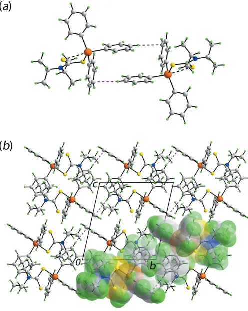
The geometric parameters characterizing the identified inter-molecular interaction operating in the crystal of (I) are collated in Table 2. A phenyl-C—H C(phenyl) contact less than the sum of the sum of the Waals radii (Spek, 2009) is noted to occur between centrosymmetrically related mol-ecules, Fig. 2(a). This is an example of a localized C—H contact whereby the hydrogen atom directed towards a single carbon atom of the ring as opposed to a delocalized inter-action where the hydrogen atom (or halide atom or lone-pair of electrons) is directed towards the centroid of the ring (Schollmeyeret al., 2008; Tiekink, 2017).
1480
Haezamet al. [Sn(C6H5)3(C7H14NS2)] Acta Cryst.(2019). E75, 1479–1485 [image:2.610.313.566.83.124.2]research communications
Table 1
Selected bond lengths (A˚ ).
Sn—S1 2.4792 (4) C1—S1 1.7587 (15) Sn—S2 2.9264 (4) C1—S2 1.7006 (16) Sn—C11 2.1446 (14) C1—N1 1.336 (2) Sn—C21 2.1349 (15) C5—N1 1.495 (2) Sn—C31 2.1754 (15) C2—N1 1.497 (2)
Table 2
Hydrogen-bond geometry (A˚ ,).
D—H A D—H H A D A D—H A
C34—H34 C24i 0.95 2.82 3.757 (3) 171
[image:2.610.44.296.91.142.2]Symmetry code: (i)xþ1;y;z.
Figure 2
[image:2.610.315.563.378.689.2]Molecular packing in the crystal of (I): (a) supramolecular dimer sustained by localized phenyl-C—H C(phenyl) interactions shown as purple dashed lines and (b) a view of the unit-cell contents in projection down thea axis. One column of dimeric aggregates is highlighted in space-filling mode.
Figure 1
[image:2.610.48.295.513.712.2]4. Hirshfeld surface analysis
The Hirshfeld surface calculations for (I) were performed employing Crystal Explorer 17 (Turner et al., 2017) and recently published protocols (Tanet al., 2019). In the absence of classical hydrogen bonds, the influence of the localized C— H interaction, Table 2, as well as interatomic H H and C H/H C contacts, Table 3, upon the molecular packing are evident as the diminutive-red spots near the participating carbon and hydrogen atoms on the Hirshfeld surfaces mapped overdnormin Fig. 3. It is also noted that with the exception of
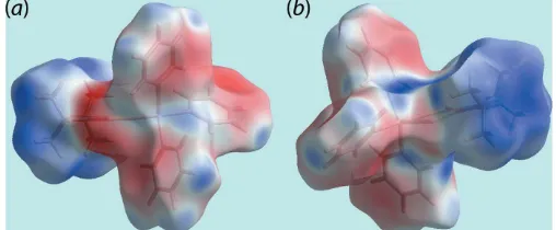
the methyl-H7Aatom, all of the specified interatomic contacts only involve the carbon and hydrogen atoms of the coordin-ated phenyl rings, Table 3. On the Hirshfeld surface mapped over electrostatic potential in Fig. 4, the light-blue and
faint-red regions, corresponding to positive and negative electro-static potential, respectively, occur about the di-iso-propyl and triphenyltin groups, respectively.
As reported recently (Pinto et al., 2019), in addition to analysing the nature and strength of intermolecular inter-actions among molecules, the analyses of Hirshfeld surfaces can also provide useful insight into metal–ligand/donor atom interactions in coordination compounds. Thus, the distance from the surface to the nearest external (de) and internal (di)
nuclei, the shape-index (S) and the curvedness (C) can also be plotted. Accordingly, Fig. 5 illustrates the Hirshfeld surfaces for the tin atom coordinated by dithiocarbamate ligand as well as by the three phenyl groups. The close proximity of the dithiocarbamate-S1 and phenyl-C11, C21 and C31 atoms to the tin centre are characterized as bright-red regions perpendicular to bond directions on the Hirshfeld surfaces mapped overde, Fig. 5(a), and dnorm, Fig. 5(b), whereas the
comparatively weak Sn—S2 interaction appears as the faint-red region. The longer Sn—C31 bond compafaint-red to other two Sn—C bonds, Table 1, is also characterized from these Hirshfeld surfaces through the curvature of the red region. The Sn—S1 and Sn—C bonds result in the large red regions on the shape-index mapping in Fig. 5(c) compared to a small red region for the Sn—S2 bond. On the Hirshfeld surfaces mapped over curvedness in Fig. 5(d), the strength of the tin– ligand bonds are characterized as the yellow areas separated by green regions. The coordination bonds for tin are also rationalized in the fingerprint plot taking into account only the Hirshfeld surface about the metal atom, Fig. 6. The distribu-tion of green points having upper short spike atde+di2.4 A˚
and the lower, long red spike atde+di2.1 A˚ are the result of
[image:3.610.312.564.70.226.2]the Sn—S and Sn—C bonds, respectively. This asymmetric distribution of points about the diagonal lacking homogeneity in colouration is due to the distorted coordination geometry about the tin atom.
Table 3
Summary of short interatomic contacts (A˚ ) in (I).
The interatomic distances are calculated inCrystal Explorer 17(Turneret al., 2017) whereby theX—H bond lengths are adjusted to their neutron values.
Contact Distance Symmetry operation
H7A H12 2.09 1x, 1y,z
H13 H26 2.26 1 +x,y,z
C12 H36 2.65 1x, 1y,z
C13 H26 2.68 1 +x,y,z
C16 H23 2.65 1x,y, 1z
C24 H34 2.68 1x,yz
Figure 3
[image:3.610.45.295.119.194.2]Two views of Hirshfeld surface for (I) mapped overdnormin the range 0.085 to +1.355 (arbitrary units), highlighting short interatomic H H and C H/H C contacts as diminutive red spots near the respective atoms.
Figure 4
Two views of Hirshfeld surface mapped over the electrostatic potential (the red and blue regions represent negative and positive electrostatic potentials, respectively) in the range0.032 to +0.035 atomic units.
Figure 5
[image:3.610.43.297.597.702.2]It is clear from the the calculation of the overall two-dimensional fingerprint plot for (I), Fig. 7(a), that the plot is asymmetric about the (de,di) diagonal in the longer distance
regions and have contributions only from the interatomic contacts involving carbon, hydrogen and sulfur atoms, Table 4. The two-dimensional fingerprint plots delineated into H H, C H/H C, C C and S H/H S contacts are shown in Fig. 7(b)–(d), respectively. In the fingerprint plot delineated into H H contacts in Fig. 7(b), the presence of the short interatomic H H interaction involving the methyl-H7Aand phenyl-H12 atoms is evident as the pair of short overlapping peaks atde+di2.1 A˚ with the other short interatomic H H
contact (Table 3) merged within the plot. The intermolecular C—H C interactions describing the localized C—H contacts are evidenced by a pronounced pair of characteristic wings around (de, di)(1.2 A˚ , 1.8A˚) and(1.8 A˚ , 1.2 A˚) in
the fingerprint plot delineated into C H/H C contacts shown in Fig. 7(c). The other short interatomic C H contacts summarized in Table 3 appear as the pair of forceps-like tips at de + di 2.7 A˚ . The fingerprint plot delineated into S H/
H S contacts in Fig. 7(d) indicate that sulfur atoms are nearly at van der Waals separation from the symmetry-related hydrogen atoms.
5. Computational chemistry
The pairwise interaction energies between the molecules within the crystal were calculated usingCrystal Explorer 17 (Turner et al., 2017) and summing up the four energy components: electrostatic (Eele), polarization (Epol),
disper-sion (Edis) and exchange-repulsion (Erep). The energies were
obtained using the wave function calculated at the HF/STO-3G level of theory. The strength and the nature of inter-molecular interactions in terms of their energies are summarized in Table 5. An analysis of these reveals that the dispersion energy component makes the greatest contribution
1482
Haezamet al. [Sn(C6H5)3(C7H14NS2)] Acta Cryst.(2019). E75, 1479–1485 [image:4.610.44.297.67.324.2] [image:4.610.313.566.102.161.2]research communications
Table 5
Summary of interaction energies (kJ mol1) calculated for (I).
Contact R(A˚ ) Eele Epol Edis Erep Etot
H7A H12i+ 6.30
H36 C12i 17.5 7.0 112.7 68.0 68.8 H13 H26ii+ 9.76
C13 H26ii 8.7 2.0 31.6 18.0 24.0
C16 H23iii 9.46 12.9 2.0 41.9 29.5 28.2
C24 H34iv 10.62 11.0 2.8 33.8 21.6 26.0
Notes: Symmetry operations: (i) 1x, 1y,z; (ii) 1 +x,y,z; (iii) 1x,y, 1z; (iv) 1x,y,z.
Table 4
Percentage contributions of interatomic contacts to the Hirshfeld surface for (I).
Contact Percentage contribution
H H 66.5
C H/H C 26.8 S H/H S 6.6 C S/S C 0.1
Figure 6
[image:4.610.313.566.207.284.2]The fingerprint plot taking into account only the Hirshfeld surface about the tin atom.
Figure 7
[image:4.610.52.567.582.724.2]to the energies in the absence of classical hydrogen (electro-static) bonds. Among the short interatomic contacts listed in Table 5, the intermolecular phenyl-C—H36 C12 contact combined with the methyl-H7A H12(phenyl) interaction, occurring between the same pair of symmetry-related mol-ecules, gives rise to the maximum total energy of interaction, compared to the other interactions, which have almost the same energy values.
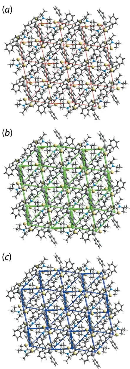
The graphical representation of the magnitudes of inter-molecular energies in Fig. 8,i.e.energy frameworks, relies on a red, green and blue colour code scheme, reflecting theEele,
Edisp and Etot components, respectively. For the direct
comparison of magnitudes of interaction energies, their magnitudes were adjusted to same scale factor of 30 with a cut-off value of 3 kJ mol1within 222 unit cells. It is clear from Fig. 8 that the green cylinders joining the centroids of molecular pairs highlighting the dispersion components make a significant contribution to the supramolecular architecture in the crystal.
6. Database survey
As indicated in a recent report (Mohamadet al., 2018), there are nearly 50 crystal structures available for molecules of the general formula Ph3Sn(S2CNRR0) and, with a number of these
having multiple molecules in the asymmetric unit, there are almost 60 independent molecules. These conform to the same structural motif. An analysis of the key geometric parameters defining the mode of coordination of the dithiocarbamate ligands showed that there were no systematic variations that could be correlated with the nature of the dithiocarbamate ligand,i.e. R/R0substituents. This observation is borne out by
DFT calculations on different organotin systems that proved the influence of molecular packing on (non-systematic) geometric parameters, including metal–sulfur/halide bonds (Buntineet al., 1998a,b, 1999). In terms of Ph3Sn(S2CNRR
0
), the mean Sn—Ssbond length is 2.47 A˚ (standard deviation =
0.013 A˚ ) and the average Sn—Sl bond length is 3.04 A˚
(0.070 A˚ ). The Sn—Ssand Sn—Slbond lengths in (I) both fall
within 2of their respective means.
The homogeneity in the molecular structures of Ph3Sn(S2CNRR0) is quite remarkable. Structural diversity is
well-established for the organotin dithiocarbamates (Tiekink, 2008; Muthalib et al., 2014) such as for molecules of the general formula R00
2Sn(S2CNRR0)2, for which four distinct
structural motifs are known (Zaldi et al., 2017). Also, the behaviour of triphenyltin dithiocarbamates contrasts the analogous chemistry of triphenyltin carboxylates (Tiekink, 1991). These are often monomeric (e.g. Basu Baulet al., 2001), as for (I), but, polymeric examples are known (e.g. Willemet al., 1997; Smyth & Tiekink, 2000). The polymeric structures occur when the carboxylate ligands are bidentate bridging, leading to trans-C3O2 trigonal–bipyramidal coordination
geometries for the tin atoms. This fundamental difference in structural chemistry arises as a result of the significant contribution of the 2S2C N
+
RR0 canonical form to the
[image:5.610.60.281.64.690.2]electronic structure of the dithiocarbamate anion, as discussed Figure 8
above. The formal negative charge on each sulfur atom makes this ligand a very efficient chelator which effectively reduces the Lewis acidity of the tin centre. Far from being a curiosity, such behaviour, i.e. dithiocarbamate ligands reducing the Lewis acidity of metal centres, when compared to related xanthate (S2COR) and dithiophopshate [(S2P(OR)2]
ligands, leads to stark differences in coordination propensities in zinc-triad element 1,1-dithiolate compounds, as has been reviewed recently (Tiekink, 2018a,b).
7. Synthesis and crystallization
All chemicals and solvents were used as purchased without purification. The melting point was determined using an automated melting point apparatus (MPA 120 EZ-Melt). Carbon, hydrogen, nitrogen and sulfur analyses were performed on a Leco CHNS-932 Elemental Analyzer.
Di-iso-propylamine (Aldrich; 1.41 ml, 10 mmol) dissolved in ethanol (30 ml) was stirred under ice-bath conditions at 277 K for 20 mins. A 25% ammonia solution (1–2 ml) was added to provide basic conditions. Then, a cold ethanolic solution of carbon disulfide (0.60 ml, 10 mmol) was added dropwise into the solution followed by stirring for 2 h. After that, triphenyltin(IV) chloride (Merck; 3.85 g, 10 mmol) dissolved in ethanol (20–30 ml) was added dropwise into the solution followed by further stirring for 2 h. The precipitate that formed was filtered and washed a few times with cold ethanol to remove impurities. Finally, the colourless
precipi-tate was dried in a desiccator. Recrystallization was carried out by dissolving the compound in a chloroform and ethanol mixture (1:1v/v). This solution was allowed to slowly evapo-rate at room temperature yielding colourless slabs of (I). Yield: 47%, m. p.: 437.8–440.2 K. Elemental analysis: Calcu-lated (%): C 57.07, H 5.51, N 2.66, S 12.19. Found (%): C 57.39, H 5.31, N 2.48, S 11.26.
8. Refinement
Crystal data, data collection and structure refinement details are summarized in Table 6. Carbon-bound H atoms were placed in calculated positions (C—H = 0.95–1.00 A˚ ) and were included in the refinement in the riding model approximation, withUiso(H) set to 1.2–1.5Ueq(C).
Acknowledgements
We gratefully acknowledge the Faculty of Health Sciences and the Faculty of Science and Technology of the Universiti Kebangsaan Malaysia for providing essential laboratory facilities and for technical support from the laboratory assis-tants. The Universiti Malaysia Terengganu is thanked for the elemental analysis. The authors also thank the Research Centre of Crystalline Materials X-ray crystallography laboratory for the X-ray intensity data.
Funding information
This work was supported by the Fundamental Research Grant Scheme (FRGS/1/2018/STG01/UKM/02/20) awarded by the Ministry of Education (MOE). Crystallographic research at Sunway University is supported by Sunway University Sdn Bhd (grant No. STR-RCTR-RCCM-001-2019).
References
Addison, A. W., Rao, T. N., Reedijk, J., van Rijn, J. & Verschoor, G. C. (1984).J. Chem. Soc. Dalton Trans.pp. 1349–1356.
Adeyemi, J. O. & Onwudiwe, D. C. (2018).Molecules,23article No. 2571.
Basu Baul, T. S., Dhar, S., Pyke, S. M., Tiekink, E. R. T., Rivarola, E., Butcher, R. & Smith, F. E. (2001).J. Organomet. Chem. 633, 7– 17.
Brandenburg, K. (2006).DIAMOND. Crystal Impact GbR, Bonn, Germany.
Buntine, M. A., Hall, V. J., Kosovel, F. J. & Tiekink, E. R. T. (1998a).J. Phys. Chem. A,102, 2472–2482.
Buntine, M. A., Hall, V. J. & Tiekink, E. R. T. (1998b).Z. Kristallogr. 213, 669–678.
Buntine, M. A., Hall, V. J. & Tiekink, E. R. T. (1999).Z. Kristallogr. 214, 124–134.
Farrugia, L. J. (2012).J. Appl. Cryst.45, 849–854.
Gielen, M. & Tiekink, E. R. T. (2005).Metallotherapeutic drugs and metal-based diagnostic agents: the use of metals in medicine, edited by M. Gielen & E. R. T. Tiekink, pp. 421–439. Chichester: John Wiley & Sons Ltd.
Hogarth, G. (2012).Mini Rev. Med. Chem.12, 1202–1215.
Khan, N., Farina, Y., Mun, L. K., Rajab, N. F. & Awang, N. (2014).J. Mol. Struct.1076, 403–410.
Khan, N., Farina, Y., Mun, L. K., Rajab, N. F. & Awang, N. (2015). Polyhedron,85, 754–760.
1484
Haezamet al. [Sn(C6H5)3(C7H14NS2)] Acta Cryst.(2019). E75, 1479–1485 [image:6.610.44.297.89.366.2]research communications
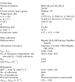
Table 6
Experimental details.
Crystal data
Chemical formula [Sn(C6H5)3(C7H14NS2)]
Mr 526.30
Crystal system, space group Triclinic,P1 Temperature (K) 100
a,b,c(A˚ ) 9.7572 (1), 11.7030 (2), 11.7602 (2)
,,() 74.419 (1), 80.114 (1), 67.285 (2)
V(A˚3) 1189.71 (4)
Z 2
Radiation type CuK (mm1) 10.25
Crystal size (mm) 0.120.110.08
Data collection
Diffractometer Rigaku XtaLAB Synergy Dualflex AtlasS2
Absorption correction Gaussian (CrysAlis PRO; Rigaku OD, 2018)
Tmin,Tmax 0.840, 1.000
No. of measured, independent and observed [I> 2(I)] reflections
28219, 4248, 4231
Rint 0.022
(sin /)max(A˚
1
) 0.597
Refinement
R[F2> 2(F2)],wR(F2),S 0.015, 0.040, 1.00
No. of reflections 4248 No. of parameters 266
H-atom treatment H-atom parameters constrained
max,min(e A˚
3
) 0.36,0.44
Computer programs:CrysAlis PRO(Rigaku OD, 2018),SHELXS(Sheldrick, 2015a),
SHELXL2014(Sheldrick, 2015b),ORTEP-3 for Windows(Farrugia, 2012),DIAMOND
Mohamad, R., Awang, N., Kamaludin, N. F. & Abu Bakar, N. F. (2016).Res. J. Pharm. Biol. Chem. Sci.7, 1269–1274.
Mohamad, R., Awang, N., Kamaludin, N. F., Jotani, M. M. & Tiekink, E. R. T. (2017).Acta Cryst.E73, 260–265.
Mohamad, R., Awang, N., Kamaludin, N. F., Jotani, M. M. & Tiekink, E. R. T. (2018).Acta Cryst.E74, 630–637.
Muthalib, A. F. A., Baba, I., Khaledi, H., Ali, H. M. & Tiekink, E. R. T. (2014).Z. Kristallogr.229, 39–46.
Pinto, C. B., Dos Santos, L. H. R. & Rodrigues, B. L. (2019). Acta Cryst.C75, 707–716.
Rigaku OD (2018). CrysAlis PRO. Rigaku Oxford Diffraction Corporation, Yarnton, England.
Schollmeyer, D., Shishkin, O. V., Ru¨hl, T. & Vysotsky, M. O. (2008). CrystEngComm,10, 715–723.
Sheldrick, G. M. (2015a).Acta Cryst.A71, 3–8. Sheldrick, G. M. (2015b).Acta Cryst.C71, 3–8.
Smyth, D. R. & Tiekink, E. R. T. (2000).Z. Kristallogr. New Cryst. Struct.215, 81–82.
Spek, A. L. (2009).Acta Cryst.D65, 148–155.
Tan, S. L., Jotani, M. M. & Tiekink, E. R. T. (2019).Acta Cryst.E75, 308–318.
Tiekink, E. R. T. (1991).Appl. Organomet. Chem.5, 1–23. Tiekink, E. R. T. (2008).Appl. Organomet. Chem.22, 533–550. Tiekink, E. R. T. (2017).Coord. Chem. Rev.345, 209–228. Tiekink, E. R. T. (2018a).Crystals,8, article no. 292. Tiekink, E. R. T. (2018b).Crystals,8, article no. 18.
Turner, M. J., Mckinnon, J. J., Wolff, S. K., Grimwood, D. J., Spackman, P. R., Jayatilaka, D. & Spackman, M. A. (2017). Crystal Explorer 17. The University of Western Australia.
Westrip, S. P. (2010).J. Appl. Cryst.43, 920–925.
Willem, R., Bouhdid, A., Mahieu, B., Ghys, L., Biesemans, M., Tiekink, E. R. T., de Vos, D. & Gielen, M. (1997).J. Organomet. Chem.531, 151–158.
supporting information
sup-1 Acta Cryst. (2019). E75, 1479-1485
supporting information
Acta Cryst. (2019). E75, 1479-1485 [https://doi.org/10.1107/S2056989019012490]
(
N
,
N
-Diisopropyldithiocarbamato)triphenyltin(IV): crystal structure, Hirshfeld
surface analysis and computational study
Farah Natasha Haezam, Normah Awang, Nurul Farahana Kamaludin, Mukesh M. Jotani and
Edward R. T. Tiekink
Computing details
Data collection: CrysAlis PRO (Rigaku OD, 2018); cell refinement: CrysAlis PRO (Rigaku OD, 2018); data reduction:
CrysAlis PRO (Rigaku OD, 2018); program(s) used to solve structure: SHELXS (Sheldrick, 2015a); program(s) used to refine structure: SHELXL2014 (Sheldrick, 2015b); molecular graphics: ORTEP-3 for Windows (Farrugia, 2012) and
DIAMOND (Brandenburg, 2006); software used to prepare material for publication: publCIF (Westrip, 2010).
(N,N-Diisopropyldithiocarbamato)triphenyltin(IV)
Crystal data
[Sn(C6H5)3(C7H14NS2)]
Mr = 526.30 Triclinic, P1
a = 9.7572 (1) Å
b = 11.7030 (2) Å
c = 11.7602 (2) Å
α = 74.419 (1)°
β = 80.114 (1)°
γ = 67.285 (2)°
V = 1189.71 (4) Å3
Z = 2
F(000) = 536
Dx = 1.469 Mg m−3
Cu Kα radiation, λ = 1.54184 Å Cell parameters from 24601 reflections
θ = 3.9–76.3°
µ = 10.25 mm−1
T = 100 K Prism, colourless 0.12 × 0.11 × 0.08 mm
Data collection
Rigaku XtaLAB Synergy Dualflex AtlasS2 diffractometer
Detector resolution: 5.2558 pixels mm-1
ω scans
Absorption correction: gaussian (CrysAlis PRO; Rigaku OD, 2018)
Tmin = 0.840, Tmax = 1.000
28219 measured reflections
4248 independent reflections 4231 reflections with I > 2σ(I)
Rint = 0.022
θmax = 67.1°, θmin = 3.9°
h = −11→11
k = −13→13
l = −13→14
Refinement
Refinement on F2
Least-squares matrix: full
R[F2 > 2σ(F2)] = 0.015
wR(F2) = 0.040
S = 1.00 4248 reflections 266 parameters 0 restraints
Primary atom site location: structure-invariant direct methods
Secondary atom site location: difference Fourier map
Hydrogen site location: inferred from neighbouring sites
sup-2 Acta Cryst. (2019). E75, 1479-1485
w = 1/[σ2(F
o2) + (0.026P)2 + 0.5977P]
where P = (Fo2 + 2Fc2)/3
(Δ/σ)max = 0.001
Δρmax = 0.36 e Å−3
Δρmin = −0.44 e Å−3
Special details
Geometry. All esds (except the esd in the dihedral angle between two l.s. planes) are estimated using the full covariance
matrix. The cell esds are taken into account individually in the estimation of esds in distances, angles and torsion angles; correlations between esds in cell parameters are only used when they are defined by crystal symmetry. An approximate (isotropic) treatment of cell esds is used for estimating esds involving l.s. planes.
Fractional atomic coordinates and isotropic or equivalent isotropic displacement parameters (Å2)
x y z Uiso*/Ueq
Sn 0.39008 (2) 0.31848 (2) 0.21713 (2) 0.01326 (4) S1 0.19166 (4) 0.50402 (3) 0.11108 (3) 0.01639 (8) S2 0.17439 (4) 0.47895 (4) 0.36811 (3) 0.01880 (8) N1 −0.00996 (14) 0.67737 (12) 0.22420 (12) 0.0176 (3) C1 0.10482 (15) 0.56682 (14) 0.23615 (14) 0.0157 (3) C2 −0.08409 (18) 0.72965 (17) 0.33124 (16) 0.0259 (4)
H2 −0.0355 0.6655 0.4017 0.031*
C3 −0.24831 (19) 0.74782 (18) 0.34878 (17) 0.0301 (4) H3A −0.3009 0.8147 0.2840 0.045* H3B −0.2902 0.7723 0.4243 0.045* H3C −0.2600 0.6682 0.3493 0.045* C4 −0.0574 (2) 0.8507 (2) 0.3257 (2) 0.0454 (5)
H4A 0.0498 0.8342 0.3141 0.068*
H4B −0.0976 0.8788 0.3998 0.068* H4C −0.1073 0.9170 0.2594 0.068* C5 −0.07259 (17) 0.75964 (14) 0.11003 (14) 0.0196 (3)
H5 −0.1546 0.8354 0.1323 0.023*
C6 −0.1481 (2) 0.69886 (17) 0.05404 (17) 0.0273 (4) H6A −0.2025 0.7616 −0.0122 0.041* H6B −0.2180 0.6691 0.1132 0.041* H6C −0.0727 0.6267 0.0250 0.041* C7 0.03571 (19) 0.81240 (16) 0.02586 (16) 0.0278 (4) H7A 0.1147 0.7437 −0.0056 0.042*
H7B 0.0798 0.8497 0.0683 0.042*
H7C −0.0173 0.8779 −0.0396 0.042* C11 0.55885 (16) 0.34926 (13) 0.28704 (13) 0.0147 (3) C12 0.68313 (16) 0.35637 (15) 0.21077 (14) 0.0191 (3)
H12 0.6918 0.3435 0.1331 0.023*
C13 0.79426 (17) 0.38199 (16) 0.24707 (15) 0.0228 (3)
H13 0.8761 0.3902 0.1931 0.027*
C14 0.78624 (17) 0.39561 (15) 0.36188 (16) 0.0220 (3)
H14 0.8633 0.4115 0.3872 0.026*
C15 0.66454 (17) 0.38591 (14) 0.43962 (15) 0.0195 (3)
H15 0.6594 0.3934 0.5188 0.023*
C16 0.55062 (16) 0.36527 (14) 0.40185 (14) 0.0167 (3)
supporting information
sup-3 Acta Cryst. (2019). E75, 1479-1485
C21 0.31244 (16) 0.16861 (14) 0.31013 (13) 0.0161 (3) C22 0.40747 (18) 0.05244 (15) 0.37020 (15) 0.0215 (3)
H22 0.5088 0.0403 0.3739 0.026*
C23 0.3558 (2) −0.04562 (16) 0.42466 (16) 0.0256 (4) H23 0.4216 −0.1239 0.4661 0.031* C24 0.20910 (19) −0.03021 (16) 0.41896 (15) 0.0227 (3) H24 0.1740 −0.0975 0.4565 0.027* C25 0.11378 (18) 0.08408 (17) 0.35814 (16) 0.0255 (4)
H25 0.0132 0.0950 0.3529 0.031*
C26 0.16529 (18) 0.18255 (16) 0.30497 (15) 0.0234 (3)
H26 0.0989 0.2610 0.2643 0.028*
C31 0.50078 (15) 0.25703 (14) 0.05537 (14) 0.0162 (3) C32 0.55917 (18) 0.12811 (15) 0.05353 (16) 0.0229 (3)
H32 0.5551 0.0668 0.1246 0.027*
C33 0.62313 (19) 0.08811 (16) −0.05083 (17) 0.0282 (4) H33 0.6622 0.0000 −0.0505 0.034* C34 0.63013 (19) 0.17592 (18) −0.15492 (16) 0.0272 (4) H34 0.6732 0.1483 −0.2262 0.033* C35 0.57407 (19) 0.30455 (17) −0.15524 (15) 0.0249 (3) H35 0.5790 0.3653 −0.2265 0.030* C36 0.51064 (17) 0.34382 (15) −0.05060 (14) 0.0197 (3) H36 0.4731 0.4319 −0.0512 0.024*
Atomic displacement parameters (Å2)
U11 U22 U33 U12 U13 U23
sup-4 Acta Cryst. (2019). E75, 1479-1485
C26 0.0182 (8) 0.0229 (8) 0.0264 (9) −0.0060 (6) −0.0006 (6) −0.0036 (7) C31 0.0131 (7) 0.0189 (7) 0.0179 (8) −0.0054 (6) −0.0003 (5) −0.0071 (6) C32 0.0225 (8) 0.0194 (8) 0.0260 (9) −0.0062 (6) 0.0008 (6) −0.0074 (7) C33 0.0265 (8) 0.0225 (8) 0.0389 (10) −0.0074 (7) 0.0034 (7) −0.0180 (8) C34 0.0241 (8) 0.0378 (10) 0.0267 (9) −0.0127 (7) 0.0062 (7) −0.0212 (8) C35 0.0270 (8) 0.0312 (9) 0.0189 (8) −0.0134 (7) 0.0027 (6) −0.0079 (7) C36 0.0201 (7) 0.0201 (8) 0.0201 (8) −0.0071 (6) 0.0001 (6) −0.0078 (6)
Geometric parameters (Å, º)
Sn—S1 2.4792 (4) C12—H12 0.9500
Sn—S2 2.9264 (4) C13—C14 1.387 (3)
Sn—C11 2.1446 (14) C13—H13 0.9500
Sn—C21 2.1349 (15) C14—C15 1.391 (2)
Sn—C31 2.1754 (15) C14—H14 0.9500
C1—S1 1.7587 (15) C15—C16 1.388 (2)
C1—S2 1.7006 (16) C15—H15 0.9500
C1—N1 1.336 (2) C16—H16 0.9500
C5—N1 1.495 (2) C21—C26 1.391 (2)
C2—N1 1.497 (2) C21—C22 1.394 (2)
C2—C3 1.517 (2) C22—C23 1.387 (2)
C2—C4 1.519 (3) C22—H22 0.9500
C2—H2 1.0000 C23—C24 1.383 (2)
C3—H3A 0.9800 C23—H23 0.9500
C3—H3B 0.9800 C24—C25 1.385 (3)
C3—H3C 0.9800 C24—H24 0.9500
C4—H4A 0.9800 C25—C26 1.387 (2)
C4—H4B 0.9800 C25—H25 0.9500
C4—H4C 0.9800 C26—H26 0.9500
C5—C7 1.515 (2) C31—C32 1.397 (2)
C5—C6 1.519 (2) C31—C36 1.396 (2)
C5—H5 1.0000 C32—C33 1.391 (3)
C6—H6A 0.9800 C32—H32 0.9500
C6—H6B 0.9800 C33—C34 1.382 (3)
C6—H6C 0.9800 C33—H33 0.9500
C7—H7A 0.9800 C34—C35 1.388 (3)
C7—H7B 0.9800 C34—H34 0.9500
C7—H7C 0.9800 C35—C36 1.390 (2)
C11—C12 1.395 (2) C35—H35 0.9500
C11—C16 1.397 (2) C36—H36 0.9500
C12—C13 1.388 (2)
supporting information
sup-5 Acta Cryst. (2019). E75, 1479-1485
C21—Sn—S2 88.10 (4) C14—C13—C12 120.24 (15) C11—Sn—S2 86.68 (4) C14—C13—H13 119.9 C31—Sn—S2 158.41 (4) C12—C13—H13 119.9 S1—Sn—S2 65.260 (11) C13—C14—C15 119.49 (15) C1—S1—Sn 95.87 (5) C13—C14—H14 120.3 C1—S2—Sn 82.36 (5) C15—C14—H14 120.3 C1—N1—C5 125.74 (13) C14—C15—C16 120.21 (15) C1—N1—C2 119.61 (13) C14—C15—H15 119.9 C5—N1—C2 114.62 (12) C16—C15—H15 119.9 N1—C1—S2 123.73 (12) C15—C16—C11 120.71 (14) N1—C1—S1 119.94 (12) C15—C16—H16 119.6 S2—C1—S1 116.33 (8) C11—C16—H16 119.6 N1—C2—C3 111.83 (14) C26—C21—C22 118.14 (14) N1—C2—C4 110.44 (15) C26—C21—Sn 119.77 (11) C3—C2—C4 112.51 (15) C22—C21—Sn 121.95 (11) N1—C2—H2 107.3 C23—C22—C21 120.74 (15)
C3—C2—H2 107.3 C23—C22—H22 119.6
C4—C2—H2 107.3 C21—C22—H22 119.6
C2—C3—H3A 109.5 C24—C23—C22 120.43 (15)
C2—C3—H3B 109.5 C24—C23—H23 119.8
H3A—C3—H3B 109.5 C22—C23—H23 119.8 C2—C3—H3C 109.5 C23—C24—C25 119.52 (15) H3A—C3—H3C 109.5 C23—C24—H24 120.2 H3B—C3—H3C 109.5 C25—C24—H24 120.2 C2—C4—H4A 109.5 C24—C25—C26 119.94 (15)
C2—C4—H4B 109.5 C24—C25—H25 120.0
H4A—C4—H4B 109.5 C26—C25—H25 120.0 C2—C4—H4C 109.5 C21—C26—C25 121.22 (15) H4A—C4—H4C 109.5 C21—C26—H26 119.4 H4B—C4—H4C 109.5 C25—C26—H26 119.4 N1—C5—C7 113.59 (13) C32—C31—C36 117.70 (14) N1—C5—C6 112.50 (13) C32—C31—Sn 120.43 (12) C7—C5—C6 114.30 (15) C36—C31—Sn 121.81 (11) N1—C5—H5 105.1 C33—C32—C31 120.96 (16)
C7—C5—H5 105.1 C33—C32—H32 119.5
C6—C5—H5 105.1 C31—C32—H32 119.5
C5—C6—H6A 109.5 C34—C33—C32 120.27 (16)
C5—C6—H6B 109.5 C34—C33—H33 119.9
H6A—C6—H6B 109.5 C32—C33—H33 119.9 C5—C6—H6C 109.5 C33—C34—C35 119.89 (16) H6A—C6—H6C 109.5 C33—C34—H34 120.1 H6B—C6—H6C 109.5 C35—C34—H34 120.1 C5—C7—H7A 109.5 C36—C35—C34 119.53 (16)
C5—C7—H7B 109.5 C36—C35—H35 120.2
sup-6 Acta Cryst. (2019). E75, 1479-1485
C5—N1—C1—S2 178.33 (11) C13—C14—C15—C16 −1.3 (2) C2—N1—C1—S2 0.0 (2) C14—C15—C16—C11 2.4 (2) C5—N1—C1—S1 −2.2 (2) C12—C11—C16—C15 −0.9 (2) C2—N1—C1—S1 179.49 (11) Sn—C11—C16—C15 −179.42 (11) Sn—S2—C1—N1 −176.75 (13) C26—C21—C22—C23 −0.9 (2) Sn—S2—C1—S1 3.78 (7) Sn—C21—C22—C23 −176.59 (13) Sn—S1—C1—N1 176.06 (11) C21—C22—C23—C24 0.8 (3) Sn—S1—C1—S2 −4.44 (8) C22—C23—C24—C25 0.1 (3) C1—N1—C2—C3 −120.82 (16) C23—C24—C25—C26 −0.9 (3) C5—N1—C2—C3 60.70 (19) C22—C21—C26—C25 0.1 (2) C1—N1—C2—C4 113.06 (18) Sn—C21—C26—C25 175.91 (13) C5—N1—C2—C4 −65.42 (18) C24—C25—C26—C21 0.8 (3) C1—N1—C5—C7 −64.0 (2) C36—C31—C32—C33 −0.8 (2) C2—N1—C5—C7 114.38 (15) Sn—C31—C32—C33 176.56 (12) C1—N1—C5—C6 67.84 (19) C31—C32—C33—C34 0.1 (3) C2—N1—C5—C6 −113.79 (15) C32—C33—C34—C35 0.5 (3) C16—C11—C12—C13 −1.6 (2) C33—C34—C35—C36 −0.3 (3) Sn—C11—C12—C13 176.97 (12) C34—C35—C36—C31 −0.4 (2) C11—C12—C13—C14 2.7 (2) C32—C31—C36—C35 1.0 (2) C12—C13—C14—C15 −1.3 (2) Sn—C31—C36—C35 −176.35 (12)
Hydrogen-bond geometry (Å, º)
D—H···A D—H H···A D···A D—H···A
C34—H34···C24i 0.95 2.82 3.757 (3) 171