Movement coordination during Sit-to-Stand in low back
persons
SHAFIZADEHKENARI, Mohsen <http://orcid.org/0000-0002-7524-1058>
Available from Sheffield Hallam University Research Archive (SHURA) at:
http://shura.shu.ac.uk/12457/
This document is the author deposited version. You are advised to consult the publisher's version if you wish to cite from it.
Published version
SHAFIZADEHKENARI, Mohsen (2016). Movement coordination during Sit-to-Stand in low back persons. Human Movement, 17 (2), 107-111.
Copyright and re-use policy
See http://shura.shu.ac.uk/information.html
1 | P a g e Movement coordination during Sit-to-Stand in low back persons
Mohsen Shafizadeh
Sheffield Hallam University, Sheffield, UK
Corresponding author: Dr. Mohsen Shafizadeh, Senior Lecturer at Academy of Sport and Physical Activity, Faculty oh Health and Wellbeing, Sheffield Hallam University, Sheffield, UK, S10 2BP.
Email: [email protected]
2 | P a g e Abstract
Purpose: The purpose of this study was to compare the inter-joint coordination during
sit-to-stand [STD] and sit-to-stand-to-sit (SIT) execution between healthy people and people with low
back pain.
Methods: Fifteen healthy adults (age= 45.14±5.18 years) and fifteen age-matched (age=
46.17±8.26) people with chronic low back pain were selected voluntarily. They performed
three repetitions of STD and SIT movement patterns in their preferred pace. Motion analysis
system was used for measuring 3-dimensional [3D] angular displacement of hip, knee and
ankle joints during execution of movement patterns. Decomposition indices were analysed
and were compared between two groups through Hotelling T2 Multivariate Analysis of
variance [MANOVA] and follow-up Analysis of Variance [ANOVA].
Results: The results showed that there is a significant difference (T2 = 18.32, F14, 5= 8.33,
p<.05) between the groups on decomposition indices. The ANOVA follow-up results showed
that there are significant differences between two groups on decomposition indices of the
whole pattern of STD (F1, 18= 7.96, p<.05), whole pattern of SIT (F1, 18= 5.37, p<.05), the
first-half phase of STD (F1, 18= 7.26, p<.05) and the first-half phase of SIT (F1, 18= 6.33,
p<.05).
Conclusions: People with low back pain have dis-coordination in the function of different
body parts, and results in pausing of one segment while the other segment moves
independently. This knowledge may help in the development of rehabilitation strategies for
movement in this population.
3 | P a g e Introduction
Low back pain is common in many developed countries (1, 2, 3, 4). According to a
national survey in the UK (1) it is reported that 40% of adults have experienced back pain
lasting more than one day in the previous 12 months. In addition, it is reported that 15% of
people with back pain said they were in pain throughout the year. The European Union
Commission study (2) in 2007 reported that 67 million people of the European countries had
experienced pain in their lower or upper back in the previous week. Strine and Hootman (3),
based on the National Health Interview Survey in 2002 in USA from adults over 18 years,
reported that 34 million people suffered from low back pain. Fernández-de-las-Peñas et al.
(4) in a recent report from Spanish population reported that 1-year prevalence of low back
pain in adults over 16 years was approximately 20 %.
Low back pain has physical, psychological, social and economical consequences for the
individual. It is believed that adults with low back pain exhibit more psychological distress,
engage in more risky health behaviours than adults without back pain (3) and are more likely
to experience depression and other physical complaints such as arthritis and osteoporosis (4,
5).
Some surveys reported that in the UK, 12.5% of all sick days were found to be related to
low back disorders. In Sweden it is estimated that 13.5% of sick days were the result of
lower back problems (6). The economic cost of back pain on society in the Netherlands has
been estimated to be 1.7% of the gross national product (7). In another survey in UK it is
reported that the direct health care cost of back pain in 1998 was 1632million, of which
approximately 35% relates to services provided in the private sector (8).
Physical and behavioural consequences of low back pain are interrelated so that
behavioural changes often are accompanied with physical limitations in painful regions. In a
severe level of back pain, it can result in movement disability that ultimately may lead to
4 | P a g e mechanical stressors in the workplace are the most important cause of low back incidence in
the developed countries and its manifestations are physical complaints in different forms such
as back ache, back pain, muscle soreness, muscle stiffness and limited joint range of motion
due to pain (10).
Keefe and Block (11) labelled the pain behaviours in low back persons into 4 categories
including guarding, bracing, rubbing and grimacing, which were later expanded by McDaniel
et al. (12) into 8 categories including guarding, bracing, grimacing, sighing, rigidity,
self-stimulation, passive and active rubbing.
Guarding is one of the observable features of pain behaviours that has attracted the
attention of scientists investigating low back pain. Keefe and Block (11) defined guarding as
abnormal stiff, interrupted, or rigid movement while moving from one position to another.
This behaviour is observable in movements such as sitting, standing, reclining, walking or
other movement patterns that require shifting from one position to another. McDaniel et al.
(12) later revised the original characteristics that were defined by Keefe and Block. They
assumed that the guarding cannot occur during a stationary position such as sitting, standing
and reclining. They included other features in their definition for guarding which were
hesitation in the execution of movement that was different from movement that when
undertaken at a slow velocity. Guarding that is considered to be an adaptive mechanism in
response to acute pain in people with low back pain (13) is accompanying with increased
muscle activity during flexion-extension tasks and walking (14, 15, 16, 17) and restricted
optimal trunk movement (18, 19). These two guarding features that are known as muscle
stiffness and joint rigidity are responsible for stabilising the spine via changes in the reflex
control of trunk muscles (20).
Coordination between different body parts or muscle groups is necessary in order to
control the multi-joint movement in a fluent manner. This synergy (21) might be deteriorated
5 | P a g e neurological problems (24) which may eventually result in the lack of coordination between
different body parts. Silfies et al. (22) in a standing reach task, demonstrated that
lumbar-pelvic coordination was more separated in time and more variable in people with chronic low
back pain compared to healthy participants. This lack of coordination was attributed to
freezing the motion of the lumbar spine in the subjects with low back pain (21, 22, 25) in
contrast to healthy people who simultaneously moved their lumbar spine and pelvis in the
same direction during trunk bending (26).
Previous studies (23, 25) have shown that inter-joint coordination is altered in the lumbar
spine and hips during sit to stand [STD] and stand to sit [SIT] in persons with LBP. The
method used to compute joint coordination in these studies was the relative phase, quantified
by subtracting the phase angle (inverse tangent of angular velocity relative / angular
displacement) of one joint from the other (29). Positive or negative values of relative phase
represent the earlier onset, or delay of movement, in one joint relative to other joint. For
example, if relative phase between hip to lumbar spine is negative, the hip movement is
delayed until after onset of the lumbar spine movement. Relative phase is an indicator of
positional changes in coordination of two joints rather than a time parameter of joint
coordination. An alternative method for representing joint coordination is the decomposition
index. This is defined as an index of dis-coordination between two segments in terms of
smooth or hesitant movement on the basis of timing (24). It shows whilst one segment is
moving another segment is stopping. This index is applicable for studying the pain
behaviours such as hesitation in guarding behaviour.
There are no previous studies which have investigated joint motion based on the
decomposition index in a population with low back pain, thus the aim of this study was to
compare movement coordination between the lumbar spine and hip joints using this method
in participants with and without low back pain.
6 | P a g e Participants
Fifteen adult (age= 46.17±8.28 years) subjects (male= 7, female=8) with chronic low back
pain and 15 age-matched (age= 45.14±5.18) asymptomatic healthy people (male=7,
female=8) were selected voluntarily. All subjects completed informed consent, Recent
Physical Activity Questionnaire [RPAQ] and Visual Analog Scale [VAS] prior to
participation in this study. They were suffered from chronic pains in low back area and
inactive in past year according to their responses in questionnaires. Research committee of
University approved all stages of study.
Instrument
An 8-camera motion analysis system (Simi motion, co) was used to calculate angular
displacement during STD and SIT according to a standard protocol. For the purpose of this
study only lumbar spine and thigh markers were analysed for calculating movement
coordination. Markers were placed on the body on the second sacral vertebra (S2), right and
left Anterior Superior Iliac Spine [ASIS]. Right and left thigh wands and markers were
placed nearly 15 cm above patella.
Procedure
Information about the execution of movement patterns was presented verbally.
Participants performed three repetitions of STD and SIT according to their preferred speed
without using their hands. They stood in the front of adjustable chair (30-40 cm height) with
neither armrest nor backrest. The height of chair was adjusted so that the knee angle in the
sitting position was 90º regardless of the person’s height. The movement was started from a
sitting position then was progressed to a standing position to complete one repetition of STD
movement. After a few seconds (2-3 seconds) the movement was continued from a standing
position and finished in a sitting position to complete one repetition of SIT movement. This
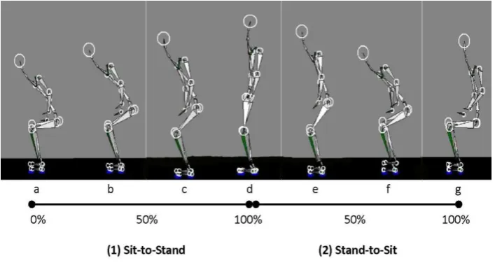
sequence was repeated 3 times in a row. Figure 1 depicts the whole sequence and two phases
7 | P a g e contraction in lumbar spine. In the first phase of both STD and SIT the eccentric contraction
and negative power are produced, whereas in the second phase of STD and SIT the
concentric contraction and positive power are produced (25).
[Figure 1- Different phases of Sit-to-Stand (STD) and Stand-to-Sit (SIT) movement
patterns]
Data analysis
Inter-joint coordination
Angular velocities of hip and lumbar spine joints were computed through dividing of
angular displacement (degree) of flexion-extension (frontal) axis to time (second). The
instantaneous velocity was computed for each frame number in order to acquire the detailed
changes in movement sequence. Decomposition index values as indicators of inter-joint
coordination were the percentage of STD and SIT time during which movement was
decomposed. A joint was considered to pause when its angular velocity dropped below 5º / s
(24). Average decomposition index values (%) were calculated for lumbar-hip joint pair in
each phase of STD, SIT and whole STD and SIT when one joint was moving while the other
joint paused.
Statistical analysis
Descriptive statistics include mean and standard deviation. Hotelling T2 MANOVA test
was used to compare movement coordination between healthy and patient groups. If the
results were significant, follow-up ANOVA tests were used to find the between group
differences on decomposition indices of STD and SIT and their phases. Confidence interval
value was set at 95% and two-sided.
Results
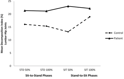
Figure 2 demonstrates the mean decomposition index changes in different phases of STD
and SIT. According to the results, decomposition index changed differently between two
8 | P a g e than the second-half phase in STD and SIT, but for healthy group the second-half phase had
higher score than the first-half phase for both STD and SIT.
The Hotelling T2 test result showed that there is a significant difference (T2 = 18.32, F14, 5=
8.33, p<.05) on decomposition indices between low back persons and healthy groups.
ANOVA follow-up results showed that there are significant differences between two groups
for decomposition indices of whole pattern of STD (F1, 28= 7.96, p<.05), whole pattern of SIT
(F1, 28= 5.37, p<.05), the first-half phase of STD (F1, 28= 7.26, p<.05) and the first-half phase
of SIT (F1, 28= 6.33, p<.05). Low back persons had significantly higher decomposition indices
relative to healthy group in whole STD (21.16 vs. 15.35), whole SIT (22.18 vs. 18.95), the
first-half of STD (21.35 vs. 16.04), and the first-half of SIT (23.04 vs. 13.18).
[Figure 2- Decomposition index of control and low back persons in different phases of STD and SIT]
Discussion
The aim of this study was to examine the effects of chronic low back pain on movement
coordination in lumbar spine and hip joints during two functional movement abilities
including STD and SIT. Our findings showed that there were significant differences between
low back persons and healthy people on decomposition indices of STD, SIT and the first-half
phases of STD and SIT. These findings are indicative of the lack of synergy among
movement of two joints that move independently due to lack of coordination. On the other
hand, while hip joint flexed lumbar joint paused and vice versa. These findings also support
the incidence of hesitation due to pain in low back persons that is demonstrated in previous
studies (11, 12).
Silfies et al. (22) showed that lumbar-pelvic coordination was more separated in time and
more variable in people with chronic low back pain. Shum et al. (23) have demonstrated that
low back persons showed different lumbar-hip coordination relative to healthy people. In
fact, the contribution of lumbar spine in STD and SIT movements was reduced due to
9 | P a g e study have revealed that muscle moment reduction in lumbar spine in sagittal plane is the
reason for changing STD and SIT strategy in low back persons because they minimise the
trunk motion and thereby reduce the muscle moment on the joint that it in turn changes
inter-joint coordination. Another study (30) showed decreased power flow from pelvic to legs in
low back persons during STD. The present findings also showed dis-coordination of joints
due to pausing in one joint whilst other joint moving.
The method of current study was different from previous studies (22, 25) that were
measured inter-joint coordination through relative phase as an indicator of phase difference
among paired-joints such as hip and lumbar spine joints. Relative phase is indicator of
positional changes in coordination (leading or lagging joint into degree) rather than time
parameter (pausing one joint into millisecond). In fact, guarding behaviour as a form of
muscle stiffness or joint freezing (14, 15, 16, 17, 18, 19, 20) that is observable in low back
persons resulted in limitation in trunk or thigh movements and it caused to inter-joint
dis-coordination.
The additional data analysis on decomposition index of lumbar and hip joints showed
different contribution of them in inducing dis-coordination in healthy and low back pain
groups. In healthy group the pausing percentage in lumbar and hip joints in entire movements
were 77% and 22% (lumbar to hip ratio: 3.5), whereas in low back pain group the pausing
percentage for lumbar and hip joints were 60% and 42% (lumbar to hip ratio:1.42). Thus, hip
joint slightly (25% less than healthy people) contributed in body weight transfer in low back
persons. These findings are important as they show to what extent hesitant movement shared
between two different body parts for executing STD and SIT.
In addition, as figure 2 shows the decomposition index for low back persons in different
phases of STD and SIT are different, so that in the first-half of STD and SIT they
demonstrated more pausing than the second-half. This pattern was different with healthy
10 | P a g e revealed that muscle powers are different in different phases of STD and SIT, so that in the
first-half phase the muscle work is negative because the type of muscular contraction is
eccentric. It seems that keeping the trunk uprightly during seat-off phase to peak lumbar
spine flexion (a, b and c in figure 1) due to painful condition deteriorates inter-joint
coordination by reducing the fluent motion and converting it into hesitant movement. Again
during SIT movement the type of muscle contraction in first-half phase is eccentric that will
interrupt the joints’ synergy which caused in more pausing during movement execution.
Reducing the angular velocity of both lumbar and hip joints during STD and SIT have
been demonstrated in previous studies (23, 31) and were explained as a preventive
mechanism against pain that are caused by muscle contraction and high levels of acceleration.
Difficulty in transferring the muscle force from pelvic to lower legs causes in an interruption
in the execution of closed kinetic chain that in turn is responsible for transferring the force
from upper to lower body parts (30). These findings suggest that reducing angular velocity in
lumbar spine same as healthy people is helpful to reduce the angular moment between two
joints and subsequently prevent the risk of losing balance. But reducing it beyond the normal
values relative to hip movement is a preventive mechanism that is observable in low back
persons that could change the mechanics of movement through increasing in hesitant
behaviours. Thus, in rehabilitation programmes of low back pain, emphasising on a constant
and fluent motion and preventing from hesitant movement reduce the pressure on the lumbar
spine through efficient utilisation of hip in coordination with lumbar spine by means of a
closed kinetic chain.
Future studies should investigate the possible mechanisms of hesitation behaviours
through electromyography [EMG] study to confirm the biomechanical findings that have
11 | P a g e In conclusion, low back pain causes dis-coordination in the function of different body
parts and results in pausing in one segment while the other segment moves independently.
Therapeutic exercises that emphasise on coordinative movement of pelvic and hip joints
could reduce disco-ordination due to freezing in movement segments.
References
1-Department of Health Statistics Division. The prevalence of back pain in Great Britain in
1998. London: Government Statistical Service, 1999.
2- European Commission, Special Eurobarometer, 272e, Nov 2007.
3- Strine, T.W. & Hootman, J.M. US national prevalence and correlates of low back and neck
pain among adults, Arthritis & Rheumatism, 2007, 57 (4): 656-665.
4- Fernández-de-las-Peñas C, Hernández-Barrera V, Alonso-Blanco C, Palacios-Ceña
D, Carrasco-Garrido P, Jiménez-Sánchez S, Jiménez-García R, Prevalence of neck and low
back pain in community-dwelling adults in Spain: a population-based national study, Spine,
2011, 36(3):213-219.
5- Sloan, T.J., Gupta, R. Zhang, W., & Walsh, D.A. Beliefs About the Causes and
Consequences of Pain in Persons With Chronic Inflammatory or Noninflammatory Low Back
Pain and in Pain-Free Individuals, Spine. 2008;33 (9):966-972.
6-Andersson GBJ. Epidemiological features of chronic low-back pain. The Lancet 1999; 354:
581-585.
7-Van Tulder MW, Koes BW, Bouter LM. A cost-illness study of back pain in the
12 | P a g e 8- Maniadakis N, Gray A. The economic burden of back pain in the UK. Pain 2000;
84:95-103.
9- Ehrlich, G.E. Bulletin of the World Health Organization 2003; 81:671-676.
10- Punnett, L., Prüss-Ustün, A., Nelson, D.I. Estimating the global burden of low back pain
attributable to combined occupational exposures, American Journal of Industrial Medicine ,
2005,48, 459-469.
11- Keefe, F.J & Block, A.R. Development of an observation method for assessing pain
behaviour in chronic low back pain patients, Behavior Therapy, 1982, 13, 363-375.
12- McDaniel, L.K., Anderson, K.O., Bradley, L.A., Young, L.D., Turner, R.A., Agudelo,
C.A., & Keefe, F.J. Development of an observation method for assessing pain behavior in
rheumatoid arthritis patients, Pain, 1986, 24, 165-184.
13- Verbunt, JA, Seelen HA, Vlaeyen JW, van der Heijden GJ, Heuts PH, Pons K, Disuse
and deconditioning in chronic low back pain: concepts and hypotheses on contributing
mechanisms, European Journal of Pain, 2003; 7, 9-21.
14- Aheren, DK, Follick MJ, Council JR, Laser-Wolston N, Litchman H, Comparison of
lumbar paravertebral EMG patterns in chronic low back pain patients and non-patient
controls, Pain 1988; 34:153-160.
15- Watson PJ, Booker CK, Main CJ, Evidence for the role of psychological factors in
abnormal paraspinal activity in patients with chronic low back pain, Journal of
Musculoskeletal Pain 1997; 5: 41-56.
16- Geisser ME, Haig AJ, Wallborn AS, Wiggert EA, Pain-related fear, lumbar flexion, and
dynamic EMG among persons with chronic musculoskeletal low back pain, Clinical Journal
13 | P a g e 17- van der Hulst, M, Vollenbroek-Hutten MM, Rietman JS, Hermens HJ, Lumbar and
abdominal muscle activity during walking in subjects with chronic low back pain: Support of
the guarding hypothesis, Journal of Electromyography and Kinesiology; 2010, 20: 31-38.
18- Hodges PW, Moseley GL, Pain and motor control of the lumbopelvic region: effect and
possible mechanisms, Journal of Electromyography and Kinesiology 2003; 13: 361-370.
19- Lund JP, Donga R, Widmer CG, Stohler CS, The pain adaptation model: a discussion of
the relationship between chronic musculoskeletal pain and motor activity, Canadian Journal
of Physiological Pharmacology 1991; 69(5): 683-694.
20-Hodges P, van den Hoorn W, Dawson A, Cholewicki J, Changes in the mechanical
properties of the trunk in low back pain may be associated with recurrence, Journal of
Biomechanics 2009; 42: 61-66.
21- Shumway-Cook A, Woollacot MH, Motor control: translating research into clinical
practice, 3rd Edition, 2007, Williams & Wilkins.
22-Silfies SP, Bhattachraya A, Biely S, Smith S, Giszter S, Trunk control during standing
reach: A dynamical system analysis of movement strategies in patients with mechanical low
back pain, Gait & Posture 2009; 29: 370-376.
23-Shum GLK, Crosbie J, Lee, RYW, Effect of low back pain on the kinematics and joint
coordination of the lumbar spine and hip during sit-to-stand and stand-to-sit, Spine 2005;
30:1998-2004.
24- Earhart GM, Bastian AJ, Selection and coordination of human locomotor forms following
cerebellar damage, Journal of Neurophysiology 2001; 85 (2):759-769.
25- Shum GLK, Crosbie J, Lee, RYW, Three-dimensional kinetics of the lumbar spine and
14 | P a g e 26-Lee RYW, Wong TKT, Relationship between the movements of the lumbar spine and hip,
Human Movement Science 2002; 21: 481-494.
27-Weiss P, Stelmach GE, Hefter H, Programming of a movement sequence in Parkinson’s
disease, Brain 1997; 120: 91-102.
28-Bennett KMB, Marchetti M, Ivoine R, Castiello U, The drinking action of Parkinson’s
disease subjects, Brain 1995; 118: 959-970.
29- Burgess-Limerick R, Abernethy B, Neal RJ, Relative phase quantifies inter-joint
coordination, Journal of Biomechanics 1993; 26:91-94.
30- Shum, GL, Crosbie, J, Lee, RY, Energy Transfer across the lumbosacral and lower
extremity joints in patients with low back pain during sit-to-stand Archives of Physical
Medicine and Rehabilitation 2009; 90 (1): 127-135.
31- Marras WS, Wongsam PE, Flexibility and velocity of the normal and impaired lumbar
15 | P a g e Figure 1- Different phases of Sit-to-Stand (STD) and Stand-to-Sit (SIT)
movement patterns.
16 | P a g e Figure 2- Decomposition index of control and low back persons in different phases of STD and SIT 0 5 10 15 20 25
STD 50% STD 100% SIT 50% SIT 100%
M ean D ec om p osi ti on In d ex (% ) Lu m b ar -H ip Joi n ts Control Patient