Original Article
Cytotoxicity of CdSe quantum dots and corresponding
comparison with FITC in cell imaging efficiency
Xiao Li1,2, Zhe Yan3, Jie Xiao1,2, Guobing Liu1,2, Yanli Li1,2, Yan Xiu1,2
1Department of Nuclear Medicine, Zhongshan Hospital, Fudan University, Shanghai, China; 2Shanghai Institute of Medical Imaging, Shanghai, China; 3School of Chemistry and Chemical Engineering, Inner Mongolia University, Hohhot, China
Received May 11, 2016; Accepted September 25, 2016; Epub January 15, 2017; Published January 30, 2017 Abstract: As a type of new emerging fluorescent nanoparticles, quantum dots applied in cellular imaging has elic
-ited broad research interests. However, the wide application of quantum dots was lim-ited by its potential bio-risk. In this study, a systematic investigation was carried out on the cytotoxicity of CdSe quantum dots, particularly fo -cusing on the aspect of particles size, plasm concentration, and incubation period. Furthermore, the comparison
between quantum dots and fluorescein isothiocyanate (FITC) on imaging efficiency was performed. MTT assay and flow cytometry proved the negligible cytotoxicity of CdSe quantum dots under the desired dyeing conditions. Given the excellent and stable optical properties of quantum dots such as high fluorescent quantum yield, broad absorp
-tion and narrow emission spectrum, the dyeing efficiency of quantum dots was superior to that of FITC. Therefore, quantum dots may be a better alternative with safe, effective and convenient labeling procedures for tissue imaging
and immunohistochemistry.
Keywords: CdSe, quantum dots, FITC, cytotoxicity, fluorescent imaging
Introduction
With the development of nano-technology, more and more nano-materials were prepared for biomedical applications, including drug
delivery, oncotherapy, artificial replaceable tis -sues and so on [1-3]. Due to its good penetrat-ing into cells and tissues, nanomaterials are prone to impact on intracellular physiological metabolism [4]. By virtue of this feature, nano-materials may be a promising agent in clinical applications. Hence, the research on improving biocompatibility of biomedical nanomaterials is necessary before extensively applying in
bio-medical fields.
As one of novel fluorescent probes, quantum
dots (QDs) composed of II-VI and III-V group ele-ments were mainly used in cellular imaging by now. QDs was also proved of the potential of invasive tissue imaging [5-10]. Compared with
the traditional organic dyes and fluorescent
proteins, QDs performed better with the respect of a wilder absorption spectrum and a
narrower emission spectrum. Meanwhile, QDs
were of high quantum efficiency, resistance to quenching, and non-sensitivity to intracellular compositions (such as enzymes) [11]. The wider exciting spectrum makes the exciting light with
a single wavelength suitable for more than one
kind of QDs. In other words, a single excitation light source can satisfy the requirement of mul
-tichannel testing. In addition, the fluorescence intensity is 10~20 times higher than equal amount of FITC, and the durability is 100~1000 times longer when compared with FITC [12]. All these above properties make QDs extensively
used in cellular imaging, and potentially useful for invasive imaging in vivo.
As a fluorescent probe with extensive applica -tion prospects, research on bio-safety of QDs are meaningful. In order to further explore the
biocompatibility and utilize the fluorescent characteristics of CdSe QDs, MTT assays and flow cytometry were conducted to measure the
Cytotoxicity and Cellular imaging efficiency of CdSe QDs
specific labeling effects on cell microtubule were compared between CdSe quantum dots and FITC via confocal calcium imaging system.
Material and methods
Agents and instruments
MTT (3-(4,5-dimethylthiazol-2-yl)-2,5-diphenyl -tetrazolium bromide) and Hoechst33258 (B2883) were purchased from Sigma. DMEM powder and Fetal Bovine Serum were
pur-chased from Gibco. Anti-α-tubulin (B-7,
sc-5-286) and goat-anti-mouse IgG (KPL, 202-1806) were purchased from Shanghai season
bio-technology co., LTD.
The 60-mm cell culture dishes, 24-well plates
and 96-well plates were purchased from Corning. Slide and cover glass were purchased from Citotest Labware Manufacturing Co., Ltd.
Disposable filters were purchased from
Millipore. Instruments included cell incubator
(Nuair, USA), centrifugal machine (Backman
Optima L-10XP), microplate reader (Bio-Rad, USA), inverted microscope (Olympus, Japan),
laser confocal fluorescence microscope
(Olympus FV1000), multi-spectrum argon gas
laser, and flow cytometer (BD FACS Calibur,
USA).
MTT assay
MTT assay as the common method to measure
cell viability was used in this study. Hela cells was utilized as the model cell. Passage between 2 and 5 was used to maintain consistency through this experiment. Cells were incubated at 37°C, 5% CO2 in DMED medium supplement with 10% fetal bovine serum, 1%
penicillin-streptomycin. Confluent cells were digested via trypsin-EDTA solution, and cells centrifugalized at 1000 rpm for 5 min. Then, the pellet was
suspended by complete medium at concentra-tion of 5×10⁴ cells/mL. Concentrations of cells were set as Table 1 in quadruplicates, and con -trols were of 180 µL cell suspension alone.
as reference. Co-incubation with QDs with dif-ferent size was performed following the same
procedures. Three kinds of QDs with different
exciting wavelength of 354 nm, 365 nm and 379 nm were selected. Incubation periods were set as 12 h, 24 h and 36 h.
Flow cytometry
For further study of the size effects, flow cytom -etry was used to evaluate the cytotoxicity as well. Hela cells in logarithmic phase were col-lected and prepared at concentration of 5×10⁴ cells/mL. In a 24 well plate, QDs with three dif-ferent sizes (CdSe-354, 365 and 379) were
added, and final concentration was set as 20
nmol/L (2 mL for each well). In the positive con-trol group, dexamethasone was added to pro-mote cell apoptosis. All cells were cultured at
37°C in a humidified atmosphere with 5% CO2
for 12 hours. The cells were collected and sus -pended in 50 µL PBS. Cells in positive control
group were divided into four parts equally. One sample was blank without any dye; the second
one was stained by 1:18 diluted Annexin V-APC; the third one was dyed with 1:50 diluted 7-AAD, and the last one was double-dyed. Experimental groups were double-dyed and then measured
in flow cytometer.
Cell fluorescent imaging
Hela cells in logarithmic phase were used in
fluorescent imaging of cell microtubule. One
milliliter Hela cells at concentration of 5×10⁴ cells/mL were added to each well of a 24-well
plate, and then cultured at 37°C in a humidified
atmosphere with 5% CO2 for 4 hours for fully
adherence. Cells were firstly rinsed by PBS and then fixed by mixture of 4% Triformol and 4%
sucrose for 20 min. Secondly, cells were washed by PBS, and then cultured with 0.25%
Triton for 15 min. Thirdly, triton were then
washed by cool PBS, and 6% BSA were then
[image:2.612.93.322.97.151.2]added for blocking for another 45 min.
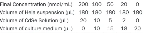
Table 1. Components of incubation samples in cyto-toxicity test of CdSe QDs
Final Concentration (nmol/mL) 200 100 50 20 0 Volume of Hela suspension (µL) 180 180 180 180 180 Volume of CdSe Solution (µL) 20 10 5 2 0 Volume of culture medium (µL) 0 10 15 18 20
After 12 h incubation, culture medium was removed, washed by cool PBS three times,
and 100 µL fresh medium and 10 µL MTT
(5 mg/mL) were added for another 4 h
incubation. Blank cells were cultured in 90
For fluorescent probe labeling, cells were first cultured with 1:100 diluted anti-α-tubulin for 1 h. PBS-rinsed cells were then cultured with FITC
or QDs labeled goat-anti-mouse IgG for another
1 hour. Cellular nucleus was finally dyed via
1:1000 diluted Hoechst33258 for 5 min and then washed by PBS. Samples were observed and photographed via confocal microscopy.
Statistics
Statistical analysis of the data was performed using the student’s t-test using SPSS 19.0, and
p-values less than 0.05 were considered as
significant.
Results
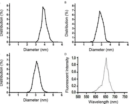
Distributions of dynamic light scattering-mea-sured hydrodynamic diameters of CdSe-354, CdSe-365 and CdSe-379 were shown in Figure
1A-C with corresponding mean diameters of 3.78, 2.8 and 3.1 nm. A typical absorption spectrum of CdSe-354 was provided in Figure 1D.
Cytotoxicity of quantum dots
Based on the results showed in Figure 2, there was a proved concentration-dependent cyto-toxicity of CdSeQDs. Cytocyto-toxicity was propor-tional to QDs concentration in the tested range.
There was a negligible cytotoxicity at concen -tration of 20 nM, which is twice of applied
con-centration of fluorescent cell imaging test. On
the other side, no size-dependent cytotoxicity was observed. For time-dependence, cytotoxic-ity increased along with prolongation of incuba-tion period, and tended to be stable after 12 h
incubation. Therefore, cytotoxicity can be neglected in a typical fluorescent cell imaging
[image:3.612.98.514.72.407.2]procedure (10 nmol/L, 12 h), and the labeling
Figure 1. Characteristics of CdSe quantum dots. Size distributions of CdSe-354 (A), CdSe-365 (B), and CdSe-379
Cytotoxicity and Cellular imaging efficiency of CdSe QDs
effect to microtubule can be guaranteed satisfactorily.
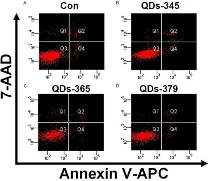
Base on the result of flow cytometry, there was no significant cytotoxicity associated to the dif -ferent sizes of QDs (Figure 3). After 12 h co-incubation with QDs-354 at the concentration of 20 nmol/L, there were 92.4% normal cells, 3.9% necrotic cells, and 0.9% apoptotic cells; For QDs-365, 90.2% normal cells, 5.6% necrot-ic cells, and 1.9% apoptotnecrot-ic cells were observed; In addition, there were 94% normal cells, 3.7% necrotic cells, and 0.7% apoptotic cells for those cultured with QDs-379.
It was proved that QDs posed great damage to macrophages through intracellular accumula-tion of QDs coupled with reactive oxygen spe-cies generation, particularly for QDs coated
with PEG-NH2 [13]. Different with macropha- ges, tumor cells with a better survivability
were used in this research. Traditionally, 16
hours were needed for one generation of Hela cell division, furthermore, there were no obvious increase of cytotoxicity detected
during 12 to 48 hours. Thus, no obviously
negative effect on cell viability will be induced. Although emission spectrum of QDs vary with distinct sizes, there are no size-dependent toxicity. For practical applications, the simul- taneous tests of multiple QDs components, which may correspond to various targets of
one or more kinds of cells, or simultaneous
physiological processes, may be realized by
kinds of emission lights resulted from single
excited light. Generally, QDs can be deemed
[image:4.612.98.516.74.413.2]as a kind of low-toxic, high-efficiency, and convenient cell fluorescent dyes.
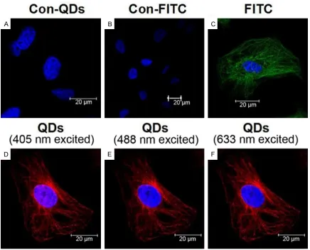
Imaging efficiency of QDs and FITC
According to the comparison of cell
microtu-bule imaging between FITC and QDs, a compa
-rable display effects were exhibited in dying microtubule of Hela cell (Figure 4). Furthermore,
the wider absorption spectrum makes QDs per
-formed better than FITC to some extent. The
narrow and symmetric emission spectrum of QDs can decrease the interference signal, so as to increase sensitivity of detection.
Discussion
With respect to the advantages also reflected
by above results of wide absorption spectrum,
and narrow emission spectrum of quantum
dots [14], peptide and antibody were
success-fully labeled with quantum dots [15, 16].
Besides, different with organic dyes, quantum
dots performed stable and exhibited resistance
to quenching, breaking the limitation of short detection period and benefiting dynamic
live-cell imaging [17, 19]. Furthermore, QDs were more resistant to enzymes in vivo and with
bet-ter biocompatibility than fluorescent
dyes-labeled proteins. Additionally, due to the sur-face effect of nanostructures, QDs were more easily to attach to other molecules [20]. All
these above items make QDs a better fluores
-cent dyes for cellular imaging and potential for
in vivo imaging.
Compared the imaging efficiency with FITC, CdSe quantum dots may be a better alternative
with safe, effective and convenient labeling
[image:5.612.98.516.75.439.2]procedures for cellular fluorescent imaging,
Figure 3. Flow cytometry for Hela cells incubated with QDs at 20 nmol/L for 12 h. A. Positive control; B. QDs-354;
C. QDs-365; D. QDs-379. For the four quadrants, Q1 is apoptotic cells; Q2 is dying cells; Q3 is normal cells, and Q4
Cytotoxicity and Cellular imaging efficiency of CdSe QDs
where the cytotoxicity was negligible under the desired dying conditions.
Acknowledgements
Supported by National Science Foundation of China: 81271608, 81201130, Shanghai Pu- jiang Program: 13PJ1401400, Science Found- ation of Shanghai: 13ZR1439200, and Shang- hai Municipal Commission of Health and Family Planning: XYQ2013106.
Disclosure of conflict of interest
None.
Address correspondence to: Dr. Yan Xiu, Depart- ment of Nuclear Medicine, Zhongshan Hospital,
Fudan University, 180 Feng Lin Road, Shanghai
200032, China. Tel: +86 21 64041990; Fax: +86 21 64041990; E-mail: [email protected]
References
[1] Ryan SM and Brayden DJ. Progress in the deliv-ery of nanoparticle constructs: towards clinical translation. Curr Opin Pharmacol 2014; 18: 120-128.
[2] Choi S, Tripathi A and Singh D. Smart nanoma -terials for biomedics. J Biomed Nanotechnol 2014; 10: 3162-3188.
[3] Walmsley GG, McArdle A, Tevlin R, Momeni A,
Atashroo D, Hu MS, Feroze AH, Wong VW,
Lo-renz PH, Longaker MT and Wan DC. Nanotech -nology in bone tissue engineering. Nanomedi-cine 2015; 11: 1253-1263.
[4] Watari F, Takashi N, Yokoyama A, Uo M, Aka
[image:6.612.89.525.72.424.2]-saka T, Sato Y, Abe S, Totsuka Y and Tohji K.
Material nanosizing effect on living organisms:
non-specific, biointeractive, physical size effects. J R Soc
Interface 2009; 6 Suppl 3: S371-388.
[5] Zhao MX and Zeng EZ. Application of functional
quantum dot nanoparticles as fluorescence probes
in cell labeling and tumor diagnostic imaging. Na-noscale Res Lett 2015; 10: 171.
[6] Zhang YP, Sun P, Zhang XR, Yang WL and Si CS.
Syn-thesis of CdTe quantum dot-conjugated CC49 and
their application for in vitro imaging of gastric ade-nocarcinoma cells. Nanoscale Res Lett 2013; 8: 294.
[7] Fan Y, Liu H, Han R, Huang L, Shi H, Sha Y and Jiang Y. Extremely High Brightness from
Polymer-Encap-sulated Quantum Dots for Two-photon Cellular and
Deep-tissue Imaging. Sci Rep 2015; 5: 9908. [8] Liu H, Tang W, Li C, Lv P, Wang Z, Liu Y, Zhang C, Bao
Y, Chen H, Meng X, Song Y, Xia X, Pan F, Cui D and Shi Y. CdSe/ZnS Quantum Dots-Labeled
Mesenchy-mal Stem Cells for Targeted Fluorescence Imaging of Pancreas Tissues and Therapy of Type 1 Diabetic
Rats. Nanoscale Res Lett 2015; 10: 959.
[9] Liu Q, Guo B, Rao Z, Zhang B and Gong JR. Strong
Two-Photon-Induced Fluorescence from Photosta -ble, Biocompatible Nitrogen-Doped Graphene
Quantum Dots for Cellular and Deep-Tissue Imag -ing. Nano Lett 2013; 13: 2436-2441.
[10] Fu A, Gu W, Larabell C and Alivisatos AP. Semicon-ductor nanocrystals for biological imaging. Curr Opin Neurobiol 2005; 15: 568-575.
[11] Cheki M, Moslehi M and Assadi M. Marvelous ap
-plications of quantum dots. Eur Rev Med Pharma -col Sci 2013; 17: 1141-1148.
[12] Zhao JJ, Chen J, Wang ZP, Pan J and Huang YH.
Double labeling and comparison of fluorescence in
-tensity and photostability between quantum dots and FITC in oral tumors. Mol Med Rep 2011; 4:
425-429.
[13] Qu G, Wang X, Wang Z, Liu S and Jiang G.
Cyto-toxicity of quantum dots and graphene oxide to
erythroid cells and macrophages. Nanoscale Res Lett 2013; 8: 198.
[14] Geszke-Moritz M and Moritz M. Quantum dots
as versatile probes in medical sciences:
syn-thesis, modification and properties. Mater Sci -Eng C Mater BiolAppl 2013; 33: 1008-1021. [15] Jamieson T, BakhshiR, Petrova D, Pocock R,
Imani M and Seifalian AM. Biological
applica-tions of quantum dots. Biomaterials 2007; 28:
4717-4732.
[16] Juzenas P, Chen W, Sun YP and Coelho MA, Generalov R, Generalova N and Christensen IL. Quantum dots and nanoparticles for photody-namic and radiation therapies of cancer. Adv Drug Deliv Rev 2008; 60: 1600-1614.
[17] Dean KM and Palmer AE. Advances in fluores -cence labeling strategies for dynamic cellular imaging. Nat Chem Biol 2014; 10: 512-523. [18] Rhyner MN, Smith AM, Gao X, Mao H, Yang
Land Nie S. Quantum dots and multifunctional nanoparticles: new contrast agents for tumor imaging. Nanomedicine (Lond) 2006; 1: 209-217.
[19] Li ZJ, Li C, Zheng MG, Pan JD, Zhang LM and Deng YF. Functionalized nano-graphene oxide
particles for targeted fluorescence imaging
and photothermy of glioma U251 cells. Int J Clin Exp Med 2015; 8: 1844-1852.
[20] Mehta VN, Kailasa SK and Wu HF. Surface
modified quantum dots as fluorescent probes