Original Article
Myelin expression factor 2 expression is associated with
aggressive phenotype in triple-negative breast cancer
Yumin Chung1, Abdul Rehman1, Hyein Ahn1, Jongmin Sim1, Min Sung Chung2, Dongho Choi2, Seung Sam Paik1, Kiseok Jang1
Departments of 1Pathology, 2Surgery, Hanyang University College of Medicine, Seoul, Korea
Received December 19, 2016; Accepted December 27, 2017; Epub April 1, 2017; Published April 15, 2017
Abstract: Triple-negative breast cancers (TNBCs) are characterized by an aggressive clinical course with frequent re-currence and short survival and an absence of molecular targets. Myelin expression factor 2 (MYEF2) is suggested to play a role in carcinogenesis by in silico analysis; however, the function of MYEF2 in cancer is largely unknown. The purpose of this study was to investigate MYEF2 expression in TNBC. Immunohistochemistry for MYEF2 was performed on tissue microarray sections from 132 patients with TNBC and interpreted using a semiquantitative im-munoreactive score (IRS). Association of MYEF2 expression with various clinicopathologic characteristics, including patient survival, was analyzed. MYEF2 expression was localized in the nucleus of tumor cells, and positive staining was observed in 90.15% of TNBCs. Expression of MYEF2 was positively correlated with tumor size (P = 0.027), AJCC stage (P = 0.013), and Ki-67 proliferation index (P < 0.001). Survival analyses using Cox proportional regression model revealed that lymphovascular invasion, lymph node metastasis, and AJCC stage were prognostic factors for disease-free and overall survival. Patients with high MYEF2-expressing tumors showed a decreased estimated sur-vival time (96 months) compared with patients with absent or low MYEF2 expression (101 months), however, the
dif-ference was not statistically significant (P = 0.178). Expression of MYEF2 is significantly correlated with aggressive
phenotypes of TNBC, such as larger tumor size, high AJCC stage, and Ki-67 proliferation index. Further functional
studies will be required to define the role of MYEF2 in cancers.
Keywords: Triple-negative breast cancer, myelin expression factor 2, immunohistochemistry, prognosis
Introduction
Triple-negative breast cancers (TNBCs), which are defined by lack of expression of estrogen receptor, progesterone receptor, and human epidermal growth factor receptor 2, constitute one of the most challenging groups of breast cancers [1, 2]. This subgroup accounts for approximately 10-15% of all breast cancers [3]. TNBCs are characterized by biologically aggres-sive tumor behavior with frequent recurrence in the first 1-3 years and a higher death rate than other subtypes of breast cancer [4, 5].
Myelin expression factor 2 (MYEF2) is a tran-scriptional repressor of the gene encoding myelin basic protein, a major component of the myelin sheath whose production is develop-mentally controlled during myelinogenesis [6]. MYEF2 is known to play a role in neuron and myotube differentiation, and is related to
human diseases, such as dilated cardiomyopa-thy and cardiac hypertrophy [6, 7]. The function of MYEF2 in cancer is largely unknown. Recent bioinformatics research suggested that MYEF2 potentially plays a role in the molecular path-way of prostate cancer development, but no direct relationship between MYEF2 and the cancer has been demonstrated [8].
In the present study, we investigated MYEF2 expression by immunohistochemistry in a se- ries of TNBCs and evaluated its association with clinicopathologic variables, including pa- tient survival.
Materials and methods
Patients and tumor samples
K5007, DakoCytomation) for 30 min at room temperature. The primary antibody was rabbit polyclonal anti-MYEF2 antibody (ab26098, Abcam, Cambridge, UK) used at a 1:100 dilu-tion. 3,3’-Diaminobenzidine was used as a chromogen for visualization, and Mayer’s hematoxylin counterstain was applied.
Interpretation of immunohistochemical stain-ing
MYEF2 expression was evaluated semiquanti-tatively by two independent pathologists who were blinded to the patients’ clinicopathologic data. Nuclear MYEF2 expression was catego-rized in terms of staining intensity and extent. The intensity score was assigned as follows: 0 (negative), 1 (weak staining), 2 (intermediate staining), and 3 (strong staining). The propor-tion score was as follows: 0 (< 10%), 1 (10%-25%), 2 (26%-50%), 3 (51%-75%), and 4 (76%-100%). The product of the intensity and proportion scores was used as the final immu -noreactive score (IRS). Thus, the maximum combined score was 12 and the minimum score was 0. Representative photomicrographs of MYEF2 immunostaining in TNBCs are shown in Figure 1.
Statistical analysis
Statistical analyses were performed using SPSS software version 19.0 (IBM Corp., Ar- Hospital, Seoul, between January 2001 and
December 2014 were retrospectively enrolled in this study.
We reviewed all hematoxylin and eosin (H&E)-stained slides, pathology reports, and other medical records to confirm the diagnosis and obtain follow-up data. The clinicopathologic parameters included American Joint Committ- ee on Cancer (AJCC) tumor stage, primary tumor size, lymph node metastasis, histologic grade, lymphovascular invasion, perineural invasion, perinodal tumor extension in cases of lymph node metastasis, Ki-67 labeling index, disease-free survival, and overall survival. This study was approved by the Institutional Review Board of Hanyang University Hospital (IRB File No 2015-12-023-001).
Tissue microarray construction
[image:2.612.90.377.71.289.2]We used a manual tissue microarrayer (Quick-Ray, Unitma, Seoul, Korea) to construct a tissue microarray (TMA) from archival formalin-fixed, paraffin-embedded tissue blocks. We selected well-fixed, non-necrotic areas by light micros -copy of H&E-stained sections, and marked the area on the corresponding paraffin blocks. Tissue cylinders of 2-mm diameter were punched from the marked region on each donor blocks and transferred to the recipient blocks.
Figure 1. Representative microphotographs of myelin expression factor 2 im-munostaining in triple-negative breast cancers. Negative (A), weak (B), inter-mediate (C) and strong (D) staining in the nuclei of tumor cells.
Immunohistochemical stain-ing
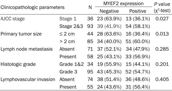
lymphovascular invasion, perineural invasion, and perinodal tumor extension. However, lar- ger tumors (2 cm or more in size) tended to have a frequent high MYEF2 expression than smaller tumors (P = 0.013, chi-square test). The tumors with higher AJCC stage (stage II or above) correlated with high MYEF2 expression (P = 0.027, chi-square test) (Table 1). In addi-tion, the high MYEF2 expression group had an increased Ki-67 labeling index (P < 0.001, t-test) (Figure 2).
Correlation between MYEF2 expression and TNBC patient survival
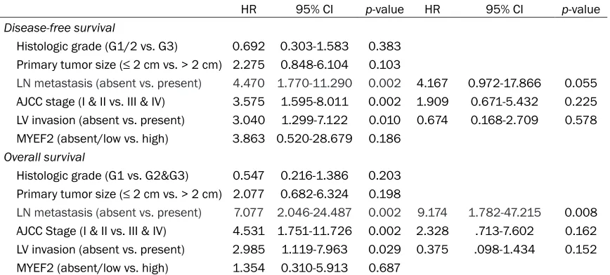
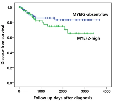
AJCC stage, lymph node metastasis, and lym-phovascular invasion showed a significant effect on overall and disease-free survival in univariable analyses (Table 2). Patients with high MYEF2-expressing tumors showed a lower estimated survival time (85.8 months) than patients with absent or low MYEF2 expression (104.7 months) but the difference was not sta-tistically significant (log rank test, P = 0.178) (Figure 3).
Discussion
Since there is no in vitro or in vivo functional study investigating the role of MYEF2 in cancer, there is no direct evidence for a role of MYEF2 in carcinogenesis or tumor progression. How- ever, several previous studies implicate poten-tial involvement of MYEF2 in cancer biology. Gene expression profiling analysis using six normal and four prostate cancer tissues identi-fied MYEF2 as one of the up-regulated tran -scription regulators in prostate cancer com-monk, NY, USA). The chi-square test was used
to compare the proportion of MYEF2 expres-sion status and various clinicopathologic parameters. The difference of Ki-67 labeling index between the groups was evaluated by t-test. The Kaplan-Meier method was used to plot survival curves. Univariable and multivari-able survival analyses using the Cox propor-tional hazards regression model were per-formed to identify prognostic relevance. Results
Clinicopathologic characteristics of TNBC patients
[image:3.612.92.389.98.256.2]Successful MYEF2 immunostaining was ach- ieved for 129 cases and the remaining 3 cases were excluded from subsequent analyses. The mean patient age was 50.9 years (range, 26 to 79 years). Twenty-four patients (18.6%) suf-fered from local recurrence or distant metasta-sis, and 18 patients (14.0%) died of breast can-cer. The mean overall survival from diagnosis was 52.3 months. The majority of TNBCs (73.6%) had histologic grade 3. By AJCC tumor staging, 37 cases were stage I (28.7%), 54 were stage II (41.9%), and 38 were stage III (29.4%). The primary tumor was evaluated as T1 in 34.4%, T2 in 53.9%, T3 in 6.3%, and T4 in 5.5% of patients. Lymph node metastasis was diag-nosed pathologically as N0 in 55.3%, N1 in 22.0%, N2 in 8.1%, and N3 in 14.6% of patients. Correlation between MYEF2 expression and various clinicopathologic parameters in TNBC MYEF2 expression was absent in only 10 cases (7.6%); most cases showed immunoreactivity of
Table 1. Correlation between MYEF2 expression and clinicopathologic factors in TNBC
Clinicopathologic parameters N MYEF2 expression (χP value2-test) Negative Positive
AJCC stage Stage 1 36 23 (63.9%) 13 (36.1%) 0.027 Stage 2&3 93 39 (41.9%) 54 (58.1%)
Primary tumor size ≤ 2 cm 44 28 (63.6%) 16 (36.4%) 0.013 > 2 cm 85 34 (40.0%) 51 (60.0%)
Lymph node metastasis Absent 71 37 (52.1%) 34 (47.9%) 0.285 Present 58 25 (43.1%) 33 (56.9%)
Histologic grade Grade 1&2 34 19 (55.9%) 15 (44.1%) 0.201 Grade 3 95 43 (45.3%) 52 (54.7%)
Lymphovascular invasion Absent 74 38 (51.4%) 36 (48.6%) 0.405 Present 55 24 (43.6%) 31 (56.4%)
noma cells revealed that MYEF2 is one of the candidate genes regulated by the Wnt/β-catenin signaling pathway that is activated in many human cancers [10]. In addition, MYEF2 has been revealed as a protein associated with SOX2, which has an oncogenic role in medullo-blastoma [11].
On the other hand, data from several other studies have suggested that MYEF2 functions as a tumor suppressor in cancers. A validation study of selected microRNAs in malignant mel-anoma revealed that microRNA-17 is one of the pared to normal prostate tissues [9]. Another
study using publically available gene expres-sion data on large number of prostate tissues (77 normal tissues, 66 primary prostate tu- mors, and 24 metastatic tumors) revealed that a set of four genes (TUBB6, MYEF2, PARM1, SLC25A22) provided the best accuracy for pre-diction of normal tissues versus metastatic tumors and primary prostate tumors versus metastatic tumors [8].
Recently, a study using high-throughput tran-scriptional analysis of HT20 colon
[image:4.612.92.525.72.263.2]adenocarci-Figure 2. Correlation between MYEF2 expression and Ki-67 proliferation index. A. The tumor with negative MYEF2 shows low Ki-67-positive tumor cells, whereas MYEF2-positive tumor has high Ki-67 labeling index. B. Mean Ki-67 labeling index of TNBCs according to MYEF2 expression status. (t-test, P < 0.001).
Table 2. Univariate and multivariate survival analyses for various clinicopathologic prognostic factors in TNBC
HR 95% CI p-value HR 95% CI p-value
Disease-free survival
Histologic grade (G1/2 vs. G3) 0.692 0.303-1.583 0.383
Primary tumor size (≤ 2 cm vs. > 2 cm) 2.275 0.848-6.104 0.103
LN metastasis (absent vs. present) 4.470 1.770-11.290 0.002 4.167 0.972-17.866 0.055 AJCC stage (I & II vs. III & IV) 3.575 1.595-8.011 0.002 1.909 0.671-5.432 0.225 LV invasion (absent vs. present) 3.040 1.299-7.122 0.010 0.674 0.168-2.709 0.578 MYEF2 (absent/low vs. high) 3.863 0.520-28.679 0.186
Overall survival
Histologic grade (G1 vs. G2&G3) 0.547 0.216-1.386 0.203
Primary tumor size (≤ 2 cm vs. > 2 cm) 2.077 0.682-6.324 0.198
LN metastasis (absent vs. present) 7.077 2.046-24.487 0.002 9.174 1.782-47.215 0.008 AJCC Stage (I & II vs. III & IV) 4.531 1.751-11.726 0.002 2.328 .713-7.602 0.162 LV invasion (absent vs. present) 2.985 1.119-7.963 0.029 0.375 .098-1.434 0.152 MYEF2 (absent/low vs. high) 1.354 0.310-5.913 0.687
HR, hazard ratio; CI, confidence interval; LN, lymph node; AJCC, American Joint Com mittee on Cancer; LV, lymphovascular;
[image:4.612.90.524.349.545.2]mor suppressor have also been reported in the previous publications. Therefore, the func-tional role of MYEF2 and its potential involve-ment in a novel mechanism in TNBC should be investigated in future studies.
Disclosure of conflict of interest
None.
Address correspondence to: Dr. Kiseok Jang, De- partment of Pathology, Hanyang University College of Medicine, 222-1 Wangsimni-ro, Seongdong-gu, Seoul 04763, Korea. Tel: +82-2-2290-8248; Fax: +82-2-2296-7502; E-mail: medartisan@hanyang. ac.kr
References
[1] Foulkes WD, Smith IE and Reis-Filho JS. Triple-negative breast cancer. N Engl J Med 2010; 363: 1938-1948.
[2] Thike AA, Iqbal J, Cheok PY, Chong AP, Tse GM, Tan B, Tan P, Wong NS and Tan PH. Triple negative breast cancer: outcome correlation with immunohistochemical detection of basal markers. Am J Surg Pathol 2010; 34: 956-964. [3] Kuo WH, Chang YY, Lai LC, Tsai MH, Hsiao CK,
Chang KJ and Chuang EY. Molecular character-istics and metastasis predictor genes of triple-negative breast cancer: a clinical study of tri-ple-negative breast carcinomas. PLoS One 2012; 7: e45831.
[4] Adamo B and Anders CK. Stratifying
triple-neg-ative breast cancer: which definition(s) to use?
Breast Cancer Res 2011; 13: 105.
[5] Lin NU, Claus E, Sohl J, Razzak AR, Arnaout A and Winer EP. Sites of distant recurrence and clinical outcomes in patients with meta-static triple-negative breast cancer: high inci-dence of central nervous system metastases. Cancer 2008; 113: 2638-2645.
[6] Muralidharan V, Tretiakova A, Steplewski A, Haas S, Amini S, Johnson E and Khalili K. Evi-dence for inhibition of MyEF-2 binding to MBP promoter by MEF-1/Pur alpha. J Cell Biochem 1997; 66: 524-531.
[7] Haas S, Steplewski A, Siracusa LD, Amini S
and Khalili K. Identification of a sequence-spe
-cific single-stranded DNA binding protein that
suppresses transcription of the mouse myelin basic protein gene. J BiolChem 1995; 270: 12503-12510.
[8] Li R, Dong X, Ma C and Liu L. Computational
identification of surrogate genes for prostate
cancer phases using machine learning and molecular network analysis. Theor Biol Med Model 2014; 11: 37.
up-regulated microRNAs. This suggests that MYEF2 might show decreased expression in melanoma, since MYEF2 is a computational predictive target of microRNA-17 [12]. The mod-ified ginseng extract (MGX) has an anti-prolifer -ative effect on A549 non-small cell lung cancer cells. MYEF2 is one of the up-regulated genes after MGX treatment, suggesting an involve-ment in anti-proliferative mechanisms [13]. Finally, in serous epithelial ovarian carcinoma, expression of the RUNX1 transcription factor contributes to cell proliferation, migration, and invasion. MYEF2 is one of the genes that are up-regulated by RUNX1 knockdown in SKOV3 ovarian cancer cells [14].
In the present study, we examined the protein expression level of MYEF2 in triple-negative breast cancer using immunohistochemistry and found that MYEF2 expression was positive-ly associated with larger tumor size, high AJCC stage and Ki-67 proliferation index. In survival analyses, patients with high MYEF2-expressing tumors had a shorter estimated survival time than patients with absent or low MYEF2 expres-sion. These results suggest that MYEF2 might be involved in the tumor progression of TNBC. This is consistent with previous microarray data indicating that MYEF2 is upregulated and relat-ed to a well-known cancer pathway.
In conclusion, our study reveals that MYEF2 expression is associated with aggressive phe-notypes of TNBCs, such as tumor size and stage and Ki-67 proliferation index. However, opposite findings implicating MYEF2 as a
[image:5.612.91.288.70.245.2][12] Li C, Wang X, Chen Y, Tan X, Li W, Yan S, Cai W, Xu X, Xu L, Yang L and Yan Y. MicroRNA-21, 204 and 125b play potential roles in tumori-genesis of melanoma. Adv Biosci Biotechnol 2015; 6: 677-692.
[13] Kim KH, Choi I, Lee YW, Cho CK, Yoo HS, Lee SB, Ho Choi S, Kwon KR and Jang JH. Target genes involved in antiproliferative effect of
modified ginseng extracts in lung cancer A549
cells. Acta Biochim Biophys Sin (Shanghai) 2014; 46: 441-449.
[14] Keita M, Bachvarova M, Morin C, Plante M, Gregoire J, Renaud MC, Sebastianelli A, Trinh XB and Bachvarov D. The RUNX1 transcription factor is expressed in serous epithelial ovarian carcinoma and contributes to cell proliferation, migration and invasion. Cell Cycle 2013; 12: 972-986.
[9] Bălăcescu L, Bălăcescu O, Crişan N, Fetica B, Petruţ B, Bungărdean C, Rus M, Tudoran O, Meurice G, Irimie A, Dragoş N and
Berindan-Neagoe I. Identifying molecular features for prostate cancer with Gleason 7 based on
mi-croarray gene expression profiles. Rom J Mor -phol Embryol 2011; 52: 1195-1202.
[10] Bol D and Ebner R. Gene expression profiling
in the discovery, optimization and develop-ment of novel drugs: one universal screening platform. Pharmacogenomics 2006; 7: 227-235.
[11] Cox JL, Wilder PJ, Gilmore JM, Wuebben EL, Washburn MP and Rizzino A. The