Int J Clin Exp Pathol 2017;10(8):9035-9041 www.ijcep.com /ISSN:1936-2625/IJCEP0021883
Original Article
Low-expression of
miR-7
promotes cell proliferation and
exhibits prognostic value in osteosarcoma patients
Shouying Liu1*, Changxi Zhou2*, Changbao Zhu3*, Qiuhe Song4*, Ming Wen5, Ye Liu6, Huaijie An7
1Department of Orthopaedics, The 253th Hospital of The Chinese People’s Liberation Army, Hohhot 010051,
China; 2Department of Nanlou Respiratory Diseases, PLA General Hospital, Beijing 100853, China; 3Department
of Orthopaedics, The 474th Hospital of The Chinese People’s Liberation Army, Urumchi 830013, China;
4Department of Dermatology, Attached Hospital of Jiujiang University, Jiujiang 332000, China; 5Department of
Human Resources, Human Resources China Communications Construction Company Ltd, Beijing 100088, China;
6Department of Anesthesiology, Beijing Obstetrics and Gynecology Hospital, Capital Medical University, Beijing
100026, China; 7Center of Basic Medical Sciences, Navy General Hospital of PLA, Beijing 100037, China. *Co-first
authors.
Received December 14, 2015; Accepted February 25, 2016; Epub August 1, 2017; Published August 15, 2017
Abstract: Aim: MicroRNAs (miRNAs) play important roles in occurrence and development of osteosarcoma. Previous studies had verified the role of microRNA-7 (miR-7)in various diseases, especially in cancers. Our purpose in this study was to investigate the values of miR-7 in development and prognosis of osteosarcoma. Methods: QRT-PCR was used to measure the expression of miR-7 in osteosarcoma tissues, adjacent tissues and healthy tissues as well as in osteosarcoma cell lines MG63, U2OS and normal osteoblastic cell line hFOB1.19. CCK-8 and siRNA assays were performed to estimate the effect of miR-7 in the process of cell proliferation. The Kaplan-Meier and Cox regression analysis were performed to detect the prognostic values of the miR-7 in osteosarcoma patients. Results: The results demonstrated that miR-7 expression decreased in osteosarcoma tissues and cell lines compared with the controls. Proliferation assay declared that the cell proliferation was accelerated by down-regulation of miR-7. Kaplan-Meier exhibited that the overall survival time of low-miR-7 expression was shorter than those with high-miR-7 expression (P=0.001). Cox regression analysis revealed that Enneking, distant metastasis and recurrence were all prognostic factors just like low-miR-7. Conclusion: The expression of miR-7 was lower in osteosarcoma tissues and cell lines and miR-7 acted as a tumor suppressor. The low-expression of miR-7 was associated with clinicopathologic charac-teristics (age, tumor site, Enneking, therapies). Moreover, miR-7 might be an independent prognostic marker and promote cell proliferation in osteosarcoma.
Keywords: Osteosarcoma, miR-7, cell proliferation, prognosis
Introduction
Osteosarcoma, a most common malignancy of bone mainly arises from the metaphysis of the long bones of adolescents and young adults. It accounts for 5% of all pediatric tumors and 8.9% of cancer-related deaths in children [1, 2]. The characteristics of osteosarcoma include easy relapse and metastasis, high malignancy and invasion. Over 50% osteosarcoma patients were diagnosed as suffering metastasis which led to low cure rate and low 5-years’ survival rate [3]. Recently, as the development of com-prehensive therapies, the 5-year’ survival rate has risen to 60-70% [4]. However, the clinical prognosis is still poor when patients develop recurrent or metastatic osteosarcoma. Mean-
while, the fundamental molecular mechanisms of the development and progress of osteosar-coma is still obscure. Therefore, it is important to explore the mechanisms and pathogenesis of osteosarcoma and develop strategies for diagnosis, treatment and prognosis of this disease.
MicroRNAs (miRNAs) which are a class of small endogenous non-coding RNA with a length of 18-25 nucleotides [5], have been confirmed to be related to many progresses of various can-cers. The differential expressions of miRNAs
spe-cific sites binding with the 3’-untranslated regions (3’-UTR) of the target-mRNAs at post transcriptional level [6]. Besides, in the impor-tant disease processes such as cell growth, cell cycle, apoptosis, migration and invasion, miRNAs have been reported to play vital roles. So far, more than 1900 human miRNAs regulat-ing about 60% of the genes in mammals have been identified [7]. MicroRNA-7 (miR-7) is an evolutionary conserved miRNA with three dif-ferent genomic loci in humans [8]. And it served as a tumor suppressor in several human can-cers [9]. However, the function of miR-7 in osteosarcoma is unknown.
In this study, we aimed to detect the expression of miR-7 in osteosarcoma tissues and cell lines, then explore the effects of miR-7 on osteosar-coma cell proliferation, further analyze the prognostic value of miR-7 expression on osteo-sarcoma patients.
Materials and methods
Clinical samples collection
The current study was conducted in Chinese PLA General Hospital and permitted by the Ethic Committee of the hospital. Patients who were collected in the research were diagnosed as osteosarcoma during 2013-2015. None of them had received any chemical and physical treatments. The clinicopathologic characteris-tics of all patients including age, gender, tumor
site, Enneking, histological type, surgical meth-od, distant metastasis and recurrence were recorded in database. Besides, written infor- med consent had been signed in advance. Tumor tissues and adjacent tissues from 87 patients with osteosarcoma were obtained, besides, 30 healthy tissue samples were obtained as control. There were 35 females and 52 males with a median age of 32.72± 16.60. The tissue samples were quickly frozen in liquid nitrogen and stored at -80°C for RNA extraction.
A 5-years’ follow-up was performed with all osteosarcoma patients. The overall survival time was defined as the diagnosed time to the last day of follow-up. The information about fol-low-up was updated each three months through telephone or questionnaire. The data were recorded in a database.
Cell culture and transfection
[image:2.612.95.519.75.264.2]The role of
miR-7
in osteosarcoma patients
all survival. The difference was considered to be significant when P<0.05.
Results
MiR-7 expression significantly decreased in osteosarcoma tissues and cell lines
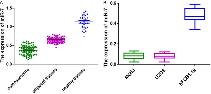
The expression of miR-7 was reported to be down-regulated in osteosarcoma in previous study [10]. To further validate this point, we detected miR-7 expression in both osteosarco-ma tissues and cell lines with qRT-PCR. The result demonstrated that the expression of
miR-7 was lower in osteosarcoma tissues than adjacent tissues and healthy tissues (Figure 1A, P<0.001). Besides, in the osteosarcoma cell lines (MG63 and U2OS), the expression of
miR-7 was significantly decreased compared mine were added in all mediums. And all cells
were incubated at 37°C in 5% CO2-humidified atmosphere.
MG63 and U2OS cell lines, hFOB1.19 cell line were seeded in 96-well plates with 5 × 10⁴ cells
per well. Then all cell lines were transfected witheither siRNAs targeting miR-7 or the nega-tive control siRNAs at a concentration of 50 nM using Lipofectamine 2000 (Invitrogen, Canada) transfection reagent.
RNA extraction and qRT-PCR
Total RNA was isolated from all tissues and cell cultures with Trizol (Invitrogen, Carlsbad, CA, USA). Reverse transcriphase was conducted to synthesize the first chain of cDNA with
TaqMan® MicroRNA Reverse Trans- cription Kit (Applied Biosystems, Fo- ster City, CA, USA). Then, qRT-PCR reaction was performed in the Applied Biosystems 7900 Fast Real-Time PCR system (Applied Biosystems, Foster City, California, USA). U6 acted as an endogenous control. The relative ex- pression quantification of miR-7 was evaluated by comparative cycle thresh-old (CT) method.
Cell proliferation assay
The ability of cell proliferation was ana-lyzed via cell Counting (CCK8) Kit-8 (Dojindo, Japan). After transfection, MG63 and U2OS cells viability at dif-ferent time points (24, 48, and 72 h) was measured at 450 nm with an enzyme immunoassay analyzer (Bio-Rad, Hercules, CA, USA). Each sample was in triple.
Statistical analysis
Statistical analysis was carried out with SPSS version 13.0 software (SPSS Inc, IL, USA). All quantified data was presented as mean ± SD. The expression level of miR-7 was ana- lyzed by T test. The relationship between clinicopathologic characteris-tics and miR-7 expression was esti-mated with chi-square test. Kaplan-Meier and cox regression analysis were taken to estimate the asso- ciation of clinicopathologic character-istics, miR-7 expression and the
over-Table 1. Relationship between clinicopathologic charac-teristics and miR-7 in patients with osteosarcoma
Clinicopathologic
characteristics n
miR-7 expression
P
High Low
Age <0.001
<30 43 37 6
≥30 44 9 35
Sex 0.510
Female 35 17 18
Male 52 29 23
Tumor site <0.001
Femur 35 28 7
Tibia 24 14 10
Humeral bone 20 1 19
Others 8 3 5
Enneking <0.001
I-II 46 35 11
III 41 11 30
Histological type 0.517 Osteoblastic 19 11 8
Chondroblastic 30 16 14 Fibroblastic 26 15 11 Telangiectatic 12 4 8
Therapies <0.001
Neoadjuvant chemotherapy 20 9 11 Resection 41 14 27 Postoperative chemotherapy 26 23 3
Distant metastasis 0.331 Absent 44 21 23
Present 43 25 18
Recurrence 0.949
[image:3.612.91.338.95.504.2]expression (high-miR-7) group and low miR-7
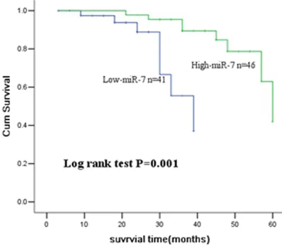
expression (low-miR-7) group according to the median expression quantity of miR-7. 46 patients were attributed to high-miR-7 group while the others belonged to low-miR-7 group. Kaplan-Meier showed that patients with
low-miR-7 expression had shorter survival time than those with high-miR-7 expression. Log rank test verified that the difference between two groups was significantly (P=0.001) (Figure
with the normal osteoblastic cell line (hFOB- 1.19) (Figure 1B, P<0.05).
Relationship between clinicopathologic char-acteristics and miR-7
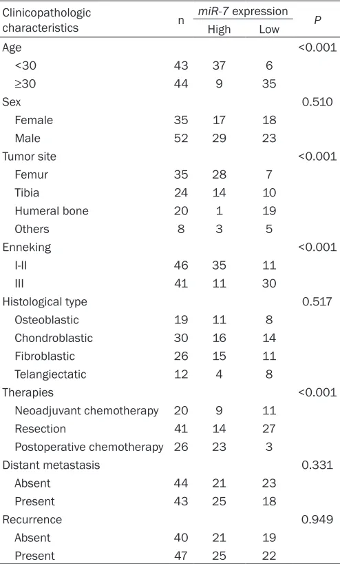
The miR-7 expression was lower in osteosarco-ma tissues and cell lines. To investigate wheth-er clinicopathologic charactwheth-eristics affected the expression of miR-7, we analyzed the rela-tionship between clinicopathologic
characteris-tics and miR-7. The results manifested that age (P< 0.001), tumor site (P<0.001), Enneking (P<0.001) and ther-apies (P<0.001) were closely related to the expression of
miR-7 (Table 1).
Down-regulation of miR-7 promoted cell proliferation
We then investigated the impact of miR-7 on osteosar-coma cell lines. The outcome revealed that down-regulation of miR7 significantly promot-ed cell proliferation in both tumor cell lines (MG63 and U2OS) compared with normal osteoblastic cell line (hFOB- 1.19) (Figure 2A, 2B).
Association between miR-7 and overall survival
[image:4.612.97.521.74.237.2]The osteosarcoma patients were divided into high miR-7
Figure 2. Cell proliferation assay of MG63 (A), U2OS (B) cell transfected with miR-7 siRNA or with control vector at an indicated time.
Figure 3. Kaplan-Meier survival curve analysis of patients with osteosarco-ma. Compared with the high-miR-7 expression group, the patients with
[image:4.612.92.369.296.537.2]The role of
miR-7
in osteosarcoma patients
3). All clinicopathologic characteristics and
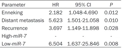
miR-7 were put into multivariate analysis to test the prognostic value of them by cox regression analysis. Enneking (HR=2.182, P=0.012), dis-tant metastasis (HR=5.623, P=0.010), recur-rence (HR=3.697, P=0.028) and low-miR-7
(HR=6.504, P=0.008) were related to the prognosis of osteosarcoma. And they might be independent prognostic markers in osteo-sarcoma (Table 2).
Discussion
With the wide study of various detecting tech-niques and further research of miRNAs, the relationships between miRNAs and the occur-rence as well as development of multiplicate tumors have attracted more and more atten-tions. The potential oncogene or antitumor effects make miRNAs expect to be molecular markers in the diagnosis and prognosis of can-cers so that provide new therapy strategies. The impact of miRNAs on osteosarcoma has been reported in many studies. For instance, down-regulation of miR-21 inhibited cell inva-sion and migration according to regulating the expression of PECK [11]. The over expression of
miR-151-3p, miR-191, miR-542-3p, miR-135b, miR-214, the decrease of miR-143, miR-199a-3p, miR-183, miR-34a, miR-218 mainly partici-pated in tumor cell proliferation, migration, cell cycle, apoptosis and so on [12-19]. Besides,
miR-223, miR-26a, miR-183, miR-9 were veri-fied to be related to the prognosis of osteosar-coma [20-23].
MiR-7 was found by Mariana et al in 2001 and then has been identified to be dysregulated in some human cancers [24]. MiR-7 has been characterized as a tumor suppressor and played roles in inhibiting cell growth,
prolifera-tion, apoptosis, migration and invasion in sev-eral types of cancer. For instance, Kefas et al, and Wang et al both found that miR-7 was down-regulated in glioblastoma, the differenc-es were that miR-7 decreased viability and invasiveness of primary glioblastoma lines in Kefas et al’ study while it inhibits cellular growth and glucose metabolism by regulating the IGF-1R/Akt signaling pathway in the study of Wang et al [25, 26]. Down-regulation of miR-7 -mediat-ed IGF1R r-mediat-educ-mediat-ed the IGF1-induc-mediat-ed activation of AKT leading to the inhibition of cell prolifera-tion and cell-cycle arrest, and an enhanced apoptotic rate in the study of Jiang et al [27]. In the study of Fang et al, miR-7 as a tumor sup-pressor could not only abolish the tumorigene-sis but reverse the metastatumorigene-sis of hepatocellu-lar carcinoma through regulating the PI3K/Akt/ mTOR-signaling pathway [28]. MiR-7 targeted different genes was considered to be an tumor suppressor and inhibited cell proliferation as well as increased apoptosis in colorectal can-cer both in the study of Zhang et al and Xu et al [29, 30]. Moreover, Li et al, found that miR-7
reduced or inhibited cell proliferation, induced cell apoptosis by suppressing the expression of both EGFR and RAF-1 oncogenes in Lewis lung cancer [31]. The over-expression of miR-7 could suppress cell viability and promoted cell apop-tosis regulated by oncogene XIAP in cervical cancer cells according to Liu et al [32]. The up-regulation of miR-7 arrested cell cycle at G1 to S transition in hepatocellular carcinoma while it inhibited cell growth and invasion in pancreatic cancer cells via Zhang et al and Ma et al [33, 34]. However, the value of miR-7 in osteosar-coma was still unclear.
In the present study, we detected the expres-sion of miR-7 in osteosarcoma tissues and cell lines. Meanwhile, we analyzed the function of
miR-7 in the progress of osteosarcoma. The result showed that miR-7 decreased in osteo-sarcoma and might serve as a tumor suppres-sor. Cell proliferation analysis revealed that
miR-7 was positive to the proliferation of osteo-sarcoma cancer cell lines MG63 and U2OS compared with normal cell line hFOB1.19. The study also determined the relationship of clini-copathologic characteristics and miR-7 expres-sion as well as the association between miR-7
[image:5.612.90.294.120.201.2]expression and overall survival. Age, tumor site, Enneking and therapies were considered to be related to the expression of miR-7 which
Table 2. Association between the expression of miR-7 and clinicopathologic characteristics with overall survival in patients with osteosar-coma
Parameter HR 95% CI P
enhanced the view that miR-7 plays a role in the progress of osteosarcoma. The results of Kaplan-Meier and cox regression analysis man-ifested that miR-7 impacted the overall survival of osteosarcoma patients and could be as an independent prognostic marker. Besides, Enneking, distant metastasis and recurrence had a predictive meaning in osteosarcoma. In conclusion, miR-7 is low-expression in osteo-sarcoma and may function as a tumor suppres-sor. Besides, miR-7 promoted osteosarcoma cell proliferation and took prognostic value in osteosarcoma patients. The suppressive func-tion of miR-7 may be helpful for the forecast of osteosarcoma and provide new therapy strategies.
Disclosure of conflict of interest
None.
Address correspondence to: Ye Liu, Department of Anesthesiology, Beijing Obstetrics and Gyneco- logy Hospital, Capital Medical University, Beijing 100026, China. Huaijie An, Center of Basic Medical Sciences, Navy General Hospital of PLA, Beijing 100037, China. E-mail: [email protected]
References
[1] Yang J and Zhang W. New molecular insights into osteosarcoma targeted therapy. Curr Opin Oncol 2013; 25: 398-406.
[2] Siegel HJ and Pressey JG. Current concepts on the surgical and medical management of os-teosarcoma. Expert Rev Anticancer Ther 2008; 8: 1257-1269.
[3] Chen L, Wang Q, Wang GD, Wang HS, Huang Y, Liu XM and Cai XH. miR-16 inhibits cell prolif-eration by targeting IGF1R and the Raf1-MEK1/2-ERK1/2 pathway in osteosarcoma. FEBS Lett 2013; 587: 1366-1372.
[4] Mirabello L, Troisi RJ and Savage SA. Osteosarcoma incidence and survival rates from 1973 to 2004: data from the Surveillance, Epidemiology, and End Results Program. Cancer 2009; 115: 1531-1543.
[5] Bartel DP. MicroRNAs: genomics, biogenesis, mechanism, and function. Cell 2004; 116: 281-297.
[6] Wang X, Wang ZH, Wu YY, Tang H, Tan L, Gao XY, Xiong YS, Liu D, Wang JZ and Zhu LQ. Melatonin attenuates scopolamine-induced memory/synaptic disorder by rescuing EPACs/ miR-124/Egr1 pathway. Mol Neurobiol 2013; 47: 373-381.
[7] Esteller M. Non-coding RNAs in human dis-ease. Nat Rev Genet 2011; 12: 861-874. [8] Kalinowski FC, Brown RA, Ganda C, Giles KM,
Epis MR, Horsham J and Leedman PJ. microR-NA-7: a tumor suppressor miRNA with thera-peutic potential. Int J Biochem Cell Biol 2014; 54: 312-317.
[9] Zhou X, Hu Y, Dai L, Wang Y, Zhou J, Wang W, Di W and Qiu L. MicroRNA-7 inhibits tumor me-tastasis and reverses epithelial-mesenchymal transition through AKT/ERK1/2 inactivation by targeting EGFR in epithelial ovarian cancer. PLoS One 2014; 9: e96718.
[10] Jones KB, Salah Z, Del Mare S, Galasso M, Gaudio E, Nuovo GJ, Lovat F, LeBlanc K, Palatini J, Randall RL, Volinia S, Stein GS, Croce CM, Lian JB and Aqeilan RI. miRNA sig-natures associate with pathogenesis and pro-gression of osteosarcoma. Cancer Res 2012; 72: 1865-1877.
[11] Ziyan W, Shuhua Y, Xiufang W and Xiaoyun L. MicroRNA-21 is involved in osteosarcoma cell invasion and migration. Med Oncol 2011; 28: 1469-1474.
[12] Duan Z, Choy E, Harmon D, Liu X, Susa M, Mankin H and Hornicek F. MicroRNA-199a-3p is downregulated in human osteosarcoma and regulates cell proliferation and migration. Mol Cancer Ther 2011; 10: 1337-1345.
[13] Li H, Liu H, Pei J, Wang H and Lv H. miR5423p overexpression is associated with enhanced osteosarcoma cell proliferation and migration ability by targeting Van Goghlike 2. Mol Med Rep 2015; 11: 851-856.
[14] Pei H, Jin Z, Chen S, Sun X, Yu J and Guo W. MiR-135b promotes proliferation and invasion of osteosarcoma cells via targeting FOXO1. Mol Cell Biochem 2015; 400: 245-252. [15] Wang X, Sun J, Fu C, Wang D and Bi Z.
MicroRNA-214 regulates osteosarcoma sur-vival and growth by directly targeting phospha-tase and tensin homolog. Mol Med Rep 2014; 10: 3073-3079.
[16] Zhang H, Cai X, Wang Y, Tang H, Tong D and Ji F. microRNA-143, down-regulated in osteosar-coma, promotes apoptosis and suppresses tu-morigenicity by targeting Bcl-2. Oncol Rep 2010; 24: 1363-1369.
[17] Zhao H, Guo M, Zhao G, Ma Q, Ma B, Qiu X and Fan Q. miR-183 inhibits the metastasis of os-teosarcoma via downregulation of the expres-sion of Ezrin in F5M2 cells. Int J Mol Med 2012; 30: 1013-1020.
[18] Zhao H, Ma B, Wang Y, Han T, Zheng L, Sun C, Liu T, Zhang Y, Qiu X and Fan Q. miR-34a inhib-its the metastasis of osteosarcoma cells by repressing the expression of CD44. Oncol Rep 2013; 29: 1027-1036.
inva-The role of
miR-7
in osteosarcoma patients
sion by down-regulating of TIAM1, MMP2 and MMP9. Asian Pac J Cancer Prev 2013; 14: 3681-3684.
[20] Zhang H, Yin Z, Ning K, Wang L, Guo R and Ji Z. Prognostic value of microRNA-223/epithelial cell transforming sequence 2 signaling in pa-tients with osteosarcoma. Hum Pathol 2014; 45: 1430-1436.
[21] Song QC, Shi ZB, Zhang YT, Ji L, Wang KZ, Duan DP and Dang XQ. Downregulation of mi-croRNA-26a is associated with metastatic po-tential and the poor prognosis of osteosarco-ma patients. Oncol Rep 2014; 31: 1263-1270. [22] Mu Y, Zhang H, Che L and Li K. Clinical signifi-cance of microRNA-183/Ezrin axis in judging the prognosis of patients with osteosarcoma. Med Oncol 2014; 31: 821.
[23] Xu SH, Yang YL, Han SM and Wu ZH. MicroRNA-9 expression is a prognostic bio-marker in patients with osteosarcoma. World J Surg Oncol 2014; 12: 195.
[24] Lagos-Quintana M, Rauhut R, Lendeckel W and Tuschl T. Identification of novel genes cod-ing for small expressed RNAs. Science 2001; 294: 853-858.
[25] Kefas B, Godlewski J, Comeau L, Li Y, Abounader R, Hawkinson M, Lee J, Fine H, Chiocca EA, Lawler S and Purow B. microRNA-7 inhibits the epidermal growth factor receptor and the Akt pathway and is down-regulated in glioblastoma. Cancer Res 2008; 68: 3566-3572.
[26] Wang B, Sun F, Dong N, Sun Z, Diao Y, Zheng C, Sun J, Yang Y and Jiang D. MicroRNA-7 directly targets insulin-like growth factor 1 receptor to inhibit cellular growth and glucose metabolism in gliomas. Diagn Pathol 2014; 9: 211. [27] Jiang L, Liu X, Chen Z, Jin Y, Heidbreder CE,
Kolokythas A, Wang A, Dai Y and Zhou X. MicroRNA-7 targets IGF1R (insulin-like growth factor 1 receptor) in tongue squamous cell car-cinoma cells. Biochem J 2010; 432: 199-205.
[28] Fang Y, Xue JL, Shen Q, Chen J and Tian L. MicroRNA-7 inhibits tumor growth and metas-tasis by targeting the phosphoinositide 3-ki-nase/Akt pathway in hepatocellular carcino-ma. Hepatology 2012; 55: 1852-1862. [29] Zhang N, Li X, Wu CW, Dong Y, Cai M, Mok MT,
Wang H, Chen J, Ng SS, Chen M, Sung JJ and Yu J. microRNA-7 is a novel inhibitor of YY1 contributing to colorectal tumorigenesis. Oncogene 2013; 32: 5078-5088.
[30] Xu K, Chen Z, Qin C and Song X. miR-7 inhibits colorectal cancer cell proliferation and induces apoptosis by targeting XRCC2. Onco Targets Ther 2014; 7: 325-332.
[31] Li J, Zheng Y, Sun G and Xiong S. Restoration of miR-7 expression suppresses the growth of Lewis lung cancer cells by modulating epider-mal growth factor receptor signaling. Oncol Rep 2014; 32: 2511-2516.
[32] Liu S, Zhang P, Chen Z, Liu M, Li X and Tang H. MicroRNA-7 downregulates XIAP expression to suppress cell growth and promote apoptosis in cervical cancer cells. FEBS Lett 2013; 587: 2247-2253.
[33] Ma J, Fang B, Zeng F, Pang H, Zhang J, Shi Y, Wu X, Cheng L, Ma C, Xia J and Wang Z. Curcumin inhibits cell growth and invasion through up-regulation of miR-7 in pancreatic cancer cells. Toxicol Lett 2014; 231: 82-91. [34] Zhang X, Hu S, Wang L, Yan B, Zhao J, Yang A