Original Article
Serum
miRNA-34a
serves as a diagnostic and
prognostic bio-marker in osteosarcoma
Chunsheng Zhi, Bo Wu
Shenyang Orthopedic Hospital, Shenyang, Liaoning, China
Received December 18, 2015; Accepted February 27, 2016; Epub March 1, 2016; Published March 15, 2016
Abstract: Background: MicroRNA-34a (miR-34a) had been reported to be decreased in osteosarcoma, but its ef-fects on diagnosis and prognosis were still unclear. The purpose of this study was to investigate the expression and clinical significance of miR-34a in osteosarcoma. Methods: The expression of miR-34a in tissues and serum of all patients and controls was detected by quantitative real-time polymerase chain reaction (qRT-PCR) assay, respective-ly. Besides, the relationship between miR-34a expression and clinicopathologic characteristics were analyzed. The association between miR-34a expression and overall survival of patients with osteosarcoma was analyzed Kaplan-Meier analysis. The prognostic value of miR-34a was estimated via cox regression analysis. Results: The expression level of miR-34a was decreased in osteosarcoma tissues and serum compared with the controls. Receiver operating characteristic (ROC) curve demonstrated miR-34a could be a diagnostic factor in osteosarcoma with an AUC of cor-responding with a sensitivity of and a specificity of. Kaplan-Meier analysis revealed that patients with high miR-34a
expression had a longer overall survival than those with low expression (Log rank test, P<0.001). According to cox regression analysis, miR-34a was considered as an independent prognostic marker in osteosarcoma. Conclusion:
miR-34a expression was down-regulated in osteosarcoma and associated with tumor progression. Moreover, its ab-normal expression made it can be an independent diagnostic and prognostic bio-marker for osteosarcoma patients.
Keywords: Osteosarcoma, miR-34a, diagnosis, prognosis
Introduction
Osteosarcoma is the most frequent primary malignant bone tumor arising from metaphysis of the long bones of adolescents and young adults, accounting for approximately 5% of all pediatric tumors [1, 2]. About 80% of patients eventually develop recurrent metastatic osteo-sarcoma following surgical treatment which lead to a low cure rate and the 5-year survival rate is only 50-60% at 20% [3, 4]. Although the treatments of osteosarcoma have achieved great improvement, the prognosis of osteosar-coma is still poor. Moreover, the molecular mechanisms involved in osteosarcoma prog-ress remains poorly understood. It is therefore of critical importance to explore the mecha-nisms that underlie the pathogenesis, thus shedding light on the complex etiology and developing strategies for the diagnosis, treat-ment and prognosis of this disease.
MicroRNAs (miRNAs) are a class of endoge-nous, small, non-coding RNAs with 18-25 nu-
demonstrated that miR-34a was decreased in osteosarcoma [15]. However, the clinical signifi -cance of miR-34a was never reported in osteosarcoma.
In our study, the expression level of miR-34a in serum and tissues samples was detected via qRT-PCR analysis, respectively. The relation-ship between miR-34a expression and clinical factors was also analyzed. Through the expres-sion level in serum, we verified its diagnosis value using ROC curve. And then we investigat-ed role of miR-34a in prognosis of
osteosarco-ma by analyzed the association between miR-34a and overall survival.
Materials and methods
Patients’ samples
50 patients who were diagnosed as osteosar-coma during 2008-2010 in Shenyang Orth- opedic Hospital were collected in our study and the study was permitted by the Ethnic Committee of the hospital. Meanwhile, 30 patients including 18 with Ewing’s sarcoma and 12 with osteomyelitis were taken as benign dis-eases controls. Besides, 50 healthy people matching ages with osteosarcoma patients we- re taken as healthy controls. Written informed consent was obtained from each participant involved in advance. None of the patients had received any chemotherapy or radiotherapy before surgery.
Osteosarcoma tissues, adjacent tissues and healthy tissues were obtained, respectively. Then they were frozen by liquid nitrogen imme-diately and stored at -80°C for RNA extraction. The serum samples from osteosarcoma pati- ents, benign diseases controls and healthy co- ntrols were severally put into blood collection tube of EDTA and stored at -80°C for use. The detailed clinical data including age, sex, site, ALP, histologic type, tumor grade, the status of metastasis and recurrence were collected and stored in a database. A 5-year follow-up was performed for all patients to evaluate its prog-nostic value. Patients who were died from unex-pected events or other diseases were excluded from our study.
RNA extraction and qRT-PCR analysis
[image:2.629.100.298.79.249.2]Total RNA was extracted from all tissues and serum using mirVana miRNA Isolation Kit (Ambion, Austin, TX, USA), respectively. Reverse transcription was conducted by TaqMan Mic- roRNA Reverse Transcription Kit (Applied Bio- systems, Foster City, CA, USA) to synthesize the first chain of cDNA. Then RT-PCR reaction was performed in the Applied Biosystems 7900 Fast Real-Time PCR system (Applied Biosy- stems, Foster City, California, USA). RNU44 was used as internal control. The relative quantity of miR-34a was calculated with the comparative cycle threshold (CT) method. Each sample was examined in triplicate.
Figure 1. The expression of miR-34a in tissues samples. It was significantly lower in osteosarcoma tissues than in adjacent tissues and healthy tissues (P<0.05).
[image:2.629.102.297.326.497.2]Statistical analysis
The SPSS version 13.0 for Windows (SPSS Inc, IL, USA) was used for statistical analysis. The difference of miR-34a expression in different tissues and serum samples were analyzed by one way ANOVA. The difference between two groups was compared by student’s t test. ROC curve was built to determine the diagnostic per-formance of miR-34a expression in patients with osteosarcoma. The overall survival of patients with different expression of miR-34a
was estimated by the Kaplan-Meier analysis. Cox regression analysis was performed to iden-tify the factor with significant influence on over -all survival. The differences were considered to be statistically significant when P<0.05.
Results
Expression of miR-34a in tissues of patients with osteosarcoma
The expression levels of miR-34a were detect-ed by qRT-PCR analysis. The results showdetect-ed that the relative expression of miR-34a in osteosarcoma tissues was obviously lower than in adjacent normal tissue and healthy con-trols (Figure 1, P<0.05).
Serum miR-34a in patients with osteosar-coma, benign diseases controls and healthy controls
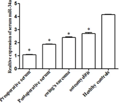
Serum miR-34a expression of preoperative and postoperative as well as benign controls and healthy controls was measured, respectively. As shown in Figure 2, serum miR-34a expres-sion of preoperative was significantly lower than that of postoperative, benign controls and healthy controls (P<0.05). These indicated that serum miR-34a expression cloud distinguish osteosarcoma patients from other patients or healthy people.
Relationship between miR-34a expression and clinicopathologic characteristics
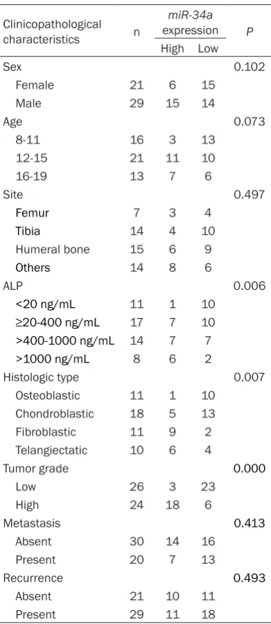
To explore whether miR-34a was related to the development of osteosarcoma, the relationship between it and clinicopathologic characteris-tics was analyzed. It was demonstrated that the expression of miR-34a was significantly influ -enced by ALP (P=0.006), histologic type (P=0.007) and tumor grade (P=0.000) which revealed that it was involved in the progression of osteosarcoma (Table 1).
[image:3.629.99.295.114.564.2]Diagnostic value of miR-34a in osteosarcoma We generated ROC curves to assess the poten-tial value of serum miR-34a in the early detec-tion of osteosarcoma. The outcome manifested that serum miR-34a expression had a high diagnostic value in discriminating patients with osteosarcoma from control subjects with an AUC value of 0.830 combing with a sensitivity of 86.7% and a specificity of 68.6% (Figure 3).
Table 1. The relationship between miR-34a expression and clinical factors of patients with osteosarcoma
Clinicopathological characteristics n
miR-34a expression P
High Low
Sex 0.102
Female 21 6 15
Male 29 15 14
Age 0.073
8-11 16 3 13
12-15 21 11 10
16-19 13 7 6
Site 0.497
Femur 7 3 4
Tibia 14 4 10 Humeral bone 15 6 9
Others 14 8 6
ALP 0.006
<20 ng/mL 11 1 10 ≥20-400 ng/mL 17 7 10
>400-1000 ng/mL 14 7 7
>1000 ng/mL 8 6 2
Histologic type 0.007
Osteoblastic 11 1 10 Chondroblastic 18 5 13 Fibroblastic 11 9 2 Telangiectatic 10 6 4
Tumor grade 0.000
Low 26 3 23
High 24 18 6
Metastasis 0.413
Absent 30 14 16
Present 20 7 13
Recurrence 0.493
Absent 21 10 11
And the ideal cut-off value was. The result veri-fied that miR-34a could be an independent bio-marker for the diagnosis of osteosarcoma.
Association between miR-34a expression and overall survival of patients with osteosarcoma
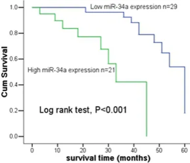
To further evaluate the association of miR-34a expression with overall survival of patients, the osteosarcoma patients were divided into high expression group (high miR-34a expression) and low-expression group (low miR-34a expres-sion) according to the median expression of miR-34a (1.259±0.126). During the follow-up,
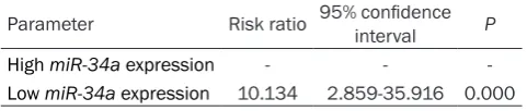
18 patients were censored. Kaplan-Meier anal-ysis demonstrated that overall survival of patients with high miR-34a expression was lon-ger compared to those with low miR-34a expression (Figure 4, log rank test, P<0.001). Multivariate analysis adjusted for clinical fac-tors and the expression of miR-34a using cox regression analysis showed the factors with statistical significance for prognosis of osteo -sarcoma were the expression of miR-34a (Table 2, HR=10.134, 95 CI%=2.859-35.916, P=0.000) and it might be an independent prog-nostic marker.
Discussion
The aberrant expression or alterations of miR-NAs are closely related to human pathologies such as cardiovascular disorders, inflammatory diseases and cancer [16, 17]. Osteosarcoma is considered to be a differentiation disease caused by genetic changes that interrupt osteo-blast differentiation from mesenchymal stem cells [18, 19]. miRNA was verified to regulate specific tissue-lineages during the differentia -tion of osteosarcoma, but their involvement is still unclear [20-22].
In recent studies, there were multiple miRNAs reported to play roles in osteosarcoma such as miR-31, miR-192, miR-215, miR-140, miR-21, miR-143, and so on. And most of these miRNAs are related to cell proliferation, cell cycle, cell invasion and migration [23-25]. miRNAs may become a new tool for the diagnosis and prog-nosis of cancers as they can exit in body fluids stably, so study about them is meaningful. miR-34a is suggested to function as a tumor suppressor gene and contributes to the inhibi-tion of invasion or metastasis in various tumor types. And it has relationship with the progres-sion and poor prognosis of many tumors. miR-34a has been confirmed to be down-regulated in many cancers such as human papillomavi-rus, hepatocellular carcinoma, prostate cancer, colon cancer, non-small cell lung carcinoma and so on [26-30]. However, the prognosis role of miR-34a in osteosarcoma was still unclear and need more comprehensive studies.
[image:4.629.101.295.79.241.2]In this study, we detected the expression level of miR-34a in osteosarcoma tissues and serum by QT-PCR. Compared the controls of tissues and serum, miR-34a expression was all decr- Figure 3. ROC curve for the diagnostic value of
miR-34a in osteosarcoma. It had a high diagnostic value with an AUC of 0.830 corresponding with a sensitivity
of 86.7% and a specificity of 68.6%.
[image:4.629.100.294.315.481.2]eased in patients with osteosarcoma. Besides, serum miR-34a expression of preoperative was lower than that of postoperative, benign dis-eases controls and healthy controls. These results showed that miR-34a might be a tumor suppressor in osteosarcoma. It was consistent with previous study in osteosarcoma [15]. Besides, its expression was considered to be influenced by ALP, histologic type and tumor grade. But the relationship between miR-34a expression and progression of osteosarcoma was not clarified in our study. The mechanism of its antitumor and its effect on progression of osteosarcoma are still needs to be further investigated. ROC curve demonstrated that miR-34a could be a diagnostic bio-marker for osteosarcoma. Meanwhile, cox regression an- alysis revealed miR-34a could be an indepen-dent prognostic marker in osteosarcoma. In conclusion, miR-34a expression is decreased in osteosarcoma tissues and serum. Its expres-sion is related to ALP, histologic type and tumor grade. miR-34a may serve as a diagnostic and prognostic marker in osteosarcoma. Further researches in a large number are warranted to further investigate the significance of miR-34a in osteosarcoma.
Disclosure of conflict of interest
None.
Address correspondence to: Dr. Bo Wu, Shenyang Orthopedic Hospital, 115 Dongbei Road, Shenyang 110044, Liaoning, China. E-mail: wubojaiyoun@ sina.com
References
[1] Siegel HJ and Pressey JG. Current concepts on the surgical and medical management of os-teosarcoma. Expert Rev Anticancer Ther 2008; 8: 1257-1269.
[2] Yang J and Zhang W. New molecular insights into osteosarcoma targeted therapy. Curr Opin Oncol 2013; 25: 398-406.
[6] Esteller M. Non-coding RNAs in human dis-ease. Nat Rev Genet 2011; 12: 861-874. [7] Liang W, Gao B, Fu P, Xu S, Qian Y and Fu Q.
The miRNAs in the pathgenesis of osteosarco-ma. Front Biosci (Landmark Ed) 2013; 18: 788-794.
[8] Wang X, Wang ZH, Wu YY, Tang H, Tan L, Gao XY, Xiong YS, Liu D, Wang JZ and Zhu LQ. Mela-tonin attenuates scopolamine-induced memo-ry/synaptic disorder by rescuing EPACs/miR-124/Egr1 pathway. Mol Neurobiol 2013; 47: 373-381.
[9] Nana-Sinkam SP and Croce CM. MicroRNAs as therapeutic targets in cancer. Transl Res 2011; 157: 216-225.
[10] Volinia S, Calin GA, Liu CG, Ambs S, Cimmino A, Petrocca F, Visone R, Iorio M, Roldo C, Ferracin M, Prueitt RL, Yanaihara N, Lanza G, Scarpa A, Vecchione A, Negrini M, Harris CC and Croce CM. A microRNA expression signature of hu-man solid tumors defines cancer gene targets. Proc Natl Acad Sci U S A 2006; 103: 2257-2261.
[11] Iwasaki Y, Arai K, Katayanagi S, Takahashi K, Yamaguchi T, Matsumoto H and Miyamoto H. [Biomarkers for neoplasmas in digestive or-gans]. Gan To Kagaku Ryoho 2004; 31: 1015-1020.
[12] Metias SM, Lianidou E and Yousef GM. MicroR-NAs in clinical oncology: at the crossroads be-tween promises and problems. J Clin Pathol 2009; 62: 771-776.
[13] He L, He X, Lim LP, de Stanchina E, Xuan Z, Li-ang Y, Xue W, Zender L, Magnus J, Ridzon D, Jackson AL, Linsley PS, Chen C, Lowe SW, Cleary MA and Hannon GJ. A microRNA compo-nent of the p53 tumour suppressor network. Nature 2007; 447: 1130-1134.
[14] Gallardo E, Navarro A, Vinolas N, Marrades RM, Diaz T, Gel B, Quera A, Bandres E, Garcia-Foncillas J, Ramirez J and Monzo M. miR-34a as a prognostic marker of relapse in surgically resected non-small-cell lung cancer. Carcino-genesis 2009; 30: 1903-1909.
[image:5.629.98.339.107.158.2][15] He C, Xiong J, Xu X, Lu W, Liu L, Xiao D and Wang D. Functional elucidation of MiR-34 in osteosarcoma cells and primary tumor sam-ples. Biochem Biophys Res Commun 2009; 388: 35-40.
Table 2. Cox regression analysis for the prognostic
value of miR-34a in patients with osteosarcoma Parameter Risk ratio 95% confidence interval P
High miR-34a expression - -
-Low miR-34a expression 10.134 2.859-35.916 0.000
[3] Marina N, Gebhardt M, Teot L and Go- rlick R. Biology and therapeutic advanc-es for pediatric osteosarcoma. Oncolo-gist 2004; 9: 422-441.
[4] Gorlick R. Current concepts on the mo-lecular biology of osteosarcoma. Cancer Treat Res 2009; 152: 467-478.
[16] Tili E, Michaille JJ, Costinean S and Croce CM. MicroRNAs, the immune system and rheumat-ic disease. Nat Clin Pract Rheumatol 2008; 4: 534-541.
[17] Wang N, Zhou Z, Liao X and Zhang T. Role of microRNAs in cardiac hypertrophy and heart failure. IUBMB Life 2009; 61: 566-571. [18] Tang N, Song WX, Luo J, Haydon RC and He TC.
Osteosarcoma development and stem cell dif-ferentiation. Clin Orthop Relat Res 2008; 466: 2114-2130.
[19] Siclari VA and Qin L. Targeting the osteosarco-ma cancer stem cell. J Orthop Surg Res 2010; 5: 78.
[20] Subramanian S, Lui WO, Lee CH, Espinosa I, Nielsen TO, Heinrich MC, Corless CL, Fire AZ and van de Rijn M. MicroRNA expression signa-ture of human sarcomas. Oncogene 2008; 27: 2015-2026.
[21] Sarver AL, Phalak R, Thayanithy V and Subra-manian S. S-MED: sarcoma microRNA expres-sion database. Lab Invest 2010; 90: 753-761. [22] Liu C and Tang DG. MicroRNA regulation of ca-
ncer stem cells. Cancer Res 2011; 71: 5950-5954.
[23] Braun CJ, Zhang X, Savelyeva I, Wolff S, Moll UM, Schepeler T, Orntoft TF, Andersen CL and Dobbelstein M. p53-Responsive micror-nas 192 and 215 are capable of inducing cell cycle arrest. Cancer Res 2008; 68: 10094-10104.
[24] Creighton CJ, Fountain MD, Yu Z, Nagaraja AK, Zhu H, Khan M, Olokpa E, Zariff A, Gunaratne PH, Matzuk MM and Anderson ML. Molecular profiling uncovers a p53-associated role for mi-croRNA-31 in inhibiting the proliferation of se-rous ovarian carcinomas and other cancers. Cancer Res 2010; 70: 1906-1915.
[25] Ziyan W, Shuhua Y, Xiufang W and Xiaoyun L. MicroRNA-21 is involved in osteosarcoma cell invasion and migration. Med Oncol 2011; 28: 1469-1474.
[26] Ogawa T, Saiki Y, Shiga K, Chen N, Fukushige S, Sunamura M, Nagase H, Hashimoto S, Mat-suura K, Saijo S, Kobayashi T and Horii A. miR-34a is downregulated in cis-diamminedichloro-platinum treated sinonasal squamous cell carcinoma patients with poor prognosis. Can-cer Sci 2012; 103: 1737-1743.
[27] Li N, Fu H, Tie Y, Hu Z, Kong W, Wu Y and Zheng X. miR-34a inhibits migration and invasion by down-regulation of c-Met expression in human hepatocellular carcinoma cells. Cancer Lett 2009; 275: 44-53.
[28] Fujita Y, Kojima K, Hamada N, Ohhashi R, Akao Y, Nozawa Y, Deguchi T and Ito M. Effects of miR-34a on cell growth and chemoresistance in prostate cancer PC3 cells. Biochem Biophys Res Commun 2008; 377: 114-119.
[29] Roy S, Levi E, Majumdar AP and Sarkar FH. Ex-pression of miR-34 is lost in colon cancer which can be re-expressed by a novel agent CDF. J Hematol Oncol 2012; 5: 58.