4-Nitro-2-phenoxyaniline
H. R. Manjunath,aM. T. Shreenivasa,bM. Mahendra,a T. M. Mohan Kumar,cB. E. Kumara Swamyband M. A. Sridhara*
aDepartment of Studies in Physics, Manasagangotri, University of Mysore, Mysore
570 006, India,bDepartment of P. G. Studies and Research in Industrial Chemistry,
Kuvempu University, Jnana Sahyadri, Shankaraghatta, Karnataka, India, and
cChemistry Department, Amrita School of Engineering, Amrita Vishwa
Vidyapeetham, Bangalore 560 035, India
Correspondence e-mail: [email protected]
Received 11 March 2010; accepted 31 March 2010
Key indicators: single-crystal X-ray study;T= 293 K; mean(C–C) = 0.004 A˚; Rfactor = 0.053;wRfactor = 0.167; data-to-parameter ratio = 12.3.
In the title compound, C12H10N2O3, the oxygen atom bridging the two aromatic rings is in a synperiplanar (+sp) conforma-tion. The dihedral angle between the aromatic rings is 71.40 (12). In the crystal, molecules are linked by
inter-molecular N—H O hydrogen bonds.
Related literature
For the pharmacological properties of nitro-2-phenoxyaniline, see: Moore & Harrington (1974); Prasadet al.(2005). For the herbicidal applications of biphenyl ether derivatives, see: Yuet al., (2008). For the applications of Schiff bases derived from aromatic amines, see: Singh et al. (1975); Cimerman et al.
(2000). For their biological and pharmacological acitvity, see: Singhet al.(1975); Cimermanet al.(2000); Shahet al.(1992); Pandeyaet al.(1999); Moreet al.(2001). For the preparation of 4-nitro-2-phenoxyaniline, see: Shreenivasaet al.(2009). For a related structure, see: Naveenet al.(2006).
Experimental
Crystal data
C12H10N2O3
Mr= 230.22 Monoclinic,P21=c
a= 10.4100 (12) A˚ b= 15.6570 (18) A˚ c= 6.9600 (17) A˚
V= 1103.5 (3) A˚3 Z= 4
MoKradiation
= 0.10 mm1
T= 293 K
0.320.30.25 mm
Data collection
MacScience DIPLabo 32001 diffractometer
3336 measured reflections
1889 independent reflections 1498 reflections withI> 2(I) Rint= 0.033
Refinement
R[F2> 2(F2)] = 0.053
wR(F2) = 0.167 S= 1.09 1889 reflections
154 parameters
H-atom parameters constrained
max= 0.13 e A˚
3
min=0.15 e A˚
3
Table 1
Hydrogen-bond geometry (A˚ ,).
D—H A D—H H A D A D—H A
N10—H10A O9i 0.86 2.17 3.023 (3) 170
Symmetry code: (i)xþ1;y1 2;zþ
1 2.
Data collection: XPRESS (MacScience, 2002); cell refinement:
SCALEPACK (Otwinowski & Minor, 1997); data reduction:
DENZO (Otwinowski & Minor, 1997) and SCALEPACK; program(s) used to solve structure: SHELXS7 (Sheldrick, 2008); program(s) used to refine structure:SHELXL97(Sheldrick, 2008); molecular graphics:PLATON(Spek, 2009) andORTEPII(Johnson, 1976); software used to prepare material for publication:
SHELXL97.
The authors are grateful to the DST and Government of India project SP/I2/FOO/93 and the University of Mysore for financial assistance. MM would like to thank the University of Mysore for awarding a project under the head DV3/136/2007– 2008/24.09.09.
Supplementary data and figures for this paper are available from the IUCr electronic archives (Reference: FJ2288).
References
Cimerman, Z., Miljanic´, S. & Galic´, N. (2000).Croat. Chem. Acta,73, 81–95. Johnson, C. K. (1976).ORTEPII. Report ORNL-5138. Oak Ridge National
Laboratory, Tennessee, USA.
MacScience (2002).XPRESS. MacScience Co. Ltd, Yokohama, Japan. Moore, G. G. L. & Harrington, J. K. (1974). US Patent No. 3840597. More, P. G., Bhalvankar, R. B. & Patter, S. C. (2001).J. Indian Chem. Soc.78,
474–475.
Naveen, S., Anil Kumar, K., Channe Gowda, D., Sridhar, M. A. & Shashidhara Prasad, J. (2006).Acta Cryst.E62, o3790–o3791.
Otwinowski, Z. & Minor, W. (1997). Methods in Enzymology, Vol. 276, Macromolecular Crystallography, Part A, edited by C. W. Carter Jr & R. M. Sweet, pp. 307–326. New York: Academic Press.
Pandeya, S. N., Sriram, D., Nath, G. & De Clercq, E. (1999).Eur. J. Pharm. Soc.9, 25–31.
Prasad, A., Sharma, M. L., Kanwar, S., Rathee, R. & Sharma, S. D. (2005).J. Sci. Ind. Res.64, 756–760.
Shah, S., Vyas, R. & Mehta, R. H. (1992).J. Indian Chem. Soc.69, 590–596. Sheldrick, G. M. (2008).Acta Cryst.A64, 112–122.
Shreenivasa, M. T., Chetan, B. P. & Bhat, A. R. (2009).J. Pharm. Sci. Technol. 1, 88–94.
Singh, P., Goel, R. L. & Singh, B. P. (1975).J. Indian Chem. Soc.52, 958–959. Spek, A. L. (2009).Acta Cryst.D65, 148–155.
Yu, H.-B., Wu, H.-F., Cui, D.-L. & Li, B. (2008).Ark. Kemi,2, 94–104.
Acta Crystallographica Section E
Structure Reports
Online
supporting information
Acta Cryst. (2010). E66, o1255 [https://doi.org/10.1107/S1600536810012237]
4-Nitro-2-phenoxyaniline
H. R. Manjunath, M. T. Shreenivasa, M. Mahendra, T. M. Mohan Kumar, B. E. Kumara Swamy
and M. A. Sridhar
S1. Comment
The phenoxy anilines are versatile intermediates for synthesizing several pharmaceutical drugs i.e. Nimesulide,
Ampxipine and Loxapine. The Nitro-2-phenoxyaniline is an intermediate for the synthesis of Nimesulide and it was
probably the first COX-2 selective non-steroidal anti inflammatory drug (NSAID) identified with this key
pharmacological properties (Moore & Harrington, 1974; Prasad et al. 2005). It is a unique molecule with twin aromatic
ring structure. The nitro-2-phenoxyaniline is a derivative of biphenyl ether. More generally, biphenyl ether derivatives
have many biological, herbicidal (Yu et al., 2008) and organic chemistry applications. Schiff bases derived from aromatic
amines have a wide variety of applications in many fields, viz., biological, inorganic and anlytical chemistry (Singh et al.,
1975; Cimerman et al., 2000). They are known to exhibit potent antibacterial, anticonvulsant, anti-inflammatory (Shah et
al. 1992), anticancer (Pandeya et al., 1999), anti-hypertensive and hypnotic (More et al., 2001) activities. With this
background, the title compound (I), was synthesized and we report its crystal structure here.
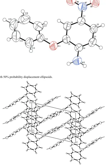
A perspective view of (I) is shown in Fig. 1. The two aromatic rings are not coplanar. This is confirmed by the dihedral
angle value of 71.38 (12)° between two six-membered rings. The oxygen atom connecting the two aromatic rings is in
syn-periplanar (sp) conformation as indicated by the torsion angle value of 13.0 (3)°. The nitro group lies in the plane of
the aniline ring as indicated by the C2—C1—N7—O8 and C6—C1—N7—O9 torsion angles of -176.1 (2)° and
-174.4 (2)°, respectively. These values are different from the values reported earlier (Naveen S. et al. 2006). The structure
exhibits both inter and intramolecular N—H···O interaction. The intermolecular N10—H10A···O9 interaction has a length
of 2.17Å and angle of 170° with symmetry codes 3/2-x,-1/2+y,1-z. The molecules exhibit layered stackings when viewd
down the 'b′ axis as shown in Fig. 2.
S2. Experimental
The 4-nitro-2-phenoxyaniline was prepared by condensation of o-chloronitrobenzene with phenol followed by acetylation
and nitration (Shreenivasa et al., 2009). The final product obtained was recrystallized using ethanol as a solvent.
Colorless crystals were appeared after 4 days by slow evaporation.
S3. Refinement
H atoms were placed at idealized positions and allowed to ride on their parent atoms with C–H distances in the range
Figure 1
[image:3.610.130.477.359.639.2]A view of (I), with 50% probability displacement ellipsoids.
Figure 2
4-Nitro-2-phenoxyaniline
Crystal data
C12H10N2O3
Mr = 230.22 Monoclinic, P21/c Hall symbol: -P 2ybc
a = 10.4100 (12) Å
b = 15.6570 (18) Å
c = 6.9600 (17) Å
β = 103.406 (4)°
V = 1103.5 (3) Å3
Z = 4
F(000) = 480
Dx = 1.386 Mg m−3
Mo Kα radiation, λ = 0.71073 Å Cell parameters from 14613 reflections
θ = 2.4–32.5°
µ = 0.10 mm−1
T = 293 K Block, colorless 0.32 × 0.3 × 0.25 mm
Data collection
MacScience DIPLabo 32001 diffractometer
Radiation source: fine-focus sealed tube Graphite monochromator
Detector resolution: 10.0 pixels mm-1
ω scan
3336 measured reflections
1889 independent reflections 1498 reflections with I > 2σ(I)
Rint = 0.033
θmax = 25.0°, θmin = 2.4°
h = −12→12
k = −18→18
l = −8→8
Refinement
Refinement on F2 Least-squares matrix: full
R[F2 > 2σ(F2)] = 0.053
wR(F2) = 0.167
S = 1.09 1889 reflections 154 parameters 0 restraints
Primary atom site location: structure-invariant direct methods
Secondary atom site location: difference Fourier map
Hydrogen site location: inferred from neighbouring sites
H-atom parameters constrained
w = 1/[σ2(F
o2) + (0.0811P)2 + 0.2121P] where P = (Fo2 + 2Fc2)/3
(Δ/σ)max < 0.001 Δρmax = 0.13 e Å−3 Δρmin = −0.15 e Å−3
Special details
Geometry. All esds (except the esd in the dihedral angle between two l.s. planes) are estimated using the full covariance matrix. The cell esds are taken into account individually in the estimation of esds in distances, angles and torsion angles; correlations between esds in cell parameters are only used when they are defined by crystal symmetry. An approximate (isotropic) treatment of cell esds is used for estimating esds involving l.s. planes.
Refinement. Refinement of F2 against ALL reflections. The weighted R-factor wR and goodness of fit S are based on F2, conventional R-factors R are based on F, with F set to zero for negative F2. The threshold expression of F2 > σ(F2) is used only for calculating R-factors(gt) etc. and is not relevant to the choice of reflections for refinement. R-factors based on F2 are statistically about twice as large as those based on F, and R- factors based on ALL data will be even larger.
Fractional atomic coordinates and isotropic or equivalent isotropic displacement parameters (Å2)
x y z Uiso*/Ueq
C1 0.4153 (2) 0.11783 (12) 0.1849 (3) 0.0575 (5)
C2 0.4872 (2) 0.04830 (14) 0.2715 (3) 0.0624 (5)
H2 0.5641 0.0561 0.3692 0.075*
C3 0.4446 (2) −0.03248 (13) 0.2129 (3) 0.0618 (5)
C4 0.3320 (2) −0.04544 (12) 0.0647 (3) 0.0578 (5)
C5 0.2615 (2) 0.02705 (13) −0.0233 (3) 0.0637 (6)
C6 0.3010 (2) 0.10777 (13) 0.0368 (3) 0.0643 (6)
H6 0.2527 0.1550 −0.0200 0.077*
N7 0.46086 (19) 0.20258 (12) 0.2446 (3) 0.0696 (5)
O8 0.39280 (19) 0.26405 (10) 0.1748 (3) 0.0929 (6)
O9 0.56703 (18) 0.21113 (11) 0.3643 (3) 0.0956 (6)
N10 0.28775 (19) −0.12433 (11) 0.0016 (3) 0.0742 (6)
H10A 0.3300 −0.1689 0.0542 0.089*
H10B 0.2174 −0.1298 −0.0909 0.089*
O11 0.15146 (19) 0.00647 (10) −0.1675 (3) 0.0991 (7)
C12 0.0845 (2) 0.06917 (13) −0.2929 (3) 0.0710 (6)
C13 −0.0425 (2) 0.08508 (17) −0.2880 (4) 0.0802 (7)
H13 −0.0804 0.0568 −0.1974 0.096*
C14 −0.1149 (3) 0.1424 (2) −0.4153 (5) 0.0973 (9)
H14 −0.2022 0.1528 −0.4110 0.117*
C15 −0.0624 (4) 0.18388 (18) −0.5465 (5) 0.1026 (10)
H15 −0.1127 0.2234 −0.6318 0.123*
C16 0.0659 (4) 0.16812 (19) −0.5551 (4) 0.1079 (11)
H16 0.1024 0.1967 −0.6469 0.129*
C17 0.1418 (3) 0.10918 (18) −0.4262 (5) 0.0934 (8)
H17 0.2287 0.0976 −0.4309 0.112*
Atomic displacement parameters (Å2)
U11 U22 U33 U12 U13 U23
C1 0.0635 (11) 0.0525 (10) 0.0538 (11) 0.0002 (9) 0.0079 (9) −0.0054 (8)
C2 0.0669 (12) 0.0677 (13) 0.0474 (10) 0.0017 (10) 0.0026 (9) 0.0018 (9)
C3 0.0713 (13) 0.0586 (11) 0.0523 (11) 0.0093 (9) 0.0079 (9) 0.0073 (9)
C4 0.0647 (12) 0.0528 (11) 0.0553 (11) 0.0008 (9) 0.0125 (9) 0.0022 (8)
C5 0.0613 (12) 0.0559 (11) 0.0657 (12) −0.0024 (9) −0.0018 (10) 0.0025 (9)
C6 0.0619 (12) 0.0545 (11) 0.0690 (13) 0.0044 (9) −0.0003 (10) 0.0015 (9)
N7 0.0731 (11) 0.0609 (11) 0.0687 (11) 0.0001 (9) 0.0042 (9) −0.0105 (9)
O8 0.0977 (12) 0.0569 (9) 0.1100 (14) 0.0061 (9) −0.0044 (10) −0.0108 (9)
O9 0.0884 (12) 0.0789 (11) 0.0993 (13) −0.0071 (9) −0.0195 (10) −0.0197 (10) N10 0.0811 (12) 0.0523 (10) 0.0814 (13) −0.0015 (8) 0.0031 (10) 0.0031 (9) O11 0.0898 (12) 0.0568 (9) 0.1187 (15) −0.0097 (8) −0.0408 (11) 0.0116 (9) C12 0.0720 (14) 0.0539 (11) 0.0716 (14) −0.0052 (10) −0.0151 (11) −0.0015 (10) C13 0.0787 (15) 0.0843 (16) 0.0693 (14) 0.0030 (13) 0.0001 (11) −0.0023 (12) C14 0.0888 (18) 0.0959 (19) 0.0906 (19) 0.0195 (15) −0.0131 (15) −0.0042 (16)
C15 0.119 (2) 0.0790 (17) 0.0801 (18) 0.0069 (17) −0.0377 (17) −0.0022 (15)
C16 0.148 (3) 0.088 (2) 0.0770 (18) −0.031 (2) 0.0041 (19) 0.0114 (15)
C17 0.0778 (16) 0.0823 (17) 0.113 (2) −0.0124 (13) 0.0075 (15) −0.0026 (16)
Geometric parameters (Å, º)
C1—C2 1.378 (3) N10—H10A 0.8600
C1—N7 1.438 (3) O11—C12 1.389 (3)
C2—C3 1.371 (3) C12—C13 1.353 (4)
C2—H2 0.9300 C12—C17 1.367 (4)
C3—C4 1.385 (3) C13—C14 1.359 (4)
C3—H3 0.9300 C13—H13 0.9300
C4—N10 1.355 (3) C14—C15 1.337 (5)
C4—C5 1.412 (3) C14—H14 0.9300
C5—C6 1.365 (3) C15—C16 1.374 (5)
C5—O11 1.375 (3) C15—H15 0.9300
C6—H6 0.9300 C16—C17 1.397 (4)
N7—O8 1.227 (2) C16—H16 0.9300
N7—O9 1.227 (2) C17—H17 0.9300
C2—C1—C6 121.29 (18) C4—N10—H10B 120.0
C2—C1—N7 119.55 (18) H10A—N10—H10B 120.0
C6—C1—N7 119.14 (18) C5—O11—C12 120.32 (16)
C3—C2—C1 119.54 (19) C13—C12—C17 121.1 (2)
C3—C2—H2 120.2 C13—C12—O11 117.8 (2)
C1—C2—H2 120.2 C17—C12—O11 121.0 (2)
C2—C3—C4 121.10 (18) C12—C13—C14 120.2 (3)
C2—C3—H3 119.4 C12—C13—H13 119.9
C4—C3—H3 119.4 C14—C13—H13 119.9
N10—C4—C3 122.70 (19) C15—C14—C13 120.9 (3)
N10—C4—C5 119.24 (19) C15—C14—H14 119.6
C3—C4—C5 118.06 (18) C13—C14—H14 119.6
C6—C5—O11 125.60 (19) C14—C15—C16 119.9 (3)
C6—C5—C4 121.45 (19) C14—C15—H15 120.1
O11—C5—C4 112.93 (18) C16—C15—H15 120.1
C5—C6—C1 118.53 (19) C15—C16—C17 120.1 (3)
C5—C6—H6 120.7 C15—C16—H16 119.9
C1—C6—H6 120.7 C17—C16—H16 119.9
O8—N7—O9 121.99 (19) C12—C17—C16 117.9 (3)
O8—N7—C1 119.21 (17) C12—C17—H17 121.1
O9—N7—C1 118.80 (18) C16—C17—H17 121.1
C4—N10—H10A 120.0
Hydrogen-bond geometry (Å, º)
D—H···A D—H H···A D···A D—H···A
N10—H10A···O9i 0.86 2.17 3.023 (3) 170