Cage peroxides having planar bridgehead
nitrogen atoms
John T. Edward, Francis L. Chubb, Denis F.R. Gilson, Rosemary C. Hynes, Françoise Sauriol, and Alain Wiesenthal
Abstract: Three new cage peroxides, 1,6-diaza-3,4,8,9-tetraoxabicyclo[4.4.2]dodecane (3a),1,6-diaza-3,4,8,9-tetraoxa-11-methylbicyclo[4.4.2]dodecane (3b), and 1,6-diaza-3,4,8,9-tetraoxatricyclo[4.4.2.411,12]hexadecane (4), have been
prepared by reaction of 1,2-diaminoethane, 1,2-diaminopropane, and trans-1,2-diaminocyclohexane, respectively, with formaldehyde and hydrogen peroxide in aqueous acidic solution. Their structures have been established by X-ray diffraction, and show the bridgehead nitrogen atoms to be predominantly sp2hybridized. The structures accord with1H
and13C NMR spectra. Variable temperature NMR studies show that the diperoxide 3a begins to undergo rapid
inversion (on the NMR time scale) at about 303 K; up to 370 K the diperoxides 3b and 4 show no conformational change.
Key words: cage compounds, formaldehyde, peroxides, amine nitrogen, hybridization.
Résumé : On a préparé trois nouveaux peroxydes en cage, les 1,6-diaza-3,4,8,9-tétraoxabicyclo[4.4.2]dodécane (3a), 1,6-diaza-3,4,8,9-tétraoxa-11-méthylbicyclo[4.4.2]dodécane (3b), et 1,6-diaza-3,4,8,9-tétraoxatricyclo[4.4.2.411,12
]hexadé-cane (4) par réaction respectivement des 1,2-diaminoéthane, 1,2-diaminopropane et trans-1,2-diaminocyclohexane avec le formaldéhyde et le peroxyde d’hydrogène en solution aqueuse acide. On a identifié les structures par diffraction des rayons X et on a montré que les atomes d’azote en tête de pont sont hybridés principalement sous la forme sp2. Les
structures sont en accord avec les spectres RMN du 1H et du 13C. Des études de RMN à températures variables
mon-trent que le diperoxyde 3a commence à subir une inversion rapide (à l’échelle de temps de la RMN) à environ 303 K; à des températures allant jusqu’à 370 K, les composés 3b et 4 ne présentent pas de changements conformationnels.
Mots clés : composés en cage, formaldéhyde, peroxydes, azote d’une amine, hybridation.
[Traduit par la Rédaction] Edward et al.
Introduction
Alder and his colleagues (1) prepared the cage compound
1 by a multistep synthesis, and the chemistry of this and
other cage compounds having bridgehead nitrogen atoms has been reviewed (2). The formally similar cage triperoxide
2 (hexamethylene triperoxide diamine or HMTD), which
also has bridgehead nitrogen atoms joined by three four-atom bridges, was prepared in 1900 by Baeyer and Villiger (3) in a one-pot synthesis (which must involve many steps) in which formaldehyde reacted with ammonium sulfate and hydrogen peroxide in aqueous solution.
At room temperature, Alder’s cage diamine 1 exists in
several conformations. In the conformation 1 represented above, the lone pairs of electrons of both tetrahedral nitro-gen atoms point inwards (in–in) into the cage; in other con-formations, the lone pairs are in–out and out–out. The tetrahedral nitrogen atoms of 1 are slightly flattened. In a typical tertiary amine, the nitrogen atom is 0.49 Å out of the plane of the three attached carbon atoms; in the diamine 1, they are 0.31 Å out of this plane (4). This flattening proba-bly takes place because of van der Waals repulsions between the two nitrogen atoms, which have van der Waals radii in the neighbourhood of 1.55 Å (5–8) and are only 2.806 Å apart in 1 (4).
On the other hand, in HMTD (2) the nitrogen atoms have been shown in crystallographic studies of Schaefer et al. (9) to be exactly planar with three-fold coordination, and with N—C distances of 1.42 Å, as compared with 1.47 Å for a Received February 23, 1999.
This paper is dedicated to Jerry Kresge in recognition of his many achievements in chemistry.
J.T. Edward,1F.L. Chubb, D.F.R. Gilson,1R.C. Hynes,1,2 F. Sauriol,1,3and A. Wiesenthal. Department of Chemistry,
McGill University, 801 Sherbrooke St. W, Montreal, QC H3A 2K6, Canada.
1Authors to whom correspondence may be addressed. 2Present address: Department of Biochemistry, University of
Toronto, Toronto, ON M5S 1A8, Canada.
3Present address: Department of Chemistry, Queen’s
typical tertiary amine. The molecule exists as a racemic mix-ture of two chiral helices having C-O-O-C torsional angles of ±129.3°, and not as the achiral cylinder 2 shown above (for ease of representation) with torsional angles of zero. Schaefer et al. have shown the nonbonded N···N distance in
2 to be 3.193 Å.
The cage compound 1 has two nitrogen atoms joined by three tetramethylene bridges, the cage 2 by three -CH2
-O-O-CH2- bridges. Is a “mixed” cage possible, having the
nitro-gen atoms bridged by one tetramethylene and two -CH2
-O-O-CH2- bridges? The attempt to prepare this compound
failed, the reaction of 1,4-diaminobutane with formaldehyde and hydrogen peroxide in acidic solution yielding no insolu-ble product. However, the reactions of some 1,2-diaminoalkanes (1,2-diaminoethane, 1,2-diaminopropane, and trans-1,2-diaminocyclohexane) yielded peroxidic prod-ucts. The structures of these three new peroxides have been established by X-ray crystallography and two-dimensional NMR, and proved to have some of the unusual features noted in the structure of HMTD (2).
Experimental
General
Melting points were recorded on a Gallenkamp apparatus and are uncorrected. Combustion analyses for C, H, and N were carried out by Galbraith Laboratories, Knoxville, Tenn. Proton magnetic resonance (1H NMR) spectra were recorded on a Varian Unity-500 spectrometer operating at 499.843 MHz for protons and at 125.697 MHz for carbon-13. Chloroform-d was used as solvent unless otherwise noted.
Various 2D experiments were carried out to assign the structures: COSY, and the two-phase sensitive experiments: HMQC and NOESY. The phase in these 2D experiments was detected using the hypercomplex mode. For COSY, the acquisition was repeated for four transients, and 256 com-plex increments were acquired. The data were processed us-ing a pseudo-echo-shaped function. The final data matrix having 1K by 1K points was symmetrized (zero filling was used only in the evolution domain). For NOESY, the data were obtained using a mixing time of 0.3 s and a relaxation delay of 1 s. The acquisition was repeated for 16 transients, and 256 complex increments were acquired. The data were processed using a Gaussian apodization function. The final data matrix had 2K by 1K points (zero filling was used only in the evolution domain).
The HMQC experiment was preceded by a BIRD nulling period. The recycling delay was set to 1 s, while the nulling period (following the BIRD pulse) was set to 0.3 s. The quisition was repeated eight times and 256 fids were ac-quired. During acquisition of the proton spectra, 13C broadband WALTZ decoupling was applied. The data were processed with Gaussian function, with zero filling in the evolution domain (13C). The final matrix size was 2K by 1K.
The spectral window in the carbon domain was about 100 ppm and in the proton domain about 7 ppm.
1,6-Diaza-3,4,8,9-tetraoxabicyclo[4.4.2]dodecane, 3a
Aqueous formaldehyde (37%, 22 mL) was added dropwise to a stirred solution of 1,2-diaminoethane (3.0 g)
in acetic acid (12 mL), followed by hydrogen peroxide (30% w/w; 30 mL), the temperature being kept below 20°C by an ice bath. The diperoxide 3a precipitated immediately and was removed by filtration, washed copiously with water to remove acidity, and dried under reduced pressure in a desic-cator: yield 8.0 g (91%), mp 79–80°C, raised to 117°C by recrystallization from ethanol.
1,6-Diaza-3,4,8,9-tetraoxa-11-methylbicyclo[4.4.2]dodecane,
3b
Aqueous formaldehyde (37%, 22 mL) was added dropwise to stirred 1,2-diaminopropane (3.7 g), followed by acetic acid (12 mL), and then hydrogen peroxide (30% w/w; 30 mL), the temperature being kept below 20°C by an ice bath. The diperoxide 3b precipated and was removed by fil-tration and washed with water: yield 6.2 g (72%), mp 80– 81°C, decomp with frothing 90°C, unchanged by recrystallization from petroleum ether.
1,6-Diaza-3,4,8,9-tetraoxatricyclo[4.4.2.411,12]hexadecane,
4
Aqueous formaldehyde (38%; 11 mL) was added to trans-1,2-diaminocyclohexane (2.82 g) in glacial acetic acid (6 mL) – water (10 mL). The solution was cooled below 20°C, and hydrogen peroxide (30%; 30 mL) was added. The peroxide precipitated immediately and was removed by fil-tration, washed with water, and dried: yield 4.76 g (83%), mp 115–116°C, raised to 123°C by recrystallization from ethanol. Anal. calcd. for C10H18N2O4: C 52.16, H 7.88, N
12.17; found: C 52.03, H 7.94, N 12.02.
1,3,5,7-Tetraazapentacyclo[3.3.2.49,10.411,12]eicosane, 5
when cool, deposited crystals of the product 5 (1.22 g, 44%), mp 238–239°C, raised to 240°C by recrystallization from cyclohexane;1H NMR (CDCl
3): 1.37 (s, 8H, alicyclic
CH2), 1.83 (s, 8H, alicyclic CH2), 2.67 (s, 4H, CH-N), 4.23
(m, 8H, N-CH2-N). Anal. calcd. for C16H28N4: C 69.52, H
10.71; found: C 69.24; H 10.33.
The monopicrate melted at 137–138°C. Anal. calcd. for C22H31N7O7: C 52.27, H 6.18; found: C 52.18, H 6.27.
Hydrolytic yields of formaldehyde
The di- or triperoxide (about 0.2 g) was hydrolyzed in 45% sulfuric acid (5 mL) for 1 h, and then a saturated solu-tion of dimedone in methanol (30 mL) – water (450 mL) was added. The pH was adjusted to 4.6 by addition of so-dium acetate, and the solution was left overnight. The pre-cipitate was removed on a weighed sintered glass filter, washed with water, and dried for more than 24 h in a desic-cator. Yields of the formal dimedone derivative were the fol-lowing: from 2, 94 and 87%; from 3a, 93 and 96%; from 3b, 95% of theoretical.
Analysis for peroxide content
The triperoxide 2 (80–100 mg) and potassium iodide (1 g) were added to 2 N sulfuric acid (5 mL), and after 20 min, the yellow solution was titrated with standard sodium thiosulfate solution using a starch indicator. Active oxygen: calcd. 23.1%; found 23.0, 22.9%.
For diperoxides 3a and 3b, the same procedure resulted in a black precipitate and erratic results, and a different
titra-tion procedure was devised. Sulfuric acid (2 N) was added dropwise to a suspension of diperoxide (80–90 mg) in a so-lution of potassium iodide (1 g) and starch indicator in water (5 mL), and after each drop, standard thiosulfate solution was added to decolourize the solution. After about 1.5 mL of acid had been added, no more colour was produced. For compound 3a, active oxygen calcd. 18.2%; found 17.7, 17.9%; for 3b, active oxygen calcd. 16.8%; found 16.6, 16.6%.
X-ray crystallography
Crystals of the cage diperoxides were obtained from sol-vents noted above, 3a and 3b as chunky prisms, 4 as rectan-gular plates. Details of crystal, data collection, and refinement parameters are given in Table 1. Data were col-lected on a Rigaku AFC6S diffractometer controlled by TEXRAY software.4 Molybdenum Kαradiation (λ= 0.7093 Å) was used. The structures were solved using direct meth-ods and refined by full-matrix least squares. Hydrogen at-oms were located in a difference Fourier map and refined isotropically. All non-hydrogen atoms were refined anisotropically. Data processing, structure solution, and structure refinement were all carried out using the NRCVAX system of crystallographic software (10). In the structure of
3a, the molecule lies across a crystallographic two-fold axis
that bisects the ethylene carbon–carbon bond. Table 2 con-tains the atomic coordinates and Beq for compounds 3a and
3b,and Table 3 the atomic coordinates and Beqfor compound
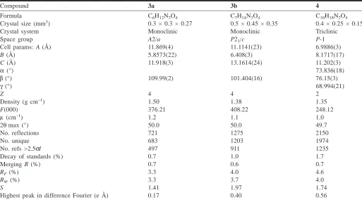
Compound 3a 3b 4
Formula C6H12N2O4 C7H14N2O4 C10H18N2O4
Crystal size (mm3) 0.3 × 0.3 × 0.27 0.5 × 0.45 × 0.35 0.4 × 0.25 × 0.15
Crystal system Monoclinic Monoclinic Triclinic
Space group A2/a P21/c P-1
Cell params: A (Å) 11.869(4) 11.1141(23) 6.9886(3)
B (Å) 5.8573(22) 6.408(3) 8.1717(17)
C (Å) 11.918(3) 13.1614(24) 11.202(3)
α(°) 73.836(18)
β(°) 109.99(2) 101.404(16) 76.15(3)
γ(°) 68.994(21)
Z 4 4 2
Density (g cm–1) 1.50 1.38 1.35
F(000) 376.21 408.22 248.12
µ (cm–1) 1.2 1.1 1.0
2θmax (°) 50.0 50.0 49.7
No. reflections 721 1275 2150
No. unique 683 1203 1974
No. refs >2.5σI 497 911 1235
Decay of standards (%) 0.7 1.0 1.7
Merging R (%) 0.7 0.6 0.7
RF(%) 3.3 4.0 4.6
RW(%) 3.3 3.7 4.0
S 1.41 1.97 1.74
[image:3.612.48.561.83.367.2]Highest peak in difference Fourier (e Å) 0.17 0.40 0.56
Table 1. Crystallographic data.
4. Anisotropic thermal parameters, torsion angles, and
least-squares planes are included as supplementary material.5
Results and discussion
Properties of the cage triperoxide 2 and of the cage diperoxides 3a, 3b, and 4
By analogy with the reaction of ammonia with formalde-hyde and hydrogen peroxide to give 2, we assumed that
re-action of 1,2-diaminoethane would give the diperoxide 3a, of 1,2-diaminopropane would give 3b, and of 1,2-diamino-cyclohexane would give 4. These assumptions proved to be true. The triperoxide 2 is a powerful explosive (11) and should be handled with extreme caution. A small amount of it on a steel plate detonates with a loud report when hit with a ham-mer. The diperoxide 3a proved to be more difficult to deto-nate, and the diperoxide 3 even more so, as expected from their progressively less favourable oxygen balances (12).
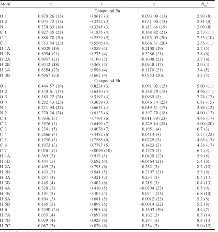
Atom x y z Beqb
Compound 3a
O 1 0.878 26 (13) 0.0617 (3) 0.093 09 (13) 2.89 (8)
O 2 0.945 72 (13) 0.2322 (3) 0.051 90 (13) 2.81 (8)
N 0.730 63 (16) 0.3345 (3) 0.113 64 (15) 2.09 (8)
C 1 0.827 35 (22) 0.1855 (4) 0.168 82 (21) 2.73 (11)
C 2 0.888 78 (20) 0.2529 (5) –0.075 05 (20) 2.55 (10)
C 3 0.753 54 (23) 0.5505 (4) 0.066 31 (20) 2.55 (11)
H 1A 0.8029 (19) 0.059 (4) 0.2100 (19) 2.7 (5)
H 1B 0.8954 (21) 0.275 (4) 0.2286 (21) 3.8 (6)
H 2A 0.8957 (21) 0.100 (5) –0.1098 (21) 3.7 (6)
H 2B 0.9442 (18) 0.368 (4) –0.0948 (17) 2.0 (4)
H 3A 0.8354 (22) 0.596 (4) 0.1176 (21) 3.4 (5)
H 3B 0.6967 (20) 0.662 (4) 0.0753 (20). 3.2 (5)
Compound 3b
O 1 0.444 57 (19) 0.8234 (3) 0.091 02 (15) 5.00 (11)
O 2 0.470 61 (17) 0.6330 (4) 0.148 79 (15) 4.96 (11)
O 3 0.185 22 (24) 0.3197 (4) 0.0935 (3) 7.74 (17)
O 4 0.292 43 (23) 0.3059 (3) 0.046 74 (23) 6.81 (15)
N 1 0.271 94 (22) 0.6634 (4) –0.019 51 (17) 3.86 (12)
N 2 0.270 24 (24) 0.6123 (4) 0.197 70 (18) 4.00 (12)
C 1 0.3836 (3) 0.7704 (6) –0.011 39 (23) 4.46 (17)
C 2 0.3976 (3) 0.6469 (7) 0.229 14 (25) 5.00 (20)
C 3 0.2261 (5) 0.4078 (7) 0.1951 (4) 6.7 (3)
C 4 0.2666 (4) 0.4482 (6) –0.0414 (3) 5.77 (22)
C 5 0.1750 (3) 0.7588 (6) 0.0229 (3) 4.65 (17)
C 6 0.1973 (3) 0.7787 (5) 0.1423 (3) 4.36 (17)
C 7 0.0761 (4) 0.8096 (10) 0.1775 (5) 6.7 (3)
H 1A 0.369 (3) 0.917 (5) –0.0420 (22) 5.9 (8)
H 1B 0.444 (3) 0.692 (4) –0.0464 (21) 5.4 (8)
H 2A 0.409 (3) 0.795 (6) 0.252 (3) 8.2 (12)
H 2B 0.433 (3) 0.541 (5) 0.2797 (21) 5.3 (8)
H 3A 0.294 (4) 0.321 (7) 0.235 (3) 10.4 (14)
H 3B 0.145 (4) 0.402 (6) 0.215 (3) 10.4 (13)
H 4A 0.328 (3) 0.410 (5) –0.0794 (23) 6.5 (9)
H 4B 0.191 (3) 0.405 (5) –0.0741 (24) 6.8 (10)
H 5A 0.104 (3) 0.681 (5) 0.0012 (22) 5.2 (8)
H 5B 0.165 (3) 0.899 (5) –0.0014 (22) 5.2 (8)
H 6 0.2490 (24) 0.908 (4) 0.1603 (19) 4.4 (7)
H 7A 0.035 (4) 0.693 (6) 0.162 (3) 8.5 (14)
H 7B 0.039 (4) 0.928 (6) 0.144 (3) 8.8 (13)
H 7C 0.087 (3) 0.829 (6) 0.254 (3) 9.0 (12)
aesds refer to the last digit printed. bB
[image:4.612.84.535.83.562.2]eqis the mean of the principal axes of the thermal ellipsoid for atoms refined anisotropically (non-hydrogens). For hydrogens, Beq= Biso.
Table 2. Final atomic coordinates (fractional) and equivalent isotropic thermal parameters.a
5Supplementary material mentioned in the text may be purchased from: The Depository of Unpublished Data, Document Delivery, CISTI,
The diperoxide 4 could not be detonated, and was submitted for combustion analysis in the usual way. The peroxides 3a and 3b were analyzed (along with 2 as a control) by decom-position of a small amount of the compound in a large ex-cess of aqueous acid to yield formaldehyde (analyzed by formation of the derivative with dimedone (13, 14)) and hy-drogen peroxide (titrated by an iodide–thiosulfate method (15)) in amounts close to theoretical. Treatment of the per-oxides with aqueous picric acid did not give their picrate salts, but rather monopicrates of the diamines from which they had been made, and treatment of a very small amount of the solid peroxide with a drop of concentrated hydrochlo-ric acid caused instantaneous decomposition to a sticky liq-uid. The reaction was particularly violent in the case of 2. The solid compounds left standing on filter paper in the lab-oratory atmosphere eventually decomposed to a sticky mass. It seems likely that this decomposition was initiated by a trace of acid furnished by a dust or aerosol particle; the formaldehyde and hydrogen peroxide produced at the initial
spot of decomposition would react to give formic acid, so that the decomposition would be autocatalytic.
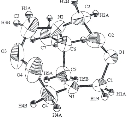
Diffraction studies
The structures 3a, 3b, and 4 were established by X-ray diffraction.5 ORTEP representations (16) are given in
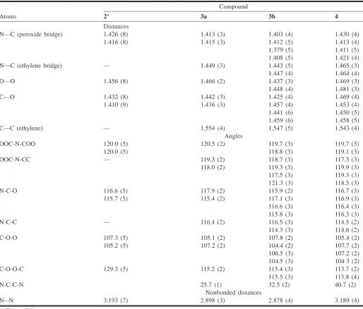
Figs. 1– 3, and in Table 4, the bond distances and angles of the three diperoxides are compared with corresponding bond distances and angles of the triperoxide 2.
The torsional angles of the peroxide groups C-O-O-C in our three diperoxides lie between 113 and 116°, close to the equilibrium torsional angle of about 112° in hydrogen perox-ide (17), while the corresponding torsional angle in HMTD is 129.3° (9). Schaefer et al. (9) point out that it takes little energy to open this angle from 112 to 129°, while closing this angle below 129° in HMTD would force nonbonded N···N atoms (3.193 Å apart) and O···O atoms (2.971 Å apart) to move closer together, with steep increases in van der Waals repulsions. Otherwise, most bond distances and
an-Atom x y z Beqb
O 1 0.1566 (4) 0.1551 (3) 0.010 54 (20) 4.41 (13)
O 2 0.2522 (4) 0.2910 (3) –0.000 24 (21) 4.70 (13)
O 3 0.1744 (4) 0.1115 (3) –0.313 73 (23) 5.31 (15)
O 4 0.3104 (4) 0.0022 (3) –0.218 19 (22) 5.14 (14)
N 1 –0.0245 (4) 0.3144 (3) –0.172 88 (22) 3.06 (12)
N 2 0.4214 (4) 0.2616 (3) –0.212 87 (23) 3.43 (13)
C 1 –0.0392 (6) 0.2540 (5) –0.0393 (3) 3.99 (18)
C 2 0.4434 (6) 0.2559 (5) –0.0919 (3) 4.28 (19)
C 3 –0.0249 (6) 0.2002 (5) –0.2470 (4) 4.33 (20)
C 4 0.4757 (6) 0.0829 (5) –0.2457 (4) 4.54 (20)
C 5 0.0578 (4) 0.4648 (4) –0.2308 (3) 2.75 (15)
C 6 0.2897 (5) 0.4099 (4) –0.2883 (3) 3.03 (15)
C 7 0.3635 (5) 0.5741 (4) –0.3203 (3) 3.66 (17)
C 8 0.2382 (6) 0.7245 (4) –0.4144 (3) 4.14 (19)
C 9 0.0078 (6) 0.7725 (4) –0.3661 (4) 4.33 (19)
C 10 –0.0683 (6) 0.6087 (4) –0.3274 (3) 3.72 (18)
H 1A –0.117 (4) 0.354 (3) 0.0019 (23) 2.8 (6)
H 1B –0.119 (4) 0.170 (4) –0.007 (3) 4.1 (7)
H 2A 0.469 (4) 0.360 (4) –0.081 (3) 4.0 (7)
H 2B 0.548 (5) 0.140 (4) –0.057 (3) 4.5 (7)
H 3A –0.077 (4) 0.260 (3) –0.329 (3) 3.5 (7)
H 3B –0.091 (5) 0.107 (4) –0193 (3) 6.4 (9)
H 4A 0.509 (5) 0.093 (4) –0.338 (3) 4.1 (7)
H 4B 0.574 (5) –0.001 (4) –0.194 (3) 3.8 (7)
H 5 0.039 (3) 0.520 (3) –0.1626 (20) 1.1 (5)
H 6 0.302 (4) 0.378 (3) –0.3685 (25) 3.1 (6)
H 7A 0.505 (4) 0.547 (3) –0.3530 (24) 3.0 (6)
H 7B 0.347 (4) 0.616 (4) –0.240 (3) 4.1 (7)
H 8A 0.273 (5) 0.684 (4) –0.499 (3) 5.5 (8)
H 8B 0.289 (4) 0.822 (4) –0.433 (3) 4.2 (8)
H 9A –0.068 (5) 0.855 (4) –0.429 (3) 5.9 (9)
H 9B –0.024 (4) 0.828 (4) –0.289 (3) 4.1 (7)
H 10A –0.206 (5) 0.639 (4) –0.296 (3) 4.1 (8)
H 10B –0.053 (4) 0.559 (3) –0.407 (3) 4.0 (7)
aesds refer to the last digit printed. bB
[image:5.612.85.526.84.479.2]gles are fairly similar in all four compounds in Table 1. However, the bridgehead nitrogen atoms of the three new diperoxides are not completely planar, lying about 0.12–0.14 Å out of the plane of the three attached carbon atoms, while in 2 the nitrogen atoms lie exactly (within experimental lim-its) in this plane. Similarly, in the three new diperoxides all except one of the C-N-C angles are slightly less than 120°, whereas in 2 all are exactly 120°.
Schaefer et al. (9) suggested that the bridgehead nitrogen atoms of 2 are sp2 hybridized because the electronegative
peroxide groups withdraw electron density and lower the
en-ergy of the electron pair in the p orbital (the anomeric effect (19)). This hybridization explains the shortened N—C dis-tances in 2. Essentially, the same explanation is offered by Whittleton et al. (19) for N—C distances in the cage diperoxide formed by reaction of formaldehyde with hydrazine and hydrogen peroxide in acidic solution (20, 21). This effect would be expected to be greater in the triperoxide 2 than in the diperoxides 3a, 3b, and 4, which might account for the less complete planarity of the bridge-head nitrogen atoms of the latter. Furthermore, the anomeric effect would explain why the N—C bonds of the peroxide bridges of 3a, 3b, and 4 are shortened, while the N—C bonds of the alkane bridges are not. It would also explain the nonbasic character of the tertiary amino groups, so that the compounds separate out of weakly acidic solutions as bases and not salts.
PMR spectrum of HMTD 2
The 1H NMR spectrum of the highly symmetrical
hexamethylenetetramine (CH2)6N4 shows a sharp singlet
atδ4.64 ppm (22), and a similar spectrum would be expected for HMTD (CH2)6N2O6 if it had the symmetrical structure
shown in 2 above, with dihedral angles of zero for the per-oxide bridges CH2-O-O-CH2. However, the dihedral angles
of ±129° of these bridges (9) result in HMTD existing as a racemic mixture of chiral helical conformers. The twist of the helix causes one hydrogen of each methylene group to point slightly inward toward the axis of the molecule, and the other slightly outward. The six methylene groups are all identical; each forms an AB system, so that the spectrum of HMTD in DMSO-d6, shown in Fig. 4, has a pair of doublets
(2J 13.3 Hz) centered atδ4.65 and 4.78 ppm. At higher
[image:6.612.329.553.105.361.2]tem-peratures, the helical conformers interconvert, as shown by the broadening of the NMR peaks, which eventually
Fig. 1. ORTEP plot (16) of compound 3a,showing numbering scheme. Ellipsoids are shown at 50% probability for non-hydrogen atoms.
[image:6.612.57.288.108.331.2]Fig. 2. ORTEP plot (16) of compound 3b,showing numbering scheme. Ellipsoids are shown at 50% probability for non-hydrogen atoms.
[image:6.612.51.294.382.608.2]collapse into a broad singlet at about 105°C, illustrated in Fig. 4.
NMR spectrum of compound 3a
This compound, like 2, is a racemic mixture of chiral con-formers. The X-ray crystallographic studies (see Fig. 1) show that the dihedral angles of CH2-O-O-CH2 are not zero, as
represented in 3a, but ±115.2° (Table 4). Again, the hydro-gen atoms of both N-CH2-C and N-CH2-O methylenes form
an AB system. However, in CDCl3 at 30°C, the 1H NMR
spectrum, shown in Fig. 5, is that of the symmetrical struc-ture 3a, having a doublet at δ3.38 (4 H) for the protons of the ethylene bridge and a doublet at δ 4.83 (8 H) for the methylene protons of the peroxide bridges. The four-bond coupling is favoured by the W-configuration of the bonds between the protons of the two different types of methylenes.
Evidently, the interconversion of enantiomeric conformers of 3a, which requires eclipsing of the bonds of two peroxide
groups, takes place at a lower temperature than the interconversion of the enantiomeric conformers of 2, which requires eclipsing of the bonds of three peroxide groups. The
Fig. 4. Effect of temperature on the PMR spectrum of HMTD (2). Compound
Atoms 2a 3a 3b 4
Distances
N—C (peroxide bridge) 1.426 (8) 1.413 (3) 1.403 (4) 1.430 (4)
1.416 (8) 1.415 (3) 1.412 (5) 1.413 (4)
1.379 (5) 1.411 (5) 1.408 (5) 1.421 (4)
N—C (ethylene bridge) — 1.449 (3) 1.443 (5) 1.465 (3)
1.447 (4) 1.464 (4)
O—O 1.456 (8) 1.466 (2) 1.437 (3) 1.469 (3)
1.448 (4) 1.481 (3)
C—O 1.432 (8) 1.442 (3) 1.425 (4) 1.469 (4)
1.410 (9) 1.436 (3) 1.457 (4) 1.453 (4)
1.441 (6) 1.450 (5) 1.459 (6) 1.458 (5)
C—C (ethylene) — 1.554 (4) 1.547 (5) 1.543 (4)
Angles
OOC-N-COO 120.0 (5) 120.5 (2) 119.7 (3) 119.7 (3)
120.0 (5) 118.8 (3) 119.1 (3)
OOC-N-CC — 119.3 (2) 118.7 (3) 117.3 (3)
118.0 (2) 119.3 (3) 119.9 (3)
117.5 (3) 119.3 (3) 121.3 (3) 118.5 (3)
N-C-O 116.6 (5) 117.9 (2) 115.9 (2) 116.7 (3)
115.7 (5) 115.4 (2) 117.1 (3) 116.9 (3)
116.6 (3) 116.4 (3) 115.8 (3) 116.3 (3)
N-C-C — 116.4 (2) 116.5 (3) 114.5 (2)
114.3 (3) 114.6 (2)
C-O-O 107.3 (5) 105.1 (2) 107.8 (2) 105.4 (2)
105.2 (5) 107.2 (2) 104.4 (2) 107.7 (2)
106.5 (3) 107.2 (2) 104.5 (3) 104 3 (2)
C-O-O-C 129.3 (5) 115.2 (2) 115.4 (3) 113.7 (2)
115.5 (3) 115.8 (4)
N-C-C-N 25.7 (1) 32.5 (2) 40.7 (2)
Nonbonded distances
N···N 3.193 (7) 2.898 (3) 2.878 (4) 3.189 (4)
[image:7.612.322.561.556.695.2]aFrom ref. 9.
interconversion of 3a requires also the eclipsing of the at-oms of the ethylene bridge, but this is already more than half complete in the ground state of the molecule: the X-ray data (Table 4) show that the dihedral angle for N-CH2-CH2-N is
not about 60°, as required for a staggered conformation, but 25.7° (Table 4).
At lower temperatures, the rate of interconversion drops, as evidenced by the changes in NMR spectra shown in Fig. 5. At –30°C, the 1H peaks of N-CH
2-O (carbon atoms
C1, C′1, C2, C′2) can be calculated as two AB quartets, one at δ= 4.66 and 4.88 ppm (2J = 13.5 Hz), the other at δ=
4.70 and 4.77 ppm (2J = 12.1 Hz). The high field doublet in
each quartet shows a long-range splitting (4J = 2 Hz) due to
coupling to one of the ethylene protons. The spectrum for ethylene (C3 and C′3) shows symmetrical multiplets at δ= 3.347 and 3.227 ppm, and additional splitting from long-range coupling.
NMR spectrum of compound 3b
This compound does not show a rapid interconversion of conformers at 30°C, so that its1H NMR spectrum has a
de-tailed structure not found in the spectrum of 3a until the temperature of the latter has been reduced to about –20°C. The methyl group of 3b is attached to the two-carbon bridge
by a bond pointing outward; a conformational inversion would end with this bond pointing inward, into a region of steric congestion, and so is not possible. This steric effect also explains why 2-methyl-1,2-diaminopropane does not re-act with formaldehyde and hydrogen peroxide to form the cage diperoxide 3c.
The 1H NMR spectrum of compound 3b (Fig. 6; see also Table 5) corroborates the details of structure established by X-ray diffraction and shown in the ORTEP diagram of Fig 2. The signal for the three methyl protons (1.16 ppm) at posi-tion 7 is split by coupling (3J 6.5 Hz) to the single proton at
position 6 (m, 3.61 ppm). This proton in turn is coupled to the A (3J 14 Hz) and B (3J 7 Hz) protons at C5. The
proxim-ity of the C5 B proton to the C6 proton is shown by the NOESY spectrum.
The signals betweenδ4.6 and 5.0 ppm in Fig. 6 are due to the protons at positions 1, 2, 3, and 4. The assignment of signals to positions 1–4 is possible because of their spin-coupling to the protons at positions 5, 6, and 7, as demon-strated by the COSY spectrum. The results are given in Ta-ble 5. The rigid cage structure of the molecule results in many pairs of protons being linked by four bonds in a W-configuration (see Table 5), and hence having measurable coupling constants.
The spectrum of 3b in DMSO-d6 has signals shifted very
slightly to higher fields, but is essentially identical to the spectrum in CDCl3.
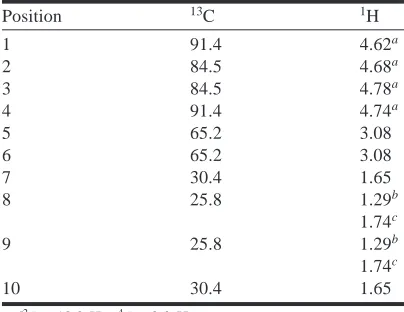
NMR spectrum of compound 4
The trans fusion of a cyclohexane ring to the diperoxide ring of 4 results in a rigid compound incapable of any conformational change. Its 1H and13C NMR (Table 6) and
COSY and NOESY spectra are those expected from the structure (Fig. 3) established by X-ray diffraction, and re-quire no comment.
Formaldehyde and cage compounds
[image:8.612.315.565.82.236.2]The results reported above illustrate the proclivity of formaldehyde to form cage compounds (see also refs. 19, 20, 23–26). A further example is given by the reaction of formaldehyde with trans-1,2-diaminocyclohexane to give the compound 5, analogous to cage compounds given by other 1,2-diamines (23, 24, 27). cis-1,2-Diaminocyclohexane does not react in this way, presumably for steric reasons. The reaction of formaldehyde with 1,2-diaminoethane gives
[image:8.612.49.294.86.470.2]Fig. 5. Effect of temperature on the PMR spectrum of 3a.
Fig. 6. The PMR spectrum of 3b.
Position 13Cδ(ppm) 1Hδ(ppm) COSY
1 89.4 A 4.689 d of d 1A, 1B, 4B B 4.868 d of d 1B, 1A, 5A
2 91.5 A 4.633 d 2A, 2B, 3B, 6
B 4.818 d 2B, 2A
3 84.3 A 4.720 d 3A, 3B
B 4.984 d of d 3B, 3A, 2A, 6
4 91.8 A 4.824 d 4A, 4B, 5B
B 4.616 d of d 4B, 4A, 1A
5 58.4 A 2.955 t 5A, 5B, 6, 1B
B 3.239 m 5B, 5A, 6, 4A
6 56.3 –3.612 m 6, 5A, 5B, 7, 3A, 2A
[image:8.612.46.297.89.258.2]7 15.6 –1.160 d 7, 6
the compound 6, which may have the two methylenes of the ethylene bridge eclipsed (as represented in 6) or staggered: the X-ray evidence is equivocal (28). However, the fused cyclohexane ring enforces staggering of the corresponding carbon atoms in 5. Dreiding-type models show that only two enantiomeric forms of this compound are possible.
General conclusions
Small cycloalkane rings of 5 or 6 carbon atoms are more stable than medium-sized rings of 7–10 carbon atoms, be-cause of steric and torsional effects. This is not true when the rings incorporate a peroxide group: medium-sized rings then become more stable than the five- or six-membered, be-cause of the differing energies attendant on change of tor-sional angle for C-O-O-C and C-C-C-C chains. This fact has not often been recognized (cf. refs. 19–21, 27, 29). The structures 3a, 3b, and 4, containing fused eight-membered rings, furnish further examples of this rule; alternative struc-tures containing the peroxide groups in five-membered rings can be written for these compounds and for HMTD (20).
Acknowledgment
The financial support of the Natural Sciences and Engi-neering Research Council of Canada is gratefully acknowl-edged. We are also grateful to Prof. R.B. Lennox and his
students for help and encouragement, to Bernadette Macdon-ald for analytical work, and to Dr. Anne-Marie Lebuis.
References
1. R.W. Alder, R.B. Sessions, A.J. Bennett, and R.E. Moss. J. Chem. Soc. Perkin Trans. 1, 603 (1982).
2. R.W. Alder. Acc. Chem. Res. 16, 321 (1983). 3. A.v. Baeyer and A. Villiger. Ber. 33, 2485 (1900).
4. R.W. Alder, A.G. Orpen, and R.W. Sessions. J. Chem. Soc. Chem. Commun. 999 (1983).
5. L. Pauling. The nature of the chemical bond. 2nd ed. Cornell University Press, Ithaca, N.Y. 1948. p. 189.
6. A. Bondi. J. Phys. Chem. 68, 441 (1964).
7. A. Gavezzotti. J. Am. Chem. Soc. 105, 5220 (1983). 8. A.I. Kitaigorodski. Molecular crystals and molecules.
Aca-demic Press, London. 1973. pp. 1–21.
9. W.P. Schaefer, J.T. Fourkas, and B.G. Tieman. J. Am. Chem. Soc. 107, 2461 (1985).
10. E.J. Gabe, Y. LePage, J.P. Charland, F.I. Lee, and P.S. White. J. Appl. Cryst. 22, 384 (1989).
11. C.A. Taylor and W.H. Rinkenbach. Army Ordnance, 5, 463 (1924).
12. W.C. Lothrop and G.R. Hendrick. Chem. Rev. 44, 419 (1949). 13. J.H. Yoe and L.C. Reid. Ind. Eng. Chem. 13, 238 (1981). 14. D. Spencer and T. Henshall. J. Am. Chem. Soc. 77, 1943
(1955).
15. E. Schmitz. Liebigs Ann. Chem. 635, 73 (1960).
16. C.K. Johnson. ORTEP - a Fortran thermal ellipsoid plot pro-gram. Technical report ORNL-5138. Oak Ridge, Tenn. 1976. 17. R.M. Hunt, R.A. Leacock, C.W. Peters, and K.T. Hecht. J.
Chem. Phys. 42, 193 (1979).
18. A.J. Kirby. The anomeric effect and related stereoelectronic ef-fects at oxygen. Springer Verlag, Berlin. 1983.
19. S.N. Whittleton, P. Seiler, and J.D. Dunitz. Helv. Chem. Acta, 64, 2614 (1981).
20. C.v. Girsewald and H. Siegens. Ber. 54, 492 (1921). 21. E. Schmitz. Liebigs Ann. Chem. 636, 73 (1960).
22. C.J. Pouchert. The Aldrich library of NMR spectra. 2nd ed. Vol. 1. Aldrich Chemical Co., Milwaukee. 1983. p. 332d. 23. G. Volpp. Chem. Ber. 95, 1493 (1962).
24. F.G. Riddell and P. Murray-Rust. Chem. Comm. 1075 (1970). 25. E. Schmitz and R. Ohme. Liebigs Ann. Chem. 635, 82 (1960). 26. M.J.S. Dewar. A semiempirical life. A.C.S., Washington, D.C.
1992. p. 103.
27. C.A. Bischoff. Ber. 31, 3248 (1898).
28. P. Murray-Rust. J. Chem. Soc. Perkin Trans. 2, 1136 (1974). 29. A.R. Katritzky, V.J. Baker, F.M.S. Brito-Palma, J.M. Sullivan,
and R.B. Finzel. J. Chem. Soc. Perkin Trans. 2, 1133 (1979).
Position 13C 1H
1 91.4 4.62a
2 84.5 4.68a
3 84.5 4.78a
4 91.4 4.74a
5 65.2 3.08
6 65.2 3.08
7 30.4 1.65
8 25.8 1.29b
1.74c
9 25.8 1.29b
1.74c
10 30.4 1.65
a2J = 13.0 Hz,4J = 2.0 Hz.
[image:9.612.71.273.81.237.2]bAxial hydrogen. cEquatorial hydrogen.