ORIGINAL RESEARCH
BRAIN
Altered Microstructure in Temporal Lobe Epilepsy:
A Diffusional Kurtosis Imaging Study
L. Bonilha, C.-Y. Lee, J.H. Jensen, A. Tabesh, M.V. Spampinato, J.C. Edwards, J. Breedlove, and J.A. Helpern
ABSTRACT
BACKGROUND AND PURPOSE: Temporal lobe epilepsy is associated with regional abnormalities in tissue microstructure, as demon-strated by DTI. However, the full extent of these abnormalities has not yet been defined because DTI conveys only a fraction of the information potentially accessible with diffusion MR imaging. In this study, we assessed the added value of diffusional kurtosis imaging, an extension of DTI, to evaluate microstructural abnormalities in patients with temporal lobe epilepsy.
MATERIALS AND METHODS: Thirty-two patients with left temporal lobe epilepsy and 36 matched healthy subjects underwent diffusion MR imaging. To evaluate abnormalities in patients, we performed voxelwise analyses, assessing DTI-derived mean diffusivity, fractional anisotropy, and diffusional kurtosis imaging– derived mean diffusional kurtosis, as well as diffusional kurtosis imaging and DTI-derived axial and radial components, comparing patients with controls.
RESULTS:We replicated findings from previous studies demonstrating a reduction in fractional anisotropy and an increase in mean diffusivity preferentially affecting, but not restricted to, the temporal lobe ipsilateral to seizure onset. We also noted a pronounced pattern of diffusional kurtosis imaging abnormalities in gray and white matter tissues, often extending into regions that were not detected as abnormal by DTI measures.
CONCLUSIONS: Diffusional kurtosis is a sensitive and complementary measure of microstructural compromise in patients with temporal lobe epilepsy. It provides additional information regarding the anatomic distribution and degree of damage in this condition. Diffusional kurtosis imaging may be used as a biomarker for disease severity, clinical phenotypes, and treatment moni-toring in epilepsy.
ABBREVIATIONS:DKI⫽diffusional kurtosis imaging; FA⫽fractional anisotropy; MD⫽mean diffusivity; TLE⫽temporal lobe epilepsy
T
he most common histologic finding in patients with medial temporal lobe epilepsy (TLE) is hippocampal sclerosis, which is defined as neuronal loss and gliosis involving the hippocam-pus.1-3Routine clinical MR imaging of patients with TLE candemonstrate signs associated with hippocampal sclerosis.4,5
Recent observations suggest that hippocampal abnormalities are not the only structural injury in TLE.6-9Imaging studies
using whole-brain quantitative morphometry have repeatedly demonstrated that TLE is associated with extrahippocampal abnormalities6,8-13involving perihippocampal and perilimbic
structures.
However, the full extent of structural abnormalities in TLE is still unclear. Therefore, the lack of a sensitive and specific marker of extrahippocampal damage may prevent the assessment of its clinical relevance. Diffusion MR imaging techniques aimed at quantifying tissue microstructure may offer more sensitive tools for determining the extent of brain pathology in TLE. A promis-ing new method called diffusional kurtosis imagpromis-ing (DKI) can provide information about cerebral microstructural abnormali-ties beyond that provided by conventional diffusion tensor imag-ing.14,15In fact, by providing a more comprehensive
characteriza-tion of water diffusion properties, DKI may be more suitable for detecting subtle brain damage in TLE.
In this study, we investigated the anatomical pattern of micro-structural abnormalities associated with TLE by contrasting the
Received July 12, 2014; accepted after revision October 19.
From the Departments of Neurology and Neurosurgery (L.B., J.C.E.) and Radiol-ogy and Radiological Science (C.-Y.L., J.H.J., A.T., M.V.S., J.A.H.), Comprehensive Epilepsy Center (L.B., J.C.E., J.B.), and Center for Biomedical Imaging (L.B., C.-Y.L., J.H.J., A.T., M.V.S.), Medical University of South Carolina, Charleston, South Carolina.
This study was supported by the American Society of Neuroradiology (grant No. 89779-01; Principal Investigator, A. Tabesh).
Please address correspondence to Leonardo Bonilha, MD, PhD, Department of Neurology and Neurosurgery, Medical University of South Carolina, 96 Jonathan Lucas St, 3rd floor CSB, Charleston, SC 29425; e-mail: [email protected]
Indicates article with supplemental on-line table.
voxel-based analyses of microstructure maps derived from DKI and from well-established DTI.
MATERIALS AND METHODS
Subjects
Thirty-two consecutive patients with left TLE were recruited from the Comprehensive Epilepsy Center at the Medical University of South Carolina, where they were diagnosed on the basis of a com-prehensive history and neurologic evaluation. The TLE diagnosis was defined in accordance with the criteria proposed by the Inter-national League Against Epilepsy,16based on a history compatible
with partial seizures with temporal onset, interictal epileptiform discharges on interictal electroencephalography, and hippocam-pal atrophy on visual inspection of MR imaging. Not all patients included in this study were surgical candidates; thus, ictal record-ing was not obtained in all patients. All patients exhibited a classic presentation for TLE, based on history, seizure semiology, epilep-tiform discharges on interictal electroencephalography, as well as routine MRI. Most patients, but not all, exhibited signs of hip-pocampal sclerosis on MR imaging, and a detailed description of the demographic and clinical information for patients included in this study is provided in the On-line Table.
The mean age of patients was 44.8⫾16.7 years; 22 patients were women. We also studied a control group of healthy individ-uals recruited from the local community who had no history of neurologic problems and no risk factors for epilepsy (mean age, 40.4⫾11.6 years; 24 women). Patients and controls were similar in sex (P⫽.21) and age (P⫽.92) distributions.
Image Acquisition
All patients and controls underwent the same imaging protocol performed on a 3T Magnetom Verio MR imaging scanner (Sie-mens, Erlangen, Germany) equipped with a 12-channel head coil. Diffusion-weighted images were obtained by using a twice-refo-cused echo-planar sequence with 3 diffusion weightings (b⫽0, 1000, and 2000 s/mm2) along 30 diffusion-encoding directions
with NEX⫽1 (NEX⫽10 forb⫽0). Other imaging parameters were TR⫽8500 ms, TE⫽98 ms, FOV⫽222⫻222 mm2, matrix
size⫽74⫻74, parallel imaging factor⫽2, no partial Fourier encoding, section thickness⫽3 mm, and 40 axial sections. Ac-quisition time was 9 minutes and 12 seconds. Structural images were obtained by using a magnetization-prepared rapid acquisi-tion of gradient echo sequence with TR⫽2250 ms, TE⫽4.18 ms, TI⫽900 ms, FOV⫽256⫻256 mm2, matrix size⫽256⫻256,
section thickness⫽1 mm, and 176 sagittal sections.
Image Processing
Tissue Volume Maps. T1-weighted images were submitted to gray and white matter tissue segmentation using the VBM toolbox (http://dbm.neuro.uni-jena.de/vbm/) for SPM8 software (http:// www.fil.ion.ucl.ac.uk/spm/software/spm8). Image normalization and segmentation were performed iteratively by using the sym-metric tissue probability map of VBM, very light regularization, and affine regularization to the ICBM space template (European Brains; http://bmap.ucla.edu/portfolio/atlases/ICBM_Template/). Spa-tial normalization was performed with affine and nonlinear transformations yielding modulated normalized gray and white
matter probability maps. All maps were transformed into stan-dard space, and their averages (gray and white matter maps sep-arately) were used to provide tissue inclusion masks for statistical analyses.
Microstructure Maps. DKI postprocessing was performed by us-ing the in-house software Diffusional Kurtosis Estimator (http:// www.nitrc.org/projects/dke),17 which performed the following
processing steps to generate diffusivity and kurtosis maps: first, motion correction through a 6-parameter rigid-body transforma-tion to spatially align all DWIs. Second, at each voxel, the diffu-sion and diffudiffu-sional kurtosis tensors were jointly fitted to the DWIs forb⫽0, 1000, and 2000 s/mm2for that voxel, and the
mean kurtosis was calculated from the tensors. The diffusion ten-sor was fitted to the DWIs forb⫽0 and 1000 s/mm2, and mean
diffusivity and (MD) fractional anisotropy (FA) were calculated from the diffusion tensor. Axial and radial diffusion and kurtosis maps were also generated.
FA maps were nonlinearly normalized to a common space by using the fMRI of the Brain Software Library, Version 4.1.7 (http://www.fmrib.ox.ac.uk/fsl) using a symmetric FA template, and the resulting transformation was applied to normalize the other maps. The spatially normalized images were then submitted to spatial smoothing with an 8-mm Gaussian filter.
Statistical Analyses. We performed whole-brain voxelwise anal-yses, comparing patients with TLE and controls regarding MD, FA, mean kurtosis, axial diffusion, radial diffusion, axial kurtosis, and radial kurtosis maps. We performed comparisons using vox-elwisettests. All results were corrected for multiple comparisons using a false discovery rate threshold ofq⬍0.01.18
RESULTS
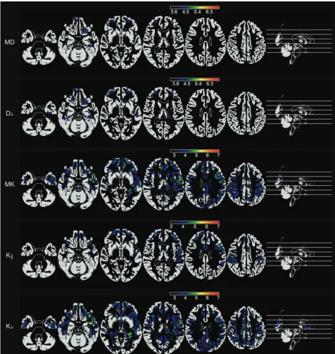
Gray Matter
Patients with TLE exhibited multiple cortical areas with signifi-cant increase in diffusion parameters, along with a concurrent decrease in kurtosis parameters.
Specifically, an increase in MD was observed within the or-bitofrontal cortices, frontal poles, medial left temporal lobe re-gion, and midposterior cingulate. No significant changes were observed with axial diffusion, but radial diffusion was increased in patients in the orbitofrontal regions, frontal poles, medial left temporal regions, left lateral temporal regions, and midposterior cingulate cortex.
In contrast, at the same statistical threshold level, a more dif-fuse pattern of significant reductions in mean kurtosis was ob-served, encompassing the temporopolar cortices, temporal and frontal opercula, medial temporal lobe regions (more pro-nounced on the left side), left lateral temporal region, prefrontal cortices and temporal poles, and cingulate and parietal regions. Overall, a higher statistical difference was noted on the left side.
Reductions in axial kurtosis were noted particularly over the frontal polar regions, left temporal pole, left lateral temporal cortex, and left precentral areas. Reductions in radial kurtosis were diffusely distributed across the entire brain but were particularly intense over the left medial temporal cortex, left temporal polar cortex, left insula, left fusiform gyrus, cingulate cortex, and left precentral regions.
There were no cortical areas of reduced diffusion or increased kurtosis measures in patients.
White Matter
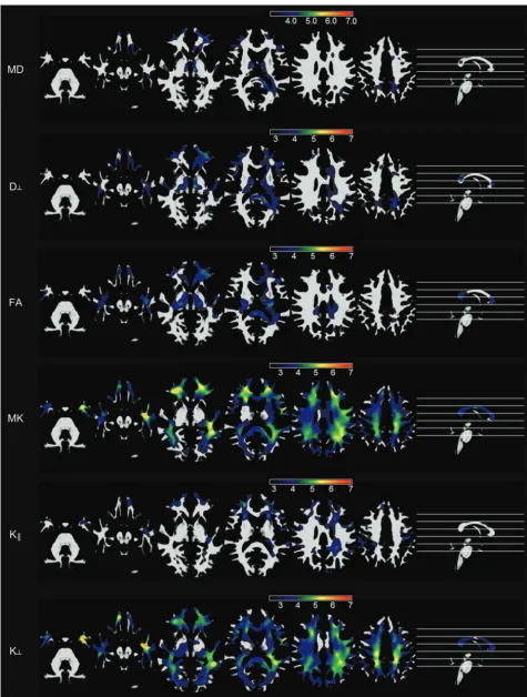
Patients with TLE demonstrated a significant increase in MD within the white matter in the frontal, left medial temporal, per-ithalamic, left occipitotemporal, and medial frontoparietal re-gions. There was also an increase in MD within the splenium of the corpus callosum. These abnormalities were also noted in areas
of radial diffusion decrement, while axial diffusion did not dem-onstrate significant abnormalities between groups.
FA decrement was noted in a widespread pattern involving the medial temporal, perithalamic, orbitofrontal, left temporopolar, left occipitotemporal, and medial frontoparietal white matter re-gions. There was also a notable decrement in FA within the sp-lenium of the corpus callosum, left side of the genu of the corpus callosum, and left uncinate fasciculus.
At the same statistical threshold, there was a remarkable
[image:3.594.55.532.47.550.2]tomical pattern of mean kurtosis reduction, which involved most of the brain white matter and was most pronounced over the left temporal, bilateral orbitofrontal, and left frontoparietal regions. These abnormalities were similar to the pattern noted with radial kurtosis decrement in patients.
These results are shown in Fig 2.
There were no areas of MD decrement, FA increase, or kurto-sis increase in patients.
DISCUSSION
In this study, we investigated microstructural gray and white mat-ter abnormalities in patients with TLE using a novel diffusion MR imaging technique called DKI. DKI is aimed at detecting changes in tissue microstructure, including those arising from non-Gauss-ian properties of water diffusion, thus providing a potentially more sensitive and specific tool for the evaluation of pathologic changes in TLE.14,15We contrasted the DKI-derived
microstruc-tural measures with the better established DTI-derived measures. We replicated findings from several previous studies using DTI to assess microstructural abnormalities in TLE. Specifically, we demonstrated areas of increased MD and reduced FA in pa-tients with TLE, which were preferentially located within, but not restricted to, the temporal lobe ipsilateral to the side of seizure onset.19-29
We observed that DKI is capable of detecting a broader ana-tomic pattern of microstructural abnormalities in patients with TLE, compared with conventional measures. Moreover, these re-sults demonstrate a distribution of network abnormalities that is in direct concordance with the hypothesized network pathology related to TLE.13,30The presence of abnormalities demonstrated
by DKI is more contiguous and anatomically plausible compared with conventional methodologies, and it is consistent with the theory of limbic and perilimbic dysfunction in TLE.31,32
In this study, we also corroborated the findings from Gao et al,33suggesting that DKI is more sensitive, compared with DTI, to
detect microstructural abnormalities in the patients with TLE. Gao et al studied a population of children with TLE; nonetheless, their results are fairly equivalent to our observations from a pop-ulation of adult subjects with TLE.
DKI and the Pathophysiology of TLE
The hypothesis that TLE is a disease that affects more than just the hippocampus has been corroborated by several imaging studies demonstrating abnormalities in TLE involving limbic struc-tures.6,8-13However, there is considerable variability across
stud-ies regarding the extent and degree of extrahippocampal damage in TLE. These variations may be directly related to the sensitivity of the methods, as further highlighted by the observation that conventional methods are not sensitive to most extrahippocam-pal histologic changes.34,35This variability in findings precludes a
better understanding of the pathophysiology of TLE.
Our results suggest that TLE is associated with extensive mi-crostructural abnormalities, encompassing a contiguous and ex-tensive network of extrahippocampal and extratemporal regions. Our results were obtained from a population of consecutive patients with TLE, most of whom demonstrated signs of hip-pocampal sclerosis on MR imaging (On-line Table). Therefore, it
is possible that the temporal and extratemporal microstructural abnormalities demonstrated by DKI are correlated with hip-pocampal cell loss. Future studies evaluating phenotypical differ-ences in the distribution of DKI abnormalities may provide fur-ther insight into this subject.
Moreover, the patient population evaluated in this study was not composed exclusively of surgical candidates (ie, it included patients with relatively well-controlled epilepsy). Most interest-ing, our strong statistical results suggest that microstructural ab-normalities were widespread across all subjects. While these find-ings support DKI as exquisitely sensitive to abnormalities associated with TLE, it is still unclear whether DKI can provide further information regarding classification into subgroups, par-ticularly as it relates to pharmacologic or surgical treatment out-comes. This study provides initial evidence of DKI as a biomarker of TLE, and we believe that future studies could address the im-pact of DKI as a clinical biomarker.
DKI as a Biomarker
The results of this study suggest that DKI may provide a sensitive and specific biomarker of TLE. Specifically, it may be a powerful tool for determining the degree of abnormality in cortical regions, which may be related to the clinical or neuropsychological pro-files, and for quantifying the burden of overall network abnor-malities on clinical progression. Specifically, extrahippocam-pal microstructural tissue abnormalities may be related to epileptogenesis in some individuals with TLE.31,36,37These
mi-crostructural changes may represent a complex interaction among multiple pathologic mechanisms such as cell loss, inflam-mation, and axonal and dendritic reorganization, which are known to occur in epilepsy.38
Clinical Feasibility of DKI
DKI provides a convenient platform for the evaluation of micro-structural abnormalities related to TLE and is one form of ad-vanced diffusion MR imaging.39However, DKI may be
advanta-geous to other methods due to its feasibility in routine clinical practice. Other modalities require significantly longer scan times and/or custom hardware. Conversely, DKI can be acquired with a high signal-to-noise ratio in approximately 6 –10 minutes, de-pending on the scanner.
Future Applications
This study provides preliminary evidence in support of the utility of DKI for characterization of microstructural abnormalities in TLE. Our results suggest that DKI– defined microstructural ab-normalities are pervasive in the hippocampal, temporal, and ex-tratemporal regions. Future studies should further investigate the histologic correlates of these abnormalities and their relationship with the clinical and neuropsychological profiles in patients with medial temporal lobe epilepsy.
CONCLUSIONS
DKI may provide complementary information regarding the lo-cation and magnitude of structural abnormalities in TLE.
Disclosures: Leonardo Bonilha—RELATED:Grant: American Society of Neuroradiol-ogy (grant No. 89779-01, Principal Investigator, A. Tabesh).* Jens H. Jensen—OTHER RELATIONSHIPS: Siemens owns a royalty-free nonexclusive license for diffusional kurtosis imaging with the pending patent held by New York University. I am one of the inventors. Maria V. Spampinato—UNRELATED:Grants/Grants Pending: Bracco,* Comments: clinical trial on 2 FDA-approved contrast agents. Ali Tabesh—RELATED: Grant: American Society of Neuroradiology,*Comments: 2013 Research Scientist Award. *Money paid to the institution.
REFERENCES
1. Sendrowski K, Sobaniec W.Hippocampus, hippocampal sclerosis and epilepsy.Pharmacol Rep2013;65:555– 65
2. Blu¨mcke I, Coras R, Miyata H, et al.Defining clinico-neuropatho-logical subtypes of mesial temporal lobe epilepsy with hippocampal sclerosis.Brain Pathol2012;22:402–11
3. Babb TL, Brown WJ.Pathological findings in epilepsy.In: Engel JJ, ed. Surgical Treatment of the Epilepsies.New York: Raven; 1987: 511– 40
4. Cendes F, Andermann F, Gloor P, et al.MRI volumetric measure-ment of amygdala and hippocampus in temporal lobe epilepsy.
Neurology1993;43:719 –25
5. Lencz T, McCarthy G, Bronen RA, et al.Quantitative magnetic res-onance imaging in temporal lobe epilepsy: relationship to neuropa-thology and neuropsychological function. Ann Neurol 1992;31: 629 –37
6. Bernasconi N, Duchesne S, Janke A, et al.Whole-brain voxel-based statistical analysis of gray matter and white matter in temporal lobe epilepsy.Neuroimage2004;23:717–23
7. Bonilha L, Kobayashi E, Rorden C, et al.Medial temporal lobe atro-phy in patients with refractory temporal lobe epilepsy.J Neurol Neurosurg Psychiatry2003;74:1627–30
8. Keller SS, Mackay CE, Barrick TR, et al.Voxel-based morphometric comparison of hippocampal and extrahippocampal abnormalities in patients with left and right hippocampal atrophy.Neuroimage
2002;16:23–31
9. McDonald CR, Hagler DJ Jr, Ahmadi ME, et al.Regional neocortical thinning in mesial temporal lobe epilepsy. Epilepsia 2008;49: 794 – 803
10. Bonilha L, Rorden C, Castellano G, et al.Voxel-based morphometry of the thalamus in patients with refractory medial temporal lobe epilepsy.Neuroimage2005;25:1016 –21
11. Bonilha L, Rorden C, Castellano G, et al.Voxel-based morphometry reveals gray matter network atrophy in refractory medial temporal lobe epilepsy.Arch Neurol2004;61:1379 – 84
12. Bernasconi N, Bernasconi A, Caramanos Z, et al.Mesial temporal damage in temporal lobe epilepsy: a volumetric MRI study of the hippocampus, amygdala and parahippocampal region.Brain2003; 126:462– 69
13. Keller SS, Roberts N.Voxel-based morphometry of temporal lobe epilepsy: an introduction and review of the literature.Epilepsia
2008;49:741–57
14. Jensen JH, Helpern JA.MRI quantification of non-Gaussian water diffusion by kurtosis analysis.NMR Biomed2010;23:698 –710 15. Jensen JH, Helpern JA, Ramani A, et al. Diffusional kurtosis
imaging: the quantification of non-Gaussian water diffusion by means of magnetic resonance imaging.Magn Reson Med2005;53: 1432– 40
16. Proposal for revised classification of epilepsies and epileptic syndromes: Commission on Classification and Terminology of the International League Against Epilepsy.Epilepsia1989;30:389 –99 17. Tabesh A, Jensen JH, Ardekani BA, et al.Estimation of tensors and
tensor-derived measures in diffusional kurtosis imaging.Magn Reson Med2011;65:823–36
18. Genovese CR, Lazar NA, Nichols T.Thresholding of statistical maps
in functional neuroimaging using the false discovery rate. Neuroim-age2002;15:870 –78
19. Keller SS, Ahrens T, Mohammadi S, et al.Voxel-based statistical analysis of fractional anisotropy and mean diffusivity in patients with unilateral temporal lobe epilepsy of unknown cause.J Neuro-imaging2013;23:352–59
20. Keller SS, Ahrens T, Mohammadi S, et al.Microstructural and volu-metric abnormalities of the putamen in juvenile myoclonic epi-lepsy.Epilepsia2011;52:1715–24
21. Concha L, Beaulieu C, Collins DL, et al.White-matter diffusion ab-normalities in temporal-lobe epilepsy with and without mesial temporal sclerosis.J Neurol Neurosurg Psychiatry2009;80:312–19 22. Gong G, Concha L, Beaulieu C, et al.Thalamic diffusion and
volu-metry in temporal lobe epilepsy with and without mesial temporal sclerosis.Epilepsy Res2008;80:184 –93
23. Gross DW, Concha L, Beaulieu C.Extratemporal white matter ab-normalities in mesial temporal lobe epilepsy demonstrated with diffusion tensor imaging.Epilepsia2006;47:1360 – 63
24. Concha L, Beaulieu C, Gross DW.Bilateral limbic diffusion abnor-malities in unilateral temporal lobe epilepsy.Ann Neurol2005;57: 188 –96
25. Thivard L, Lehericy S, Krainik A, et al.Diffusion tensor imaging in medial temporal lobe epilepsy with hippocampal sclerosis. Neuro-image2005;28:682–90
26. Kimiwada T, Juhasz C, Makki M, et al.Hippocampal and thalamic diffusion abnormalities in children with temporal lobe epilepsy.
Epilepsia2006;47:167–75
27. Focke NK, Yogarajah M, Bonelli SB, et al.Voxel-based diffusion tensor imaging in patients with mesial temporal lobe epilepsy and hippocampal sclerosis.Neuroimage2008;40:728 –37
28. Nilsson D, Go C, Rutka JT, et al.Bilateral diffusion tensor abnor-malities of temporal lobe and cingulate gyrus white matter in chil-dren with temporal lobe epilepsy.Epilepsy Res2008;81:128 –35 29. Bonilha L, Edwards JC, Kinsman SL, et al.Extrahippocampal gray
matter loss and hippocampal deafferentation in patients with tem-poral lobe epilepsy.Epilepsia2010;51:519 –28
30. Spencer SS.Neural networks in human epilepsy: evidence of and implications for treatment.Epilepsia2002;43:219 –27
31. Bonilha L, Martz GU, Glazier SS, et al.Subtypes of medial temporal lobe epilepsy: influence on temporal lobectomy outcomes? Epilep-sia2012;53:1– 6
32. Richardson MP.Large scale brain models of epilepsy: dynamics meets connectomics.J Neurol Neurosurg Psychiatry2012;83:1238–48 33. Gao Y, Zhang Y, Wong CS, et al.Diffusion abnormalities in
tempo-ral lobes of children with tempotempo-ral lobe epilepsy: a preliminary dif-fusional kurtosis imaging study and comparison with diffusion tensor imaging.NMR Biomed2012;25:1369 –77
34. Eriksson SH, Free SL, Thom M, et al.Quantitative grey matter his-tological measures do not correlate with grey matter probability values from in vivo MRI in the temporal lobe.J Neurosci Methods
2009;181:111–18
35. Eriksson SH, Thom M, Symms MR, et al.Cortical neuronal loss and hippocampal sclerosis are not detected by voxel-based morphome-try in individual epilepsy surgery patients. Hum Brain Mapp
2009;30:3351– 60
36. Rorden C, Bonilha L, Nichols TE.Rank-order versus mean based statistics for neuroimaging.Neuroimage2007;35:1531–37 37. Wennberg R, Arruda F, Quesney LF, et al.Preeminence of
extrahip-pocampal structures in the generation of mesial temporal seizures: evidence from human depth electrode recordings.Epilepsia2002; 43:716 –26
38. Thom M, Eriksson S, Martinian L, et al.Temporal lobe sclerosis associated with hippocampal sclerosis in temporal lobe epilepsy: neuropathological features. J Neuropathol Exp Neurol 2009;68: 928 –38
39. Wedeen VJ, Hagmann P, Tseng WY, et al.Mapping complex tissue architecture with diffusion spectrum magnetic resonance imaging.