University of New Orleans University of New Orleans
ScholarWorks@UNO
ScholarWorks@UNO
University of New Orleans Theses and
Dissertations Dissertations and Theses
12-19-2008
Preparation, Characterization, And Application of Liposomes in
Preparation, Characterization, And Application of Liposomes in
the Study of Lipid Oxidation Targeting Hydroxyl Radicals
the Study of Lipid Oxidation Targeting Hydroxyl Radicals
Chanel Fortier
University of New Orleans
Follow this and additional works at: https://scholarworks.uno.edu/td
Recommended Citation Recommended Citation
Fortier, Chanel, "Preparation, Characterization, And Application of Liposomes in the Study of Lipid
Oxidation Targeting Hydroxyl Radicals" (2008). University of New Orleans Theses and Dissertations. 889. https://scholarworks.uno.edu/td/889
Preparation, Characterization, And Application of Liposomes in the Study of Lipid Oxidation
Targeting Hydroxyl Radicals
A Dissertation
Submitted to the Graduate Faculty of the University of New Orleans
in partial fulfillment of the requirements for the degree of
Doctor of Philosophy in
The Department of Chemistry
by
Chanel Angelique Fortier
B.S. Chemistry, Louisiana State University, Baton Rouge, 2000
ACKNOWLEDGEMENTS
I would first, like to thank God for his many blessings and giving me the
preparation I needed to complete this degree.
This dissertation is dedicated to my mother, Lorraine K. Fortier and my late
father, Alfred J. Fortier, Jr., who sacrificed so much and gave me the foundation I
needed to complete this degree. I share this milestone in my life with my sisters
Doretha Williams, Sharon Barnes, and my brothers Alfred Fortier III, Dwayne Fortier,
and Jason Fortier and my extended family that always encouraged me to complete this
degree even when times were challenging.
I would like to thank Dr. Matthew Tarr, in a special way for his unconditional
support, guidance and mentoring of me during my acquisition of this degree. I would
also like to thank my committee members, Drs. Matthew Tarr, Guijun Wang, Yang Cai,
and Edwin Stevens for assistance and helpful discussions. I am forever indebted to Drs.
Richard Cole and Bing Guan for their partnership in the success of the MALDI-MS
studies.
I would like to thank Drs. Gabriela Blagoi and Thuvan Piehler for teaching me
how to prepare liposomes and characterize them with Digital Fluorescence Microscopy
and use the Fluorescence spectroscopy instrumentation, their help was invaluable.
Thanks to my group members past and present, Sourav Chakraborty, Anindya Pradhan,
ingenious knowledge of instrumentation allowed me to get back up and running as
soon as possible.
In addition, to my friends who traveled this road with me Jeanelle Jenkins,
Zakhia Moore, Kelly Morten, Kimari Slaughter, April Noble, Elisha Josepha, Arriel Wicks
Bryant Jones, and Drs. Leyte Winfield, Brandi Darensbourg, Crystal Benjamin and
Florastina Payton when one of us succeeds all of us succeed. Also, to my mentor Dr.
Isiah Warner, who always believed that I could earn a PhD. in chemistry no matter
what, I share this achievement with you.
With deepest gratitude, I thankfully acknowledge Dr. Vicki Colvin and her group
at Rice University for welcoming me and the Tarr group following Hurricane Katrina so
that we could continue our research.
Finally, I thank the Board of Regents, Health Excellence Grant, and the National
TABLE OF CONTENTS
LIST OF TABLES AND FIGURES………..…...vi
ABSTRACT……….………….……ix
CHAPTER 1 INTRODUCTION……….………..1
SECTION 1.1. REACTIVE OXYGEN SPECIES ……….….…...1
SECTION 1.2. FENTON CHEMISTRY ….………...………….…..2
SECTION 1.3. FENTON CHEMISTRY IN BIOLOGICAL SYSTEMS……….…3
SECTION 1.4. TECHNIQUES TO MEASURE HYDROXYL RADICALS AND OTHER REACTIVE SPECIES……….………4
SECTION 1.5. LIPIDS, MICELLLES, AND LIPOSOMES……….……...5
SECTION 1.6. LIPOSOMES AS MODELS FOR CELL MEMBRANES………..8
SECTION 1.7. CHARACTERIZATION OF LIPOSOMES...13
SECTION 1.8. LIPOSOMES: PHASE TRANSITION TEMPERATURE…...13
SECTION 1.9. LIPID OXIDATION……….16
SECTION 1.10. FREE RADICAL REACTIONS...16
SECTION 1.11. ANTIOXIDANTS:ENDOGENOUS AND EXOGENOUS……….19
SECTION 1.12. CHOLESTEROL AND ITS OXIDATION...21
SECTION 1.13. METHODS TO DETERMINE AMOUNT OF LIPID OXIDATION..24
SECTION 1.13A. PEROXIDE VALUE………24
SECTION 1.13D. 2-THIOBARBITURIC ACID (TBA) VALUE……….25
SECTION 1.14A. FLUORESCENCE AND FLUORESCENCE-BASED METHODS….26
SECTION 1.14B. FLUORESCENCE SPECTRA……….29
SECTION 1.15. EFFECT OF CHELATOR ON FENTON CHEMISTRY...33
SECTION 1.16. MALDI TOF MASS SPECTROMETRY OF PHOSPHOLIPIDS……33
SECTION 1.17 INITIAL RATES……….36
CHAPTER 2 FLUORESCENT PROBES AS REPORTERS FOR HYDROXYL RADICAL
PENETRATION INTO LIPOSOMAL MEMBRANES AND LIPID
OXIDATION………..……….……..37
CHAPTER 3 EVALUATION OF HYDROXYL RADICAL OXIDATION OF UNSATURATED LIPID MEMBRANES
……….……...86
CHAPTER 4. CONCLUSIONS AND FUTURE DIRECTIONS……….112
List of Tables and Figures
Figure 1.1 Example of fatty acid, triacylglycerol, and phospholipids………..6
Figure 1.2 Phospholipid, lipid bilayer, and unilamellar and multilamellar liposomes……12
Figure 1.3 The Phase Transition of Phosphatidylcholine……….………..….15
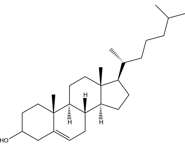
Figure 1.4 Structure of Cholesterol……….23
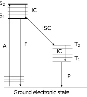
Figure 1.5 Jablowski Diagram………28
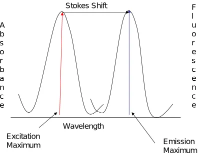
Figure 1.6 Absorbance, Fluorescence emission and the Stokes shift………..………30
Figure 1.7. General schematic of a fluorimeter with 90º setup……….32
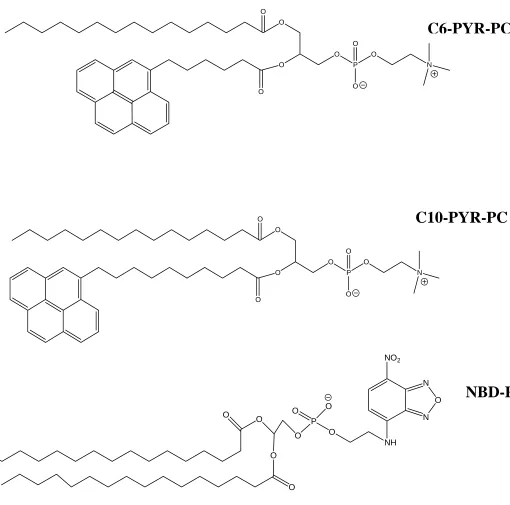
Figure 2.1. Structure of Fluorescent Phospholipids………..42
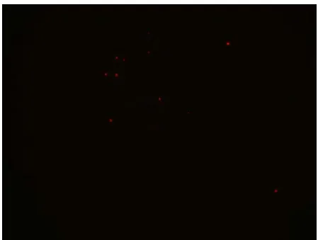
Figure 2.2. Digital Fluorescence Microscopy Image of C6-PYR-PC Liposomes…….…….50
Table 2.1. Approximate initial rates of fluorescence loss for fluorescent membrane probes degraded with high dose of hydroxyl radical……….……….52
Table2.2. Observed pseudo first order rate constants for aqueous phenol degradation with Fenton reagent under steady state hydroxyl radical concentration……….54
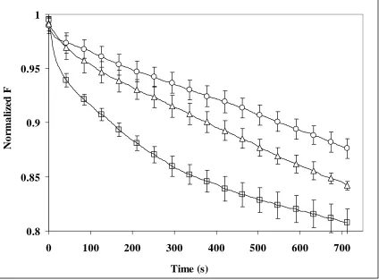
Figure 2.3a.1% NBD-PE liposomes with 0.2mM Fe+2-EDTA and H 2O2 addition at 0.8mL/hr with initial concentration at 1.5M. Curves were normalized to 1 where liposomes were added……….………..55
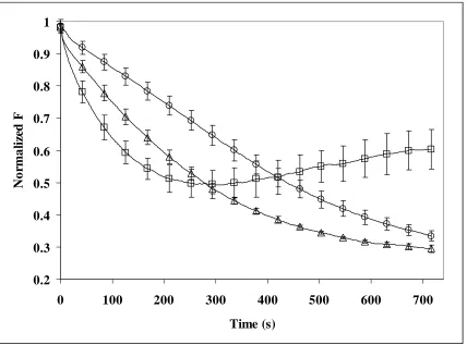
Figure 2.3b. 1% C6-PYR-PC Liposomes with 0.2mM Fe+2-EDTA and H 2O2 addition 0.8mL/hr with an initial concentration of 1.5M……….56
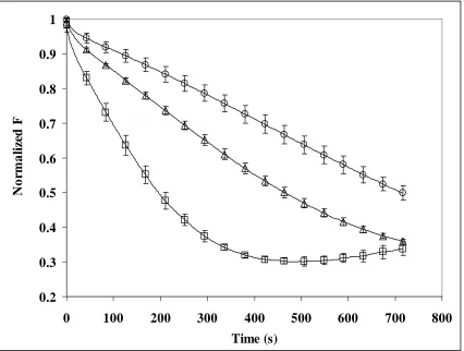
Figure 2.3c. 1% C10-PYR-PC liposomes with 0.2mM Fe+2-EDTA and H 2O2 addition at 0.8mL/hr with initial concentration at 1.5M……….…..57
Figure 2.4. Chemiluminescence Background Signal from Unlabelled liposomes at 37ºC……….……….59
List of Tables and Figures cont’d.
Figure 2.7a. 1% C6-PYR-PC Liposomes with no cholesterol and 0.3µM Fe+2-EDTA and
H2O2 addition at 0.8mL/hr with initial concentration at 3µM………..65
Figure 2.7b. 1% C6-PYR-PC liposomes with cholesterol and 0.3µM Fe+2-EDTA and H2O2 addition at 0.8mL/hr with initial concentration at 3µM………66
Figure 2.8a. 1% C10-PYR-PC Liposomes with no Cholesterol and 0.3µM Fe+2-EDTA and H2O2 addition at 0.8mL/hr with initial concentration at 3µM………..67
Figure 2.8b. 1% C10-PYR-PC Liposomes with Cholesterol and 0.3µM Fe+2-EDTA and 2.4nM/hr H2O2 addition……….………..68
Table 2.3. Observed rates of fluorescence loss for fluorescent membrane probes degraded with low dose of hydroxyl radical………69
Figure 2.9a. Effect of varying biphenyl concentration with C6-PYR-PC liposomes at 35ºC with bolus addition of 0.2mM Fe+2-EDTA with H 2O2 addition at 0.8mL/hr with initial concentration at 1.5M………..72
Figure 2.9b. Effect of varying biphenyl concentration with C10-PYR-PC liposomes at 35ºC with bolus addition of 0.2mM Fe+2-EDTA with addition of H 2O2 at 0.8mL/hr with initial concentration at 1.5M……….73
Figure 2.10. Blank cholesterol with no Fenton Reagent: m/z 369 is cholesterol water loss product and m/z is protonated cholesterol product……….. 76
Figure 2.11. Cholesterol with Fenton reagent and protonated ketone………77
Figure 2.12. Blank DMPC with no Fenton Reagent; protonated DMPC at m/z 678……….………78
Figure 2.13. DMPC with Fenton reagent………..………..79
Figure 3.1.Unsaturated Phospholipid Structures……… 92
List of Tables and Figures cont’d.
Figure 3.3a. Egg Yolk Phosphatidylcholine liposomes with C6-PYR-PC with and without
cholesterol: ◊ = without cholesterol and ∆ = with cholesterol………96
Figure 3.3b. Egg Yolk Phosphatidylcholine liposomes with C10-PYR-PC with and without
cholesterol:◊ = without cholesterol and ∆ = with cholesterol……….97
Figure 3.3c. Egg Yolk Phosphatidylcholine liposomes with C12-PYR-PC with and without
cholesterol:◊ = without cholesterol and ∆ = with cholesterol……… 98
Figure 3.4a. The effect of alpha-tocopherol on the degradation of C6-PYR-PC in Cis6PC
liposomes without cholesterol: o = 0µM alpha tocopherol, ∆ = 30µM alpha-tocopherol,
x = 50µM alpha-tocopherol………...101
Figure 3.4b. The effect of alpha-tocopherol on the degradation of C10-PYR-PC in Cis6PC
liposomes without cholesterol.: o = 0µM alpha tocopherol, ∆ = 30µM alpha-tocopherol,
x = 50µM alpha-tocopherol………102
Figure 3.4c. The effect of alpha-tocopherol on the degradation of C12-PYR-PC in Cis6PC
liposomes without cholesterol: o = 0µM alpha tocopherol, ∆ = 30µM alpha-tocopherol,
x= 50µM alpha-tocopherol……….103
Figure 3.5. Effect of gamma tocopherol on C6-PYR-PC cis6PC liposomes with no
cholesterol: o = 0µM gamma-tocopherol, ∆ = 30µM gamma-tocopherol, x = 50µM
ABSTRACT
In the onset of many chronic illnesses including Parkinson’s, Alzheimer’s, and
cardiovascular diseases, there is evidence to support the delicate balance between
pro-oxidant and antipro-oxidant species is shifted in favor of the former. Under these
conditions, many reactive oxygen species (ROS) including hydroxyl radicals, are
generated. Hydroxyl radicals formed in close proximity to DNA, nucleotides, proteins,
and lipids rapidly oxidize these biological molecules in a nonspecific way. However, their
toxicity is limited by their short lifetimes.
Currently, the mechanism by which hydroxyl radicals are involved in the onset of
many illnesses, particularly with regard to lipid peroxidation, has yielded some
controversy in the literature. Conventional studies which generate hydroxyl radicals with
Fenton chemistry through bolus additions of iron and hydrogen peroxide do not mimic
conditions found physiologically because there is a steady-state concentration of
hydrogen peroxide concentration found in normal cellular systems. Also, former reports
that used fluorescent fatty acids or free probes intercalated within liposomal
membranes did not have the probes covalently attached to the phospholipids making
up the liposomes. Thus, the actual placement of the probes over the analysis time may
vary with experimental conditions.
The objective of this research project was to prepare, characterize, and employ
Also, compared to the popularly-used technique of electron spin resonance,
(ESR), our aim was to use a fluorescence-based approach which yielded the advantages
of high sensitivity, fast analysis time, and less expensive equipment requirements.
Degradation of fluorescently-tagged liposomes with probes covalently bound to
the phospholipids was correlated with the ability of hydroxyl radicals and other possible
reactive oxygen species to penetrate into the liposomes to deeper into the lipophilic
layer. However, alone this experimental setup may not fully define the mechanistic role
of hydroxyl radicals in lipid oxidation. Thus, a complementary approach embracing the
use of MALDI-TOF mass spectrometry, lipophilic scavenger studies, and the effects of
cholesterol and temperature allow a deeper understanding of the radically-driven
oxidation of lipids. It was determined that hydroxyl radicals were generated and reacted
Chapter 1. INTRODUCTION
Section 1.1. Reactive Oxygen Species
A free radical is defined as any species that has one or more unpaired electrons.
This definition includes the hydrogen atom, some transition elements and oxygen,
which itself is a biradical.1 If molecular oxygen is reduced by a single electron it
generates superoxide. The formation of superoxide is very favorable owing to the
biradical formation of molecular oxygen. Once molecular oxygen abstracts an electron
during the aerobic events constantly occurring in the human body, it can go on to help
generate hydrogen peroxide through the Haber-Weiss reaction.2 If superoxide picks up
another electron, it generates the peroxide ion which is not a radical. This peroxide ion
quickly gets protonated to generate hydrogen peroxide. In the course of these serial
reactions, hydrogen peroxide can undergo dismutation generating superoxide and
water or it can react with available iron(II) to generate the toxic hydroxyl radical
through Fenton chemistry.3
Hydroxyl radicals are characterized as being a type of reactive oxygen species
(ROS). Other reactive oxygen species include singlet oxygen and peroxynitrite. The
resulting hydroxyl radical has been reported to initiate lipid peroxidation through
reactions beginning with abstraction of an allylic hydrogen from a polyunsaturated fatty
acid generating a lipid radical. Subsequent addition of oxygen to the carbon-centered
radical can generate a peroxyl radical which can generate further lipid peroxidation
Section 1.2 Fenton Chemistry
The discovery of Fenton chemistry, involving the generation of hydroxyl radicals
through the oxidation of iron (II) by hydrogen peroxide and a host of other related
reactions have been credited to Henry J. Fenton 5, 6with further definition of this
chemistry being investigated by Haber and Weiss.2 The Fenton reaction,
Fe+2 + H2O2 Fe+3 + OH• + OH- Equation 1.1
given by Equation 1.1 has been used to describe the generation of hydroxyl radicals,
but the Fenton process is actually a set of reactions that occur together. Some of the
additional reactions that occur include:7
Fe+3 + H
2O2 Fe+2 +H+ + HO2• Equation 1.2
Fe+3 + HO
2• Fe+2 +H+ + O2 Equation 1.3
Fe+2 + HO
2• Fe3+ + HO2- Equation 1.4
Fe+2 + OH• Fe+3 + OH- Equation 1.5
H2O2 + OH• H2O + HO2• Equation 1.6
Equations 1.2 and 1.3 show how iron is catalytically regenerated. Equations 1.5 and 1.6
show what happens at high concentrations of iron (II) and H2O2 where both effectively
scavenge the hydroxyl radical. Hydroxyl radical scavengers can also affect the rates of
the above reactions by changing the concentration of reactants. Some of these
Section 1.3. Fenton Chemistry in Biological Systems
Reactive oxygen species can be formed at various places in the human body, but
the mitochondria are the primary source of superoxide and hydrogen peroxide in cells.
Superoxide in the mitochondria will generally undergo superoxide dismutation due to
action of the enzyme superoxide dismutase. The two main factors controlling oxygen
radical production is oxygen tension and concentration of partially reduced
mitochondrial components. Oxidative stress occurs when electrons “leak” outside of the
mitochondrial membrane.4, 8 Physiologically much of the iron required to catalyze
Fenton-generated hydroxyl radicals are bound to storage proteins such as transferrin,
ferritin or hemoglobin and do not exist “freely” 8. However, “free” iron also referred to
as non-transferrin bound iron (NTBI) may exist. Chelation of this “free” iron by citrate
or albumin usually stabilizes the iron, and the concentration is usually 1-10µM. 910, 11
Recent reports have shown that under oxidative stress “bound” iron can be
released from these storage proteins and even at low concentrations, less than 10µM
iron can act as the catalyst needed to generate hydroxyl radicals in the presence of a
steady-state concentration of hydrogen peroxide, which itself has been shown to
penetrate cell membranes freely due to its uncharged state and can participate in cell
Section 1.4. Techniques to measure hydroxyl radicals and other reactive
oxygen species
Hydroxyl radicals have been measured in lipid phases using a variety of
techniques. Measuring hydroxyl radicals directly has proved to be challenging because
of their short lifetimes. Electron paramagnetic resonance has been touted as one of the
most direct methods to measure hydroxyl radicals. 12 Hydroxyl radical formation has
previously been measured with the spin trap 5,5-dimethyl-1-pyrroline-N-oxide (DMPO)
while observing the ability of iron chelates to penetrate into bulk lipid phases. Although
they were certain that hydroxyl radicals were formed in oleic acid, in lipid esters the
formation of hydroxyl radical adducts was uncertain. It was also shown that due to the
short lifetimes of hydroxyl radicals that they were most likely to exist within lipid phases
if they were formed there instead of the exterior environment. Hay and co-workers
designed new spin traps that would be anchored at specific depths within the
membrane of large unilamellar vesicles.13 The spin traps were highly lipophilic and were
used to position the nitrone trap deep within the lipid bilayer. However, the spin traps
were not covalently attached to the phospholipids so some “wagging and bobbing” of
the spin traps were detected.
Braughler and co-workers14 studied whether ferrous iron was oxidized in the
presence of various medium compositions and in the presence of lipid. Their results
revealed that ferrous iron oxidation was significant in the presence of phosphate or
However, it was shown that in the presence of liposomes, membranes, micelles and
fatty acids, the recoverable efficiency of ferrous iron was reduced to less than 50%. It
is interesting to note the result that commercial lipids are unavoidably contaminated
with lipid hydroperoxides on the order of 0.02 to 0.1%. Moreover, the authors
suggested that oxidation of ferrous iron by lipid hydroperoxide and also hydrogen
peroxide is more complex than simple ferric iron which can not be easily reduced.14
Section 1.5 Lipids, Micelles, and Liposomes
Lipids are separated into three general classes, free fatty acids, triacylglycerols
and phospholipids. The free fatty acids are long chain aliphatic carboxylic acids which
can be saturated or unsaturated. The common types of saturated fatty acids contain an
even number of carbon atoms ranging from 12 to 22 15. Phospholipids are the primary
components of cell membranes. They are composed of a dialkyl-, diacyl-, or
acylalkyl-glycerol esterified to phosphate which in turn is bonded to a variety of polar groups,
such as choline16.Their structures include variations in the nature of the polar head
group and acyl chains. Myristic, palmitic and stearic acids are three common saturated
free fatty acids that are denoted 14:0, 16:0, and 18:0 because they contain 14, 16 and
18 carbon atoms with no double bonds.16 Some common unsaturated free fatty acid
which contain double bonds are oleic acid denoted (18:1) because it has 18 carbon
atoms and one double bond, linoleic acid (18:2), linolenic acid (18:3), arachidonic acid
Figure 1.1 Example of fatty acid, triacylglycerol, and phospholipids, respectively
a.) palmitic acid (fatty acid)
CH3-(CH2)14-COOH
b.) triacylglycerol
C
O
O
CH
2HC
O
C
CH
2R
R
O
O
C
O
R
c.) phospholipid, dimyristoylphosphatidylcholine
O
O
O
P
O
O
-O
N
+O
The unsaturated free fatty acids commonly occur in the cis formation separated by a
single methylene group. However, some trans unsaturated fatty acids do occur to a
small extent such as elaidic acid which has the same denotation as oleic acid except
that it occurs in the trans rather than cis formation. Some of the unsaturated free fatty
acids, such as linoleic and linolenic acids, are noted as being essential nutrients because
they are not synthesized in the body. Moreover, docosahexaenoic acid from fish and
algae has been included in infant formula to improve visual and brain functions 17.
Triacylglycerols are composed of fatty acids esterified to glycerol. They are the
main constituent of vegetable oils and food lipids and are important storage lipids. In
contrast, phospholipids are fatty acids esterified to glycerol which contain phosphoric
acid and organic bases. Phospholipids constitute the major component of cell
membranes and liposomes15.
Micelles are composed of an aggregate of surfactant molecules where the
surfactant is composed of a single polar headgroup and a single fatty acid chain.
Micelles were used as a substrate to study the oxidation of methyl linoleate in
tetradecyltrimethylammonium bromide (TTAB) and sodium dodecyl sulfate (SDS)
solutions. Results were measured using UV spectroscopy and electrospray mass
spectroscopy. 18
Interestingly enough, Fe(II) was able to catalyze oxidation of methyl linoleate in
the acidic TTAB media but not the basic SDS media. Moreover, Fe(II)/H2O2 was able to
In addition, Fe(II)/ascorbic acid was able to catalyze methyl linoleate in SDS but
not in TTAB micellar solutions. In summary, the Fe(II)/Fe(III) couple was necessary to
oxidize the methyl linoleate at the acidic pH of the TTAB micellar solutions. Also,
ascorbic acid can behave as an antioxidant and pro-oxidant and the formation of an
iron/ascorbate complex was suggested.18
In a related study19, the oxidation of methyl linolenate based on its physical state
in octadecane oil-in-water emulsions was investigated. Also, the effect of pH, iron
concentration, emulsifier type and cooling rate on the oxidation of methyl linolenate
was determined. The results revealed that oxidation rates were higher with solid
octadecane compared to the liquid emulsions, In addition, higher iron concentrations
and lower pH were more favorable for oxidation rates of octadecane. The authors
attributed the behavior to the migration of methyl linolenate to the surface of the oil in
water crystalline octadecane where it could interact with the iron in the aqueous media
to a greater extent.19
Section 1.6. Liposomes as models for cell membranes
Liposomes were first introduced by A.D. Bangham and co-workers in 1965 and
have been used frequently in experimental studies to garner a deeper understanding
into the mechanisms governing lipid peroxidation 20
The suitability of using liposomes as models for cell membranes is proven by
their composition of one or more phospholipids which is found to constitute the bulk of
Liposome systems are convenient to use because the effect of pH and
temperature can easily be monitored 26 In addition, incorporating hydrophobic and
hydrophilic probes within the hydrophobic bilayer or encapsulated aqueous interior is
straight-forward. The advantages of a liposomal model also include a simplified
construct without the complexity of authentic cell membranes which also contain
proteins, DNA, and other important biomolecules which could make interpretation of
experimental findings complicated by their interferences. Obviously the simplified model
is limited in that it may not represent a typical cellular membrane in all respects and the
liposomes should always be prepared fresh to avoid autoxidation of the lipids.
However, liposomal systems can be used to give introductory insight towards and
explanation of the complex mechanisms that govern lipid oxidation and may also be
used to create benchmarks for experimentation involving complex cellular systems27.
Many synthetic methods are currently available to generate liposomes.28, 29 In a
method first introduced by Bangham,20 liposomes are formed spontaneously when the
acyl chains of a lipid film, created by evaporation of the solvent by a gentle stream of
nitrogen or rotoevaporation, interact through hydrophobic interactions forming a bilayer
which encapsulates an aqueous interior.
One set of polar headgroups are simultaneously directed toward the aqueous
interior while the other set is directed toward the outer aqueous medium. One
advantage of this method is the absence of any alcohols which could affect the
Initially, multilammelar vesicles (MLVs) are formed which are characterized by
multiple concentric bilayers with each bilayer encapsulating aqueous interiors at
increasing diameters, though liposomes have also been shown to adopt tubular shapes
as well. The spacing between each bilayer is established by a balance of the Van der
Waals, electrostatic and hydration forces between adjacent bilayers.20 However in most
applications employing lipsomes these MLVs are subjected to an extrusion process to
generate unilamellar liposomes which have a single bilayer 29. During the extrusion
process, the MLVs are taken up by a gas-tight syringe (in many cases having a volume
of 1mL) and this suspension is passed back and forth through a filter housing
containing a polycarbonate membrane which is size exclusive (in many cases having a
diameter of equal or lesser value than 100nm) to a second gas-tight syringe of the
same volume as the first syringe. Multiple odd passes are performed to ensure
unilamellarity without contamination from larger vesicles on even passes 27. These
unilamellar liposomes are labeled small (diameter of 0.02-0.05µm) or large (diameter of
0.06µm or greater.
Liposomes, also known as vesicles, have been previously used as models for cell
membranes due to their similarity in composition and behavior to cells. Liposomes are
generated spontaneously when phospholipids are added to an aqueous environment
Entropically driven forces cause the phospholipids to line up tail-to-tail generating
a spherical hydrophobic bilayer structure which encapsulates water where one set of
the hydrophilic polar head groups are directed to the center and the other polar head
groups are directed to the outer aqueous medium. The hydrophobic bilayer of the
liposome, in addition to phospholipids, may contain cholesterol, proteins or other
additives to mimic conditions commonly found in cells. Both the lipid bilayer and the
aqueous interior have been previously labeled with fluorescent probes in drug-delivery,
and biosensor studies.27
Figure 1.2 Phospholipid, lipid bilayer, and unilamellar and multilamellar
liposomes.
Polar
Headgroup
Fatty
acyl
chains
Lipid Bilayer
Multilamellar
Liposome
Section 1.7. Characterization of Liposomes
Typical characterization methods used to evaluate lamellarity of liposomes include
electron microscopy, 1H, 13C and 31P NMR, dyanamic light scattering, and digital
fluorescence microscopy.
Electron microscopy can be used to yield information on the size, size distribution,
volume distribution and the lamellarity of the liposomes by bombarding the sample with
electrons26, 29. In NMR studies information on the orientation of the fatty acids and the
polar headgroups, packing density, and distribution of lipids in the inner and outer
layers can be obtained.32, 33. In dynamic light scattering34, also known as
photo-correlation spectroscopy, a laser source is used to measure the scattering of a dilute
suspension of liposomes in which the particles are assumed to be moving but are
independent entities which do not interact with each other. Upon taking a series of
measurements, a histogram can be generated showing the size distribution of the
liposomes. A unimodal histogram suggest unilamellar liposomes are present, with the
average hydrodynamic radius (RH) calculated over a series of measurements signifying
the average radius of the particles and the polydispersity term (polyd) giving a measure
of the standard deviation in the size distribution. In digital fluorescence microscopy,
whether or not the liposomes are aggregated is assessed.Images can be taken of the
fluorescent liposome at their excitation and emission wavelengths.
Section 1.8. Liposomes: Phase Transition Temperature
Specifically, a phase transition temperature (Tm) exists separating a more-ordered
solid-gel phase below Tm from a more fluid liquid-crystalline temperature above Tm.
In the solid-gel phase, the head groups are closely packed with very little spacing
between the acyl chains which are positioned in a tilted position. In contrast, the
liquid-crystalline phase is characterized by more spacing between the polar head groups and
acyl chains.
At the phase transition temperature, a combination of the solid-gel and liquid-crystalline
phases exist with large mass defects within the bilayer. It was previously reported by
Kraske and co-workers36 that an uncharged molecule, glucose, was able to penetrate
dimyristoylphosphatidylcholine liposomes to the greatest extent at the phase transition
Figure 1.3 The Phase Transition of Phosphatidylcholine
Below Tm At Tm Above Tm
Solid-Gel Phase Liquid-Crystalline Phase
Section 1.9. Lipid Oxidation
Lipid Oxidation is a topic that spans many fields including biology, free radical
chemistry, nutrition, and biochemistry. It has been studied for over sixty years and yet
many problems still remain unresolved. In vivo mechanisms of lipid peroxidation, the
effects of antioxidants including phytochemicals, and the mechanisms of
polyunsaturated lipid nutrition still remain a mystery. Lipid oxidation refers to the
oxidation of unsaturated fatty acids and their derivatives. These oxidation reactions
generally occur when the lipids are exposed to air and there is an imbalance between
pro-oxidant species such as hydroxyl radicals, superoxide, hypochlorous acid, hydrogen
peroxide, and singlet oxygen compared to antioxidant species such as vitamins A, D, E,
and K.
In biological systems, only hydroxyl radicals,37-39 and high oxidation states of iron
such as the ferryl iron, 40 peroxynitrite,3 and nitrogen dioxide41 have been shown to
initiate lipid oxidation. Superoxide and hydrogen peroxide have not been shown to
initiate lipid oxidation unless through the Haber-Weiss mechanism which includes
formation of hydroxyl radical.
Section 1.10. Free Radical Reactions
The free radical reactions that lipids engage in are largely due to the pi electrons of
unsaturated lipids.15 This reactivity is due to the pi electrons being of lower energy
compared to sigma electrons. The chain reaction mechanism of free radical reactions
The lower energy of pi electrons makes them accessible for hydrogen abstraction,
addition and elimination reactions, fragmentation reactions, rearrangement reactions,
disproportionation reactions, and oxidation-reduction reactions, all of which can be the
initiating start of free radical reactions. Propagation reactions occur when free radicals
react to form new free radicals. The free radical reactions are ended when two radicals
combine to form non-radical products.15
Taking a look at these reactions more closely, initiators (I) such as singlet oxygen,
superoxide, and hydroxyl radicals can abstract a hydrogen from unsaturated lipids (LH)
generating a lipid free radical (L•).
LH +I IH + L• Equation 1.7
The propagation reaction where oxygen (O2) reacts with the lipid free radical is very
fast which generates a lipid peroxyl radical (LOO•).
L• + O2 LOO• Equation 1.8
Then this peroxyl radical can react to form hydroperoxides (LOOH) and another lipid
radical (L•).
LOO• + LH LOOH + L• Equation 1.9
The termination reaction occurs when two free radicals react to form non-radical
products such as two lipid peroxyl radicals reacting together.
LOO• + LOO• non-radical products Equation 1.10
LOO• + A LOOH + A• Equation 1.11
Antioxidants are defined as substances which can attenuate or prevent the oxidation of
a substrate when it is present in small amounts relative to the amount of the substrate.
There are numerous ways an antioxidant can affect lipid peroxidation including having a
decrease in oxygen concentration, scavenging initial radicals, chelating metal catalysts,
decomposing radical products and breaking the chain reactions that abstract hydrogens
or electrons.10, 42 These former reactions are by no means all of the chain reactions
because alkoxyl radicals, alkyl radicals and other radicals and products can be
generated during this free radical process.
The kinetics of the above reaction scheme are complicated in the presence of trace
metals such as copper or iron because metals can catalyze the decomposition of
unsaturated lipids by electron transfer,
M(n+1) + LH Mn++ L• + H+ Equation 1.12
or by one-electron redox reactions,
Mn + ROOH Mn+1 + RO• +OH- Equation 1.13
M n+1 + ROOH Mn +ROO• +H+ Equation 1.14
Another complicating factor exists in heterogeneous systems where metals and lipid
SECTION 1.11. Antioxidants: Endogenous and Exogenous
In the human body there are endogenous enzymes and water and lipid soluble
antioxidants which maintain the delicate balance between pro-oxidant and antioxidant
species. Peroxisomes are endogenous enzymes located in the mitochondria which
convert oxygen radicals into non-deleterious species.
Superoxide dismutase, which is in the intracellular and extracellular media, controls the
amount of superoxide in the body converting it into hydrogen peroxide. In addition,
catalase and glutathione decompose hydrogen peroxide and hydroperoxides,
respectively. 43
Some non-enzymatic antioxidants also exist and are classified as being
water-soluble and lipid-water-soluble. Some water-water-soluble antioxidants include ascorbic acid, uric
acid, or glutathione. Some lipid-soluble antioxidants include vitamin E and
beta-carotene. Both the water and lipid-soluble antioxidants’ main role is to scavenge free
radicals43. Vitamin E is composed of different tocopherols and tocotrienols which have
been shown to behave as an antioxidant and scavenge two molecules of peroxyl
radicals. However, there is also evidence to support the pro-oxidant activity of Vitamin
E under certain reaction conditions. 44 Moreover, though Vitamin C is a poor antioxidant
alone, in the presence of Vitamin E, a synergistic affect occurs where the tocopheroxyl
radicals formed during radical scavenging is reduced. This allows for propagation of
chain reactions to be reduced.
Flavonoids, another type of exogenous antioxidants, usually react by free radical
scavenging. However, flavonoids can also become pro-oxidants under certain
experimental conditions by greatly accelerating the production of hydroxyl radicals from
H2O2 in the presence of Fe+3-EDTA at physiological pH.
Fukuzawa et al.46 investigated the oxidation of alpha-tocopherol in micelles and
liposomes by the hydroxyl, peroxyl, and superoxide free radicals. . In this study,
samples underwent radiolysis of the aqueous solutions with gamma rays. Micelles and
liposomes were both charged and uncharged. Hydroxyl radicals were effective oxidants
of both the micelles and the liposomes.The perhydroxyl ion was also effective as an
oxidant of as long as the pH was acidic. The alpha tocopherol, due to its low water
solubility was found in the micelles and the liposomes. The alpha tocopherol in the
positively charged micelles and liposomes were oxidized as a higher rate by superoxide
than in uncharged or negative liposomes or micelles.
Alessi and co-workers47 investigated the similarities and differences between the
kinetics of peroxidation of vitamin E-containing liposomes and human low-density
lipoprotein. Interestingly enough, it was found that for tocopherol-mediated
peroxidation (TMP) to occur, the liposomes must exists as isolated systems to protect
the tocopherol radical. This premise was supported by the highly lipophilic character of
Section 1.12. Cholesterol and its oxidation
Cholesterol has been widely studied due to its oxidation products that have been
implicated in adverse health effects. Cholesterol is an apolar lipid constituent of cell
membranes. It is present in large amounts in the lipoproteins of blood and is the
precursor for the generation of glucocorticoids, sexual hormones, and vitamin D48. The
oxidation products of cholesterol are commonly called oxysterols or cholesterol oxides.
The oxidation products can be generated enzymatically or non-enzymatically in
biological systems.
The allylic 7-position carbon is susceptible to oxygen attack to produce its
hydroperoxides. These oxides have been observed in low-density lipoprotein (LDL),
oxidized in vitro, rat skin, marine animals and plant tissues. Cholesterol oxidation in
solution or in aqueous media is initiated by hydrogen abstraction to produce a
delocalized three carbon allylic radical followed by oxygen attack mainly at carbon 7 to
produce the epimeric hydroperoxy products at 7-alpha and 7-beta, with the
7-beta-hydroperoxide dominating.
It is interesting to note that oxygen attack at carbon position 4 is not favored
due to steric hindrance by the trialkyl-substituted carbon-5 and the neighboring effect
of the OH group on carbon-3.
Cholesterol oxidation can also occur in the solid or crystalline phase generating the
25-hydroperoxycholesterol product through a side chain oxidation scheme involving the
Moreover, two dihydroperoxides, 3-beta-hydroxy-20R- and 20S-cholesterol, have been
isolated in autooxidized bulk cholesterol as well. The monohydroxides of cholesterol
also decompose into complex mixtures of hydroxyl, keto, epoxyene and ketodiene
secondary products that have received considerable attention because of their biological
HO
H H
H
Section 1.13. Methods to Determine Amount of Lipid Oxidation
Section 1.13A. Peroxide Value
The peroxide value, or the amount of peroxides in a sample, can be determined
empirically using an iodometric titration, colorimetrically or electrometrically. This
method requires typically a gram or more of potassium iodide to perform the titration.
Its results are affected by light, air, and iodine reaction with double bonds. In addition
there is a ferric thiocyanate method to determine oxidation which only requires 0.1g
sample sizes. This method typically yields higher values than the iodometric technique
on the order of 1.5 to 2 times the relative peroxide value. The peroxide value technique
is very sensitive to the sample history with the peroxide value reaching a maximum with
degree of oxidation according to temperature, degree of polyunsaturation, exposure to
light and metals.15
Section 1.13B. Conjugated Dienes
The conjugated diene method of measuring lipid oxidation relies on the strong
absorption of conjugated dienes at a maximum of 234nm. It offers the advantage of
being a direct, sensitive method to follow lipid oxidation, and is suitable for monitoring
early stages of lipid oxidation. However, it is of limited utility at high stages of lipid
oxidation due to secondary products absorbing in the ultraviolet range, such as
In addition, high temperatures, metals and light can yield low values due to
decomposition of the conjugated dienes. Moreover, interferences such as partially
hydrogenated fats are a limitation of this method. 15
Section 1.13C. Carbonyl Compounds
The amount of total carbonyls in a sample can be determined by its reaction with
2,4-dinitrophenylhydrazine (2,4-DNPH), which yields colored hydrazone derivatives that
are measured by an absorbance of 430-460nm, and expressed as nmol hexanal/kg
sample.15
Section 1.13D. 2-Thiobarbituric acid (TBA) value
The 2-thiobarbituric acid value test is an older, more widely used test than some
of the other techniques. This test is a colorimetric assay to measure oxidation products
in biological systems. To perform this test, it is necessary to observe the pink product
that absorbs in the range of 532-535 nm. This signal is due to the product formed
upon reaction of TBA with oxidation products of polyunsaturated lipids. One limitation
of this method is that it is not specific, meaning a positive signal is produced by a large
number of secondary oxidation products, these products are referred to as TBA-reactive
substances or TBARS. The test is standardized by malondialdehyde. 15
A variety of factors affect the production of the pink color in the TBA test including
temperature and heating time, pH, metal ions, and antioxidants. The selectivity of the
Section 1.14A. Fluorescence and Fluorescence-Based Methods
Fluorescence is a luminescent analytical technique which involves analytes
absorbing a photon of light followed by emission of a photon of light at a longer
wavelength.49 Molecules which undergo fluorescence are called fluorophores.
Fluorescence usually occurs within aromatic and heterocyclic compounds.
The three luminescent techniques include fluorescence, phosphorescence and
chemiluminescence all of which are characterized by the excited state. Fluorescence
involves molecules absorbing energy from a ground state to an excited singlet state,
partially relaxation, and then emission of a photon to return the molecules back to the
ground electronic state. In phosphorescence, the molecule returns to the ground state
following emission of a photon from the spin-forbidden triplet excited state, which
involves a change in spin. In chemiluminescence, a chemical reaction generates an
electronic excited state which subsequently emits a photon to return to the ground
state 49Fluorescence is short-lived with a lifetime existing at less than 10-5 seconds. In
the case of phosphorescence the change of spin leads to longer lifetimes at several
seconds or longer. An advantage of fluorescence is the high sensitivity with detection
limits one to three orders of magnitude lower than absorption spectroscopy. Moreover,
fluorescence has long linear concentration ranges compared to absorption
The Jablowski diagram outlines the possible pathways a molecule can take to return to
the ground state following absorption of electromagnetic radiation. Absorption of light is
signified by A from a ground singlet state to an excited singlet state signified by S1 or
S2.. Each of the singlet excited states is composed of a number of vibrational energy
levels. When a molecule returns to a lower excited state of the same spin such as S1
from S2, without emitting a photon the process is called internal conversion, signified by
IC. Fluorescence emission signified by F occurs when light is emitted as a molecule
returns to the ground state from a singlet state of the same spin, So, the singletground
state. The light is emitted at a longer wavelength due to energy loss during the excited
state lifetime. This process is known as the Stokes shift.
Phosphoresence signified by P involves a molecule returning to a singlet ground
state, So from a spin forbidden excited triplet electronic state signified by T1. Since
fluorescence is spin favored over phosphorescence, it takes place over a shorter time
interval (1E-5 to 1E-8 seconds) compared to (1E-4 seconds to hours). Vibrational
relaxation is another radiationless phenomenon which occurs when excited molecules
transfer energy to other molecules through collisions over a short time scale (<1E-12
Figure 1.6 Jablowski Diagram
S
1S
2A
F
IC
P
T
2T
1ISC
IC
Section1.14B. Fluorescence Spectra
A fluorescence emission spectrum is taken when the excitation wavelength is held
constant (often at the absorbance maximum), and the emission wavelength is scanned.
The excitation spectrum is taken when the emission wavelength is held constant (often
at its maximum), and the excitation wavelengths is scanned. The Stokes shift is the
peak-to-peak distance of the maxima of the excitation and emission spectra.
Figure 1.6 Absorbance, Fluorescence emission and the Stokes shift
A
b
s
o
r
b
a
n
c
e
F
l
u
o
r
e
s
c
e
n
c
e
Wavelength
Stokes Shift
Excitation
Maximum
Emission
There is an inherent benefit of fluorescence over absorbance because the fluorescence
method is based on a dark background, whereas in absorbance, the background is the
highest observed light intensity. In a typical fluorescence spectrometer, the detector is
positioned at a 90 degree angle relative to the light source. This configuration
minimizes transmitted and reflected light reaching the detector. The signal to noise
enhancement is significant and the detection limit is lowered by a factor of 10000
compared to the 180 degree setup. The excitation and emission monochromators are
wavelength filters which allowed specific wavelengths to be selected before and after
fluorescence events50. In addition, the sensitivity of the fluorescence instrument is
high because the process of fluorescence is cyclical, with the same fluorophore being
excited and detected. In fact, a single fluorophore can yield thousands of detectable
photons. The cycling does not help the absorbance technique because the detector is in
line with the light source.
The excitation and emission spectra are sensitive to the type of solvent,
Figure 1.7. General schematic of a fluorimeter with 90º setup.
Excitation Source
Excitation
Monochromator
Sample
Emission
Monochromator
Section 1.15. Effect of Chelator on Fenton Chemistry
One of the most commonly used chelators with Fenton chemistry is
ethylenediaminetetraacetate or (EDTA). Like many other chelators, the reactivity of
EDTA is strongly dependent upon the chelator-to-metal ion ratio. Thus, it can inhibit or
enhance the lipid oxidation of detergents and liposomes. The chelation of iron can alter
its redox potential, solubility, and reactivity in reactions leading to the initiation of lipid
peroxidation51. It was determined that chelation of iron (II) by EDTA led to a rapid
autoxidation of iron (II). The rate of autoxidation increased up to equimolar
concentrations of EDTA-to-iron (II).However, when iron (II) was chelated by
(Adenosine diphosphate) ADP it did not result in a rapid oxidation of iron (II).
Section 1.16. MALDI-TOF Mass Spectrometry of Phospholipids
GC/MS methods have been used to study phospholipids but often require
time-consuming, labor-intensive derivitization methods which do not maintain the structural
integrity of the phospholipids. With the introduction of soft ionization methods,
phospholipids can now be studied without prior chemical alteration of the analyte.
Phospholipids have been previously studied using matrix-assisted laser desorption
ionization-time of flight mass spectrometry (MALDI-TOF MS) and electrospray ionization
(ESI MS). The advantages afforded by using MALDI-TOF MS compared to electrospray
ionization MS are fast sample preparation and analysis time, ease of use, simpler
A note of interest is that the ion yield from MALDI-TOF studies are not highly
dependent on the degree of saturation of the phospholipids as opposed to ESI. Also,
compounds with a lower molecular weight are more sensitively detected with MALDI.
With ESI the interpretation of the low mass region is not complicated by matrix peaks
and it is easily coupled to chromatographic columns. However, quantitative studies with
MALDI-MS are sensitive to the homogeneity of the matrix-analyte co-crystallization.
Thus, statistical studies are essential to determine the reproducibility of the
method.48Moreover, when using MALDI-TOF MS, it was found that the signals were
highly dependent on the nature of the matrix, and whether or not water was a part of
the matrix. Some of the best matrices to use include alpha-cyano-4-hydroxycinnamic
acid, esculetin, and 2,5-dihydroxybenzoic acid.
It was determined that the negative ion mode yielded mainly (M-H)- ions and were
more simpler to interpret compared to the positive ion mode which led to the formation
of the protonated, sodiated, and potassiated adducts.16
Post-source decay MS by Al-Saad in the positive ion mode 52of phospholipids
showed that the nature of the cation coordinated at the negatively charged oxygen
position of the choline headgroup had a dramatic effect on the subsequent products
formed. The sodiated ion produced many fragment ions while the protonated ion
This difference in fragmentation is thought to stem from the binding of the proton only
to the negatively charged oxygen while the sodium was found to associate with several
regions of the phosphatidylcholine molecule. In addition, phosphatidylcholines were not
detected in the negative ion mode.
MALDI-TOF MS has been previously used to study lipid oxidation. Oborina and
Yappert53 compared the oxidation of stearoyl-arachidonyl phosphatidylcholine in
sphingomyelin (SM) versus dipalmitoylphosphatidylcholine (DPPC). The results revealed
that oxidation was faster with the DPPC compared to the SM. The data was explained
by the fact that SM had the slower oxidation by the hydrophobic tails of SM being more
disordered and the SM vesicles had a tight network of H-bonds that bridge neighboring
SM molecules and poses a stronger interfacial barrier to the passage of oxidants.
Shadyro and co-workers54 coupled radiation, thin layer chromatography and
MALDI-TOF MS to study the fragmentation of cardioplin in model membranes.
Cardioplin is found in the inner mitochondrial membrane and is a proton trap which is a
mitochondrial enzyme involved in oxidative phosphorylation.
When cardioplin levels decrease it can lead to programmed cell death. A mechanism
was determined in this study from gamma-radiation on a model membrane. Two
fragmentation products were isolated from this free radical mechanism, phosphatidic
acid and phospatidylhydroxyacetone. This work was followed up by the iron-mediated
The free radical mechanism involved hydroxyl radical attack on the polar headgroup of
two starting substrates, cardioplin and galactocerebroside to generate phosphatidic acid
and ceramide.
Using 31P-NMR and MALDI-TOF MS a study was done to compare the analysis of
high-density and low-density lipoprotein. The motivation behind the study was to
further the understanding of atherosclerosis. It was determined that
lysophosphatidylcholine could be monitored by both techniques but only 31P-NMR could
be used to detect chlorohydrines. The limitations of 31P-NMR was that it had low
sensitivity and provided only a small amount of information on fatty acid composition. 56
MALDI-TOF MS was also used to study phosphorylated lipids in biological fluids
using immobilized metal affinity chromatography with a solid ionic crystal matrix.57 This
study employed a novel ionic crystal matrix composed of p-nitroaniline with butyric acid.
Moreover, a new extraction, isolated and cleanup procedure was developed using
ZipTip which had an immobilized metal ion affinity stationary phase.
Section 1.17. Initial Rates
When complicated kinetic profiles are observed in reactive oxygen species
studies, calculating the initial rates is often a straight-forward way to analyze the
data. The initial rate is calculated by taking a measurement over the beginning of
the data where the data can be analyzed with linear regression since all data is
Fluorescent Probes as Reporters for Hydroxyl Radical Penetration into
Liposomal Membranes and Lipid Oxidation
Abstract
The ability of hydroxyl radicals to penetrate into liposomal model membranes
(dimyristoylphosphatidylcholine, DMPC) has been demonstrated. Liposomes were
prepared then characterized with digital fluorescence microscopy and dynamic light
scattering following extrusion to determine liposomal lamellarity, size, and shape.
Hydroxyl radicals were generated in the surrounding aqueous media using
modified-Fenton reagent (hydrogen peroxide and Fe2+) with the water-soluble iron-chelator,
ethylenediaminetetraacetic acid (EDTA). High and low doses of radical were used, and
the low dose was achieved with physiologically relevant iron and peroxide
concentrations. Fluorescent probes covalently bound to the membrane phospholipid
were used, including two lipophilic pyrenyl probes within the membrane bilayer and one
polar NBD probe at the water-membrane interface. Radical reactions with the probes
were monitored by following loss of fluorescence and by observing oxidation products
via matrix-assisted laser desorption ionization-time of flight mass spectrometry
(MALDI-TOF MS). Differences in the probe position within the membrane were correlated with
the reactivity of the probe in order to assess radical access to the site of the probe. For
all probes, reaction rates increased with increasing temperature. Within the membrane
bilayer, reaction rates were greater for the probe closest to the membrane-water
Kinetic models, scavenger studies, and product identification studies indicated that
hydroxyl radical reacted directly with the in-membrane probes without the mediation of
a secondary radical.
Introduction
Hydroxyl radicals are a class of reactive oxygen species (ROS) which have been
implicated in the onset of many illnesses including Parkinson’s1 and Alzheimer’s2
diseases, cancer3 and atherosclerosis.4 Their toxicity is manifested physiologically
through attack on biomolecules5, 6 in close proximity to the site of their formation. A
recent review by Pignatello7 and co-workers outline the typical reaction scheme of
hydroxyl radicals with organic compounds. The first step of hydroxyl radical attack on
organic molecules typically involves either hydrogen atom abstraction or addition to
double bonds. After initial attack by one of these mechanisms, additional reactions
occur, often involving molecular oxygen. However, the role of hydroxyl radicals in lipid
oxidation of cells and liposomes has yielded some controversy.8 Previous reports have
suggested that the high reactivity and short lifetime of hydroxyl radicals generated in
the exterior aqueous phase of liposomes or cells exclude these radicals from being the
direct initiators of lipid peroxidation9 and that hydroxyl radicals instead create secondary
radicals, including peroxyl radicals, which subsequently oxidize cells.10, 11In contrast,
other authors have proposed that Fenton-generated hydroxyl radicals can occur in lipid
Thesepotential pathways for hydroxyl radical oxidation of lipids are of interest since the
impact of hydroxyl radicals formed in various cellular regions depends on the
mechanisms by which these radicals react with cellular material.
The ability of hydroxyl radicals to penetrate into lecithin liposomal membranes
intercalated with pyrene has been previously studied.16 It has been shown that hydroxyl
radicals created in the surrounding aqueous media with pulse radiolysis can in fact
penetrate into the liposomal membranes and react with the pyrene and lecithin in the
hydrophobic membranes in a competitive manner. In addition, it was shown that
hydroxyl radicals react near the phospholipid headgroup. However, these experiments
employed pyrene as a free probe within the liposomal membrane, and therefore the
pyrene could diffuse readily within the lipid layer. Consequently, the specific site of
pyrene-hydroxyl radical reaction within the membrane was not known. In addition,
varying the concentration of pyrene within the membrane in the millimolar range could
have lead to the formation of pyrenyl dimmers within the membrane.17
Lipid peroxidation has been formerly investigated using fluorescent pyrenyl
probes in the presence of liposomes and lipoproteins.18, 19 A pyrenyl fatty acid and
pyrenyl sphingosine were both shown to degrade by observing loss of fluorescence
when incubated with iron (II) sulfate and ascorbic acid or copper (II) sulfate only. In
addition, the thiobarbituric acid approach was used to measure lipid oxidation. Though
this approach is commonly used, it can be complicated by the presence of other species
In addition, one of these investigations also added 0.1 mol% hydroperoxides (relative
to the phospholipids) to the experimental system. The presence of these
hydroperoxides complicated the interpretation of hydroxyl radical impact in these
systems.
Phospholipids covalently labeled with N-(7-nitrobenz-2-oxa-1,3-diazol-4-yl) (NBD)
have been widely used to study biochemical and biophysical lipid phenomena including
membrane organization and dynamic properties.20 Moreover, the NBD probe has been
shown to be highly polar. Thus, this probe was used in our study for probing the
reactivity of hydroxyl radicals at the aqueous interface of the phospholipid.21
The fluidity of the membrane may also affect the ability of hydroxyl radicals to
penetrate into the liposomal membrane. Kraske previously reported that DMPC
liposomes have maximum permeability to small molecules at the phase transition
temperature (Tm)22 compared to above and below this temperature. Thus, the effect of
temperature on this system was also investigated in the current study.
In this study, we utilized small, unilammellar liposomes as models for cell
membranes due to their similar composition and structure compared to cellular
membranes.23 The work reported here differs from previous research in that we used
fluorophores that were covalently bound to the phospholipids. This approach allowed
for monitoring the probe reaction with hydroxyl radical as a function of probe position
within the membrane. In addition, our system used a Fenton approach to generate
Aqueous hydrogen peroxide was continuously added at low flow rates to liposome
suspensions in order to generate a steady-state concentration of hydroxyl radicals, as
similarly performed previously.2526, 27
In comparison to the popularly-used ESR technique,28, 29 the fluorescence
approach used in our study has the advantages of high sensitivity, fast analysis time,
ease of use, and less expensive equipment requirements. A multi-faceted approach
embracing the enhanced specificity of MALDI-TOF-MS30, 31 and scavenger studies32
coupled to the high sensitivity of the fluorescence studies was used to provide a deeper
understanding of the in-membrane oxidation by hydroxyl radicals. A better
understanding of hydroxyl radical attack on membranes can help elucidate the
mechanistic role this radical plays in the development of some diseases.
Experimental Methods
Materials
Three separate fluorescent probes, each purchased from Invitrogen, were used
in this study: N-(7-nitrobenz-2-oxa-1,3-diazol-4-yl)-1,2-dihexadecanoyl-sn
-glycero-3-phosphoethanolamine (NBD-PE),
1-hexadecanoyl-2-(1-pyrenehexanoyl)-sn-glycero-3-phosphocholine (C6-PYR-PC), and
1-hexadecanoyl-2-(1-pyrenedecanoyl)-sn-glycero-3-phosphocholine (C10-PYR-PC). Each of these probes has a fluorescent label covalently
attached to the phospholipid molecule. For NBD-PE, the probe is attached to the
headgroup, while C6-PYR-PC and C10-PYR-PC have the fluorophore attached to the tail
Ferrous sulfate heptahydrate and phosphate-buffered saline (PBS, 0.9% NaCl) were
obtained from Sigma. Hydrogen peroxide (30% aqueous) was purchased from Fisher
and its concentration was measured iodometrically.33Cholesterol was purchased from
Aldrich. Chloroform and EDTA were purchased from EM Science. All aqueous solutions
were prepared using Nanopure UV (Barnstead/Thermolyne, Dubuque, IA) deionized
water with a distilled water feed to the water purifier. All reagents were used as
received without further purification.
Preparation of Liposomes
Our pyrenyl-labeled liposomes characterized the penetration depth of hydroxyl
radicals into the hydrophobic membrane. The NBD-labeled probe 34 was chosen to
characterize its reaction with hydroxyl radical at the aqueous interface due to its high
hydrophilicity. NBD-PE or pyrenyl-labeled DMPC liposomes were prepared using a
modified version of the procedure reported by McNamara and Rosenzweig.35 A 40 µL
aliquot of a 50 mM lipid cocktail composed of a 1:1 molar ratio of DMPC:cholesterol was
placed under a stream of nitrogen overnight. To incorporate the fluorescent label, one
of the fluorescent probes was included in the mixture prior to drying at 1 mol% relative
to the unlabelled lipid content. Next, the film was hydrated while vigorously vortexing
with 1 mL of 1 mM PBS. The final lipid concentration was 2 mM. The liposomes were
extruded 9 times using an Avanti Mini-extruder with a 100 nm polycarbonate filter in
Dynamic Light Scattering
All dynamic light scattering experiments were carried out with a Protein Solution
Dynamic Light scattering instrument and analyzed using Dynamics Software. Settings
on the instrument were for aqueous buffer conditions. For each sample, 40
measurements were taken at a total lipid concentration of 0.2 mM.
Digital Fluorescence Microscopy
All digital fluorescence images of pyrenyl and NBD-labeled liposomes were
obtained using an Olympus IX71 inverted fluorescence microscope as described
previously.35 A 5 µL sample of labeled liposomes with a total lipid concentration of 3.3
µM was pipetted onto a glass coverslip and then covered with a second coverslip. The
filter cube for imaging the C6-PYR-PC liposomes consisted of an excitation bandpass
filter at 330-385 nm with dichroic mirror at 400 nm and emission wavelength at 429
nm. The filter cube for imaging the NBD liposomes consisted of excitation at 460 nm
with a 50 nm bandpass, dichroic mirror set at 500 nm, with emission wavelength at 515
nm. The liposome images were collected using a 10X objective with a neutral density
filter (0.55 o.d.). A CCD camera (Olympus DP70) was employed for digital imaging of
the liposomes. The liposome sample was vortexed before imaging with five, 5 second
pulses to disperse the sample. An exposure time of 0.3 s was used to acquire the
images. ImagePro software was used for image collection. Adobe Photoshop v7.0 was
Fluorescence Studies
The fluorescence experiments were carried out on a Perkin Elmer LS 55
Luminescence Spectrometer equipped with a magnetic stirrer set on low. The quartz
cuvette path length was 10 mm (Starna Cells, Atascadero, CA). Degradation of the
fluorescent probes was followed by monitoring fluorescence emission at fixed excitation
and emission wavelengths as a function of time. For pyrene, the excitation and
emission wavelengths were set at 345 and 377 nm, respectively. For NBD, the
excitation and emission wavelengths were set at 463 and 536 nm, respectively. For
these time-based emission studies, two separate reaction protocols were used.
For degradation studies using high iron and peroxide loadings, a 3 mL aqueous
solution of 0.2 mM Fe+2 and 0.2 mM EDTA was added to the cuvette. After 300 seconds
of stirring, 1.5 M H2O2 (aq) was added continuously at 0.8 mL/hr with a syringe pump
(kd Scientific Model 100). The total volume of H2O2 delivered during a 16 minute
experiment was 0.23 mL. This volume was sufficiently small to not cause substantial
changes in solution concentrations due to increased volume. Addition of peroxide was
continued for 300 s, then a bolus addition of 300 µL of liposomes was added to the
cuvette so that a final lipid concentration of 0.2 mM was achieved in the cuvette. All
figures for these experiments are presented so that time zero corresponds to the time
Additional experiments were conducted with iron and peroxide concentrations
near physiological conditions. For these studies, 3 mL of a buffered solution of
liposomes (3.3 µM lipid) was added to the cuvette.
To the cuvette were then added Fe+2 and EDTA to achieve a concentration of 0.3 µM
for each. The solution was stirred for 600 s, then a 3 µM solution of H2O2 (aq) was
continuously added at 0.8 mL/hr until the final volume of 0.23 mL was delivered. For
these experiments, all figures are labeled time zero when H2O2 addition began.
For all experiments, the temperature of the cuvette and its contents were
controlled by a thermostated cuvette holder. Temperatures used in this study include
15, 23, and 35ºC.
Biphenyl Scavenger Studies
Pyrenyl-labeled liposomes with varying concentrations of biphenyl were prepared
in order to assess the effect of biphenyl as a scavenger in the lipid layer. A 40 µL
aliquot of a 50 mM lipid cocktail consisting of a molar ratio of 1:1 DMPC/cholesterol with
0-3 mol% biphenyl per mol of the lipid was added to a test tube and the pyrenyl probe
(either C6-PYR-PC or C10-PYR-PC) was added at 1 mol% of the total lipid content. The
solution was allowed to evaporate under a stream of nitrogen. The liposomes were
reconstituted in 1 mL of 1 mM PBS buffer and extruded as described above in order to