R E S E A R C H
Open Access
Klf8 regulates left-right asymmetric
patterning through modulation of Kupffer
’
s
vesicle morphogenesis and
spaw
expression
Che-Yi Lin
1,6†, Ming-Yuan Tsai
2,6†, Yu-Hsiu Liu
3, Yu-Fen Lu
4, Yi-Chung Chen
4, Yun-Ren Lai
1, Hsin-Chi Liao
1,
Huang-Wei Lien
5, Chung-Hsiang Yang
5, Chang-Jen Huang
5and Sheng-Ping L. Hwang
1,3,4*Abstract
Background:Although vertebrates are bilaterally symmetric organisms, their internal organs are distributed asymmetrically along a left-right axis. Disruption of left-right axis asymmetric patterning often occurs in human genetic disorders. In zebrafish embryos, Kupffer’s vesicle, like the mouse node, breaks symmetry by inducing asymmetric expression of the Nodal-related gene, spaw, in the left lateral plate mesoderm (LPM). Spaw then stimulates transcription of itself and downstream genes, including lft1, lft2, and pitx2, specifically in the left side of the diencephalon, heart and LPM. This developmental step is essential to establish subsequent asymmetric organ positioning. In this study, we evaluated the role of krüppel-like factor 8(klf8) in regulating left-right asymmetric patterning in zebrafish embryos.
Methods: Zebrafishklf8expression was disrupted by both morpholino antisense oligomer-mediated knockdown and a CRISPR-Cas9 system. Whole-mount in situ hybridization was conducted to evaluate gene expression patterns of Nodal signalling components and the positions of heart and visceral organs. Dorsal forerunner cell number was evaluated in Tg(sox17:gfp)embryos and the length and number of cilia in Kupffer’s vesicle were analyzed by immunocytochemistry using an acetylated tubulin antibody.
Results:Heart jogging, looping and visceral organ positioning were all defective in zebrafishklf8morphants. At the 18– 22 s stages,klf8morphants showed reduced expression of genes encoding Nodal signalling components (spaw,lft1,lft2, andpitx2) in the left LPM, diencephalon, and heart. Co-injection ofklf8mRNA withklf8morpholino partially rescuedspaw expression. Furthermore,klf8but notklf8△zfoverexpressing embryos showed dysregulated bilateral expression of Nodal signalling components at late somite stages. At the 10s stage,klf8morphants exhibited reductions in length and number of cilia in Kupffer’s vesicle, while at 75% epiboly, fewer dorsal forerunner cells were observed. Interestingly,klf8mutant embryos, generated by a CRISPR-Cas9 system, showed bilateralspawexpression in the LPM at late somite stages. This observation may be partly attributed to compensatory upregulation ofklf12b, becauseklf12bknockdown reduced the percentage ofklf8mutants exhibiting bilateralspawexpression.
Conclusions:Our results demonstrate that zebrafish Klf8 regulates left-right asymmetric patterning by modulating both Kupffer’s vesicle morphogenesis andspawexpression in the left LPM.
Keywords:Zebrafish, Klf8, Spaw, L-R patterning, Kupffer’s vesicle
* Correspondence:[email protected]
†Equal contributors
1Department of Bioscience and Biotechnology, National Taiwan Ocean
University, Keelung, Taiwan
3Department of Life Science, National Taiwan University, Taipei, Taiwan
Full list of author information is available at the end of the article
Background
Despite the outward appearance of bilateral symmetry in vertebrates, internal organs exhibit substantial left-right asymmetry. In humans, genetic disorders that affect left-right asymmetric patterning may result in organ hetero-taxy [1], complex congenital heart disease, and asplenia/ polysplenia [2]. In order to study the various processes that establish left-right asymmetry in a laboratory set-ting, several vertebrates, including mice and zebrafish, have been utilized. Largely based on these animal stud-ies, the major developmental processes which establish asymmetry are known to include: symmetry-breaking in the node, the transfer of asymmetric Nodal expression from the node to the left lateral plate mesoderm (LPM), asymmetric expression ofNodal and downstream genes in the left LPM, and the completion of left-right asym-metric organ morphogenesis [3, 4].
Clockwise rotation of nodal cilia creates a directional nodal flow, which is responsible for the preferential acti-vation of Nodal expression on the left side of the em-bryo [5]. Not surprisingly, mutations in genes involved in ciliogenesis [6] or its regulation [7, 8] have been found to disrupt normal left-right patterning. Leftward nodal flow generates an initial accumulation of NODAL pro-tein on the left side of the embryo. Subsequently, self-enhancement and lateral-inhibition systems involving NODAL, LEFTY1 and LEFTY2 reinforce the asymmetric distribution and restrict Nodal gene expression to the left side of the organism [9].
Nodal signalling is initiated by the binding of NODAL to the ACTIVIN receptor and EGF-CFC co-receptor, which results in the formation of an intracellular regula-tory complex. This complex consists of phosphorylated SMAD2, SMAD4 and FoxH1, and directly activates tar-get gene transcription [10]. Left-side specific enhancers (ASEs) with FoxH1 binding motifs are present in the murine Nodal and Lefty2 genes [11]. Thus, NODAL amplifies its own expression in the left LPM via SMADs/FoxH1 interaction with the ASE. Simultan-eously, NODAL induces Lefty2 expression, which in-hibits low-level NODAL signalling, and thereby restricts
Nodal expression to the left LPM. This asymmetric
NODAL activation induces expression of Pitx2 in the left LPM, via its ASE. PITX2 is a homeodomain tran-scription factor implicated in left-right asymmetric organ morphogenesis [12], and loss of Pitx2 expression has been shown to affect the asymmetric distribution of in-ternal organs in several vertebrates [13].
In zebrafish embryos, Kupffer’s vesicle (KV) performs a similar role to the mouse node in initiating left-right asymmetric patterning [14]. KV is derived from dorsal forerunner cells (DFCs), which are formed via a Nodal signalling-dependent ingression of surface enveloping layer cells from the dorsal blastoderm margin. This
ingression occurs at the blastula stage, when embryonic epiboly initiates [15]. DFCs then migrate toward the vegetal pole and organize into multiple rosette-like, thelial structures at the end of gastrulation. These epi-thelial rosettes then merge into a single epiepi-thelial rosette and differentiate into the ciliated KV, with the vesicle lumen arising from apical membrane expansion during early somite stages. Tilted cilia are positioned with the basal body at the posterior pole of DFCs, and these mo-tile cilia are asymmetrically distributed along the anterior-posterior axis of KV. Furthermore, cilia asym-metry is established by the Rho kinase, Rock2b, and is essential to generate an anti-clockwise swirling flow that commences asymmetric Nodal signalling [16–18]. In addition torock2b, deficiencies in several genes involved in the movement, formation or positioning of cilia, such
as dnah9,ift88, andvangl2, have been shown to disrupt
normal left-right patterning [14, 19, 20].
Three nodal-related genes, namely ndr2 (cyc), ndr1
(sqt), and southpaw (spaw), have been identified in zeb-rafish [10]. Among these genes,spaw exhibits the earli-est expression in the left-side of the LPM, and stimulation of its own transcription during somitogen-esis shifts its expression domain from the posterior to the anterior left LPM [21, 22]. Furthermore, morpholino knockdown of spaw decreases expression of genes en-coding Nodal signalling components, including spaw,
lefty1(lft1), lefty2(lft2), and pitx2, in the left LPM [22], affecting the left-right asymmetric distribution of heart, pancreas, and diencephalon. Together, these studies demonstrate the essential role of Spaw, and underscore its relevance as a NODAL homolog in establishing left-right asymmetry of teleosts [22, 23].
Similar to mouse embryos, different repressors of the Nodal signalling pathway have been reported to modu-late the induction or maintenance of asymmetric Nodal signalling in teleosts. At the 4–6 s stages, spaw is expressed bilaterally in KV, while charonis expressed in a region adjacent to KV, where it antagonizes Spaw ac-tivity and contributes to biased spaw expression in the left LPM [24]. Furthermore, repression of Spaw activity in the right LPM or cardiac field by Lft1 or Lft2 is also essential in the establishment of left-right patterning. Notably,lft1 expression in the notochord is induced via binding of BMP4 with BMP receptor 1aa at the early segmentation stage, while lft2expression in the left car-diac field is activated by Spaw in the anterior LPM [25–27]. Despite this detailed knowledge about Spaw repressor proteins, it is still unknown whether asym-metric spaw expression in the left LPM can be regu-lated by transcription factors.
contain C-terminal zinc finger DNA binding motifs, and distinct N-terminal regulatory elements. KLF8, like KLF3 and KLF12, possesses a regulatory domain that interacts with C-terminal binding protein (CtBP) [30]. Interaction of KLF8 with the co-repressor CtBP inhibits embryonic
Gamma-Globingene expression [31, 32], a role confirmed
inklf8-deficient mice [33]. KLF8 also functions as a medi-ator of focal adhesion kinase to activatecyclin D1 expres-sion, modulating cell cycle progression [34]. Recently, we used morpholino knockdown and rescue experiments to show that zebrafish Klf8 has a novel function in cerebellar development. In this context, Klf8 modulates the expres-sion ofp53andmetto maintainptf1a-expressing neuronal progenitors, which are required for proper development of cerebellar Purkinje and granule cells [35]. In addition, we noted thatklf8morphants often exhibited a no-loop heart at 48 h post fertilization (hpf).
In this study, we demonstrate that zebrafish Klf8 plays an additional role in regulating left-right asymmetric patterning. Heart jogging, looping and visceral organ positioning were defective inklf8morphants. At 18–22 s stages, expression levels of spaw, lft1, lft2, and pitx2
were decreased or eliminated in the left LPM, dienceph-alon, and heart of the majority of klf8 morphants. In contrast,klf8overexpression resulted in bilateral expres-sion of spaw and its downstream target genes in these tissues. Both dorsal forerunner cell number, and the length and number of cilia in KV were also affected in
klf8 morphants. However,klf8 CRISPR-Cas mutant em-bryos showed bilateral spaw expression in the LPM, which may have been partly due to compensatory upreg-ulation ofklf12b.
Methods
Ethics approval
All animal procedures were approved by the Institu-tional Animal Care and Use Committee of Academia Sinica (Protocol ID: 15–12-918).
Zebrafish maintenance and staging
Adult AB zebrafish,Tg(sox17:gfp)s870/+ and klf8mutants (klf8d25, klf8i17), generated by a CRISPR-Cas9 system were maintained in high density, self-circulation systems (Aqua Blue), or 20 L aquaria supplied with filtered fresh water and aeration under a photo period of 14 h light and 10 h dark. Embryos were maintained at 28.5 °C, and morphological criteria were defined as described [36].
Plasmid construction, morpholino and mRNA injection The full-lengthklf8coding sequence orklf8lacking zinc finger domain (klf8△zf) was cloned into the T7TS vector, and used as template to synthesize capped mRNA with the mMESSAGE mMACHINE T7 Kit (Ambion). Previ-ously published morpholinos (MOs) were used [35],
including two MOs that prevent Klf8 protein translation:
klf8-MO1atg(2.2 ng) andklf8-MO2atg(1.9 ng), two MOs that prevent klf8 mRNA splicing: MODO (0.73 ng) and MOAC (0.73 ng), and one control MO:klf8-4 mm MO1 (2.2 ng). An additional MO to prevent klf8 mRNA spli-cing: MODO2 (0.73 ng; 5′- TGGGTCACATCCATCT CACCTGATC -3′; targets the donor site of exon 3) was used. A 1.5-fold greater dosage of P53 MOsp [37] was injected, as compared to the co-injected klf8-MOs. To verify upregulation ofklf12bin homozygousklf8d25mutant F6 embryos,klf12bMO (5 or 10 ng) was used.klf12bMO (5′-ATTCCGTCTAGCATTAACATCCTGT-3′), which is complementary to 20 bases of the coding region including the ATG start codon (underlined) and five bases of the 5′ untranslated region, was used. The indicated MO or mRNA was microinjected into one- or two-cell zygotes using a Nanoject II automatic injector (Drummond).
Whole mount in situ hybridization, whole mount immunohistochemistry, and photography
Whole mount in situ hybridization was performed on embryos treated with 0.003% phenylthiocarbamide, using digoxigenin-antisense RNA probes and alkaline phosphatase-conjugated anti-digoxigenin antibody. Various templates derived from pGEMT or pGEMT-Easy vectors were linearized, and the following anti-sense RNA probes were generated (restriction site and promoter in parentheses): bmp2b (BamHI/T7),
charon (BamHI /Sp6), myl7 (NcoI /SP6), gata5(SacII/
SP6), gata 6 (SalI/T7), lft1 (SalI/T7), lft2 (HindIII/ SP6), ntl (XhoI/T7), oep (NcoI/SP6), pitx2c (EcoRI/ T7), and spaw (SpeI/T7). To produce the dvr1 anti-sense RNA probe, PCR product that was generated using M13 forward and M13 reverse primers was used as a template and transcribed by T7 RNA polymerase.
To detect changes in KV cilia, one- or two-cell zygotes
of Tg(sox17:gfp) were microinjected with different klf8
-MOs. The 10s stage embryos were fixed in 4% parafor-maldehyde for 2 h at room temperature (RT) and dehy-drated in methanol at −20 °C. After dehydration, the embryos were permeabilized using acetone at−20 °C and washed by Phosphate-buffered saline with tween 20 (PBST) followed by blocking in 5% serum. The embryos were incubated with anti-acetylated tubulin antibody (1:250, Sigma-Aldrich) for 2 h, at RT, followed by mouse Alexa Fluor 568 for 1 h, at RT (1:250, Invitrogen).
To investigate DFC number alteration, one- or two-cell zygotes of Tg(sox17:gfp) were microinjected with different
in 1% blocking solution (Roche) at RT for 2 h. The em-bryos were incubated with anti-GFP antibody (1: 200) at 4 °C overnight, followed by rabbit Alexa Fluor 488 (1:200, Invitrogen) incubation for 1 h, at RT. Nuclei were then stained with Hoechst 33,341 (1:1000 in PBST, Invitrogen).
Brightfield embryo images were taken using an Axio-Cam HRC camera mounted on a Zeiss Imager M1 microscope. Fluorescent images were taken using a Leica TCS-SP5-MP confocal microscope.
Generation ofklf8mutants using CRISPR-Cas9 system
klf8 mutants were generated using a CRISPR-Cas9 sys-tem targeting exon 2. sgRNA was designed by ZGENE BIO BIOTECH INC. (Taipei, Taiwan) and DNA template was amplified from pZGB plasmids containing klf8
sgRNA. PCR was conducted using forward (5′-ACA CAGGAAACAGCTATGACCATG-3′) and reverse (5′ -GATCCG CACCGACTCGGTGCCACTTT-3′) primers, andklf8sgRNA were synthesized using the MEGAshort-script T7 TranMEGAshort-scription Kit (Ambion, Austin, TX, USA).
klf8 sgRNA (86.3 pg) and capped nls-zCas9-nls mRNA (34.5 pg, Addgene) [38] were co-injected into one-cell zygotes. Genomic DNA was isolated from pools of 10 embryos at 24 hpf. PCR was conducted using forward (5′- TCTTTCTACTCCTCCCCCAACTAA-3′) and re-verse (5′- CCACACCCCTTTCCAATAACTCTA-3′) primers and amplified DNA was then digested with T7 endonuclease I to evaluate insertion and deletion effi-ciency. The rest of the embryos were reared to adult-hood. Injected fish were designated as the F0 generation. To detect the DNA sequence alterations induced byklf8
sgRNA, genomic DNA was isolated from clipped tail fin of adult F1 fish, and high resolution melt analysis was performed. PCR was conducted in a 20μL reaction com-prising 8–12 ng genomic DNA, 3.5 mM MgCl2, 1× Master
Mix containing Taq DNA polymerase, dNTP mix and LightCycler 480 ResoLight dye, and 5 pmol each of forward (5′- ATCTCAGAACTCGGGTCACTTTTT-3′) and re-verse (5′- CCACCATACACTCCACCTCCTC-3′) primers. The PCR conditions were 95 °C for 300 s, 1 cycle for pre-incubation, 95 °C for 10 s, 65 °C to 53 °C with a 0.5 °C gra-dient decline for 10 s, and 72 °C for 10 s, for 70 cycles of amplification, and 95 °C for 60 s, 40 °C for 60 s, 65 °C to 95 °C for 1 s melting interval for high resolution melting. DNA sequencing was conducted to confirm F1 adult fish with induced DNA sequence alterations. Twoklf8F1 mu-tants includingklf8d25with a 25 bp deletion andklf8i17with a 17 bp insertion in the exon 2klf8sgRNA target site were crossed with wild type fish to produce the F2 generation. Subsequently, homozygous F4 klf8d25 and klf8i17 mutants were generated by intercrossing of respective heterozygous F3 mutants and maintained to the F5 generation.
Reverse transcription PCR (RT-PCR) and reverse transcription quantitative real-time PCR (RT-qPCR) RT-PCR was used to evaluate the efficacy of sp.-MOs. cDNA from 24 hpf, forward primer (5′- ATCAAGCC GGAGCCAGAGGAGGTG-3′) and reverse primer (5′ -GCCGTCGGTGAAGTGCCAGGTG-3′) were used. RT-qPCR [39] was used to compare expression levels ofklf3,
klf12aor klf12b in wild type and homozygous F5 klf8d25
and klf8i17 mutant embryos. cDNA was generated by a GoScript Reverse transcription system (Promega) using total RNA isolated from 10 to 12 s stage wild type or two homozygousklf8d25andklf8i17mutant embryos. RT-qPCR was conducted in a 10μL reaction containing 1× LightCy-cler 480 SYBR Green I Mix (Roche), respective primer pairs (5μg) and 1/10 cDNA from wild type or mutant em-bryos. PCR conditions were 95 °C for 10 min, then 55 cycles of, 95 °C for 10 s, 60 °C for 10 s, and 72 °C for 10 s, followed by a 4 °C pause. Primer pairs forklf3were forward (5′-TATCCAAGTGGACATTACTGTGGG-3′) and re-verse (5′-CAGTGGGCAACACAGAACGGCAG-3′). Pri-mer pairs for klf12a were forward (5′-GAGCGGGTCTC TTTCTGCCAGTG-3′) and reverse (5′ -CAATAAACCG-TATGAGGGAAAGGC-3′). Primer pairs for klf12b were forward (5′-GGCAATCCCTGCTCCTCAGAAAC-3′) and reverse (5′-CCACATCGTAGACTCCAAAATGCG-3′).
Quantification of cilia number and length and lumen of Kupffer’s vesicles as well as DFC number
The cilia length and number were quantified using LAS AF and MetaMorph software according to the following steps: (i) merge images with LAS AF for MetaMorph analysis; (ii) “Threshold Image” was set to demarcate cilia and KV cell locations, and the image was converted to grayscale; (iii) from the Arithmetic menu, “Logical AND” was selected, and KV cilia regions were defined; (iv) “Calibrate Distances” was set to define units of length (μm); and (v) the length and number of cilia were quantified by selecting “Integrated Morphometry Ana-lysis” in the Measure menu. The area of KV lumen was quantified using ImageJ software as follows: (i) The merged grayscale images from MetaMorph were loaded in ImageJ; (ii) “Elliptical selections” was selected to de-marcate cilia area; (iii)“Set scale” was selected to define units of area (μm2); and (iv) the area was determined by selecting“Measure”in the Analyze menu.
In order to evaluate the DFC number, immunofluores-cence confocal images of Tg(sox17:gfp) 75% epiboly em-bryos were merged using ImageJ software using the following steps: (i) Images were loaded into ImageJ; (ii)
Statistics
Two-tailed Student’s t-tests with unequal variance were conducted to compare number of cilia, cilia length, lumen area and DFC. To compare theklf8mRNA rescue effect, Fisher’s Exact Test was used. Statistical tests were performed with Excel software. Differences withp< 0.05 were considered to be statistically significant.
Results
klf8morphants display abnormal heart jogging, looping and visceral organ positions
During a previous study investigating the role of Klf8 in cerebellar development [35], we noted that klf8 mor-phants often exhibited a no-loop heart at 48 hpf. Because cardiac development is asymmetric, this observa-tion suggested that Klf8 may regulate the general process of left-right patterning. Thus we performed klf8 knock-down experiments with previously validated klf8-MOs and systematically evaluated heart morphogenesis by whole-mount in situ hybridization, using amyl7antisense RNA probe. A phenotypically normal, L-jog heart tube was readily detected in wild type and klf8-4 mm
MO1-injected embryos, while embryos MO1-injected with klf8 -MO1atg or klf8-MO2atg often showed no-jog (32% for MO1atg, 33% for MO2atg) or right-jog (4% for MO1atg, 3% for MO2atg) heart tubes at 24 hpf (Fig. 1a). Consequently, embryos injected with klf8-MO1atg or klf8-MO2atg fre-quently developed a no-loop heart (53% for MO1atg, 40% for MO2atg) as compared to the vast majority of wild type or klf8-4 mm MO1-injected embryos showing a D-loop heart at 48 hpf (Fig. 1b). At 72 hpf, a similar percentage of
klf8-MO1atg- orklf8-MO2atg-injected embryos displayed a no-loop heart (40% for MO1atg, 38% for MO2atg) as com-pared to wild type or klf8-4 mm MO1-injected embryos, which almost invariably had a D-loop heart (Fig. 1c). Em-bryos showing a delayed heart cone phenotype were also identified in theklf8-MO1atg(13%) or klf8-MO2atg(27%) groups at 24 hpf (Fig. 1a). However, this delayed pheno-type was not observed in the 48 hpf or 72 hpf time points. By these stages, the delayed morphants caught up devel-opmentally and displayed either D-loop, no-loop or L-loop hearts (Fig. 1b&c). We also examined the position of digestive organs inklf8morphants by whole-mount in situ hybridization, using a gata6 antisense RNA probe. In
Fig. 1Knockdown of zebrafishklf8caused defects in heart jogging and looping, and visceral organ positions.aklf8-MO1atgorklf8-MO2atg-injected
embryos stained withmyl7exhibited left (L)-jog, no-jog or right (R)-jog and were compared to stained wild-type or 4 mm-MO1-injected control embryos at 24 hpf.bmyl7stained embryos injected withklf8-MO1atgorklf8-MO2atgdisplayed D-loop, no-loop or L-loop heart and were compared to wild type and
control embryos at 48 hpf.cmyl7stained embryos injected withklf8-MO1atgorklf8-MO2atgdisplayed D-loop, no-loop or L-loop heart and were compared
to wild type and control embryos at 72 hpf.dAt 54 hpf,gata6stained wild type or embryos injected withklf8-4 mm MO1,klf8-MO1atgorklf8-MO2atg
klf8-MO1atg- or klf8-MO2atg-injected embryos, fre-quent occurrence of organ dysmorphologies were ob-served. Reversal of liver and pancreas position (20% for MO1atg, 11% for MO2atg), only intestine develop-ment (15% for MO1atg, 3% for MO2atg), and bilateral liver and pancreas (1% for MO1atg, 1% for MO2atg) were all frequently detected in klf8 morphants, but were rare events in wild type or control embryos at 54 hpf (Fig. 1d). These results confirmed that klf8
loss-of-function affects left-right patterning in zebra-fish embryos.
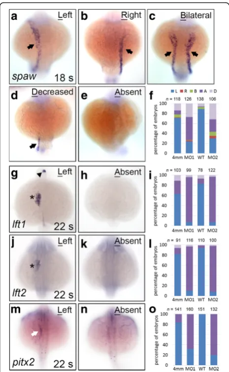
klf8deficiency affects the level and pattern of expression for genes in the Nodal signalling pathway
Genes encoding Nodal signalling components, including
spaw,lft1,lft2, andpitx2, were asymmetrically expressed in the left side of the diencephalon, heart, or lateral plate mesoderm (LPM) during the 18–22 s stages in zebrafish embryos (Fig. 2a–m). Disruption in the expression of left-side specific Nodal signalling genes results in organ heterotaxy. We observed thatspawexpression in the left LPM was either decreased (no expression in the anterior LPM and low expression in the posterior LPM, 27%) or absent (46%) in many klf8-MO1atg-injected embryos (Fig. 2d–f ). A similar effect (32% decreased, 25% absent, 8% bilateral, 5% right) was observed following klf8 -MO2atg injection (Fig. 2b-f ). Consistent with these re-sults, the expression of genes downstream of spaw(lft1,
lft2, and pitx2) was also absent or decreased in the left diencephalon, heart, and LPM of most klf8-MO1atg or
klf8-MO2atg-injected embryos (Fig. 2h–o). Additionally, we injected three splicing MOs (MODO2, MOAC, and MODo) to block splicing of klf8 mRNA (Additional file 1: Figure S1, A). RT-PCR indicated that splicing of klf8
mRNA was effectively disrupted in MOsp-MOs-injected embryos at 24 hpf (Additional file 1: Figure S1, B). Ex-pression of spaw in the left LPM was either decreased (8%) or eliminated (63%) in most 18 s stage embryos injected with MOsp-MOs(Additional file 1: Figure S1, C).
Klf8 was previously shown to repress p53 expression and inducemetexpression to modulate the development of Purkinje cells and proliferation of granule cells [35]. To confirm that defective left-right patterning did not arise from induction of p53 due to klf8 deficiency, we analysed heart looping and gene expression of Nodal sig-nalling components in embryos that were co-injected withp53-MOsp, alongsideklf8-MO1atgorklf8-MO2atg.
Of the embryos co-injected with p53-MOsp, together with klf8-MO1atg or klf8-MO2atg, 35–39% exhibited a no-loop heart at 72 hpf, similar to embryos injected with
klf8 MOs alone (Additional file 2: Figure S2, A). Like-wise, expression levels ofspaw,lft1, lft2, andpitx2 were reduced or eliminated in the left LPM, diencephalon, and heart of high percentages of co-injected embryos
during the 18–22 s stages (Additional file 2: Figure S2, B-E). We also found that the expression levels of gata5
and oep (which are known to be expressed in the LPM at the 22 s stage) were unaffected by klf8 knockdown (Additional file 2: Figure S2, F-M). Together these data clearly indicate that increased p53 expression and apop-tosis are not responsible for the decreased expression of genes involved in Nodal signalling.
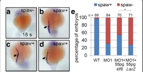
To further confirm that the decrease in spaw expres-sion is a consequence of klf8 loss-of-function, we per-formed rescue experiments by co-injecting embryos with
klf8-MO1atg and klf8 mRNA. Approximately 69% of
klf8-MO1atg-injected embryos exhibited eliminated or
Fig. 2Genes encoding Nodal signalling components exhibited
decreased or abolished expression inklf8morphants. The majority of
decreased spaw expression in the left LPM at the 18 s stage (Fig. 3a, e). However, this proportion showed a sta-tistically significant reduction to 44% for embryos co-injected with klf8-MO1atg and klf8 mRNA (Fig. 3b-e). Taken together, these results demonstrate that klf8 loss-of-function causes downregulation of Nodal signalling component genes.
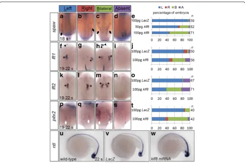
Overexpression ofklf8mRNA causes bilateral expression of genes involved in Nodal signalling
Since klf8 knockdown reduced expression of genes in-volved in Nodal signalling, we hypothesized thatklf8 over-expression may have the opposite effect. While the majority of embryos injected with 100 pg ofLacZmRNA expressed spaw exclusively in the left LPM at the 18 s stage, 24% of embryos injected with 50 pg and 51% of em-bryos injected with 100 pg ofklf8mRNA expressedspaw
bilaterally in the LPM (Fig. 4c, e). Thus, a dose-dependent effect ofklf8expression was revealed. Moreover, embryos overexpressing klf8also frequently exhibited bilateral ex-pression patterns of lft1, lft2, and pitx2in the dienceph-alon, heart, and LPM at 19–22 s stages (Fig. 4h–t). On the other hand, ntl expression in the notochord was not al-tered in 22 s stage embryos overexpressing klf8as com-pared to LacZ-overexpressing embryos (Fig. 4u-w). In order to investigate whether the zinc finger DNA binding domain of Klf8 is involved in regulating the expression pattern of spaw or its downstream genes, we injected mRNA for klf8lacking the zinc finger DNA binding do-main (klf8△zf). We found that injection of 100 pgklf8△zf
only induced a low percentage of embryos to exhibit bilat-eral expression of spaw (6.3%), lft1(3.6%), lft2 (1.6%) or
pitx2(11.9%), compared to higher rates of bilateral spaw
(48.3%),lft1(31.6%), lft2(37.5%) or pitx2(44.3%) expres-sion in klf8 injected embryos at 18 s or 19–22 s stages (Additional file 3: Figure S3). These results demonstrate
that overexpression of klf8 does not affect the midline structure, but induces ectopic expression of spawand its downstream genes when the Klf8 zinc finger DNA binding domain is intact.
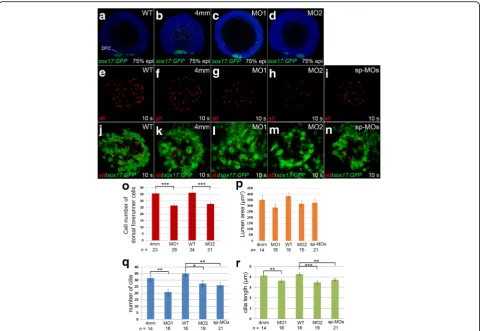
klf8deficiency affects morphogenesis of Kupffer’s vesicle and asymmetriccharonexpression
Since asymmetric flow, generated by rotation of cilia within KV, is essential to initiate left-right asymmetric patterning, and KV is derived from dorsal forerunner cells (DFCs), we then investigated whether klf8 knock-down affected cilia or DFC number during KV morpho-genesis. Individual klf8-MOs or control MO were microinjected into one- or two-cell Tg(sox17:gfp) zy-gotes, and immunofluorescence was conducted using anti-GFP antibody. We found that the number of DFCs at the dorsal margin was significantly reduced in klf8 -MO1atg- (average of 26.6 DFCs) orklf8-MO2atg- (average of 27.6 DFCs) injected Tg(sox17:gfp) embryos as com-pared to wild type (average of 36.2 DFCs) orklf8-4 mm MO1- (average of 35.7 DFCs) injected embryos at 75% epiboly (Fig. 5a-d, o). Cilia were then detected by im-munofluorescence staining of 10s stage embryos using an anti-acetylated tubulin antibody. KV lumen size was smaller, but not significantly so, inTg(sox17:gfp)embryos injected with klf8-MO1atg, klf8-MO2atg, or MOsp-MOsas compared to wild type orklf8-4 mm MO1-injected con-trol embryos at 10s stage (Fig. 5e-n, p). Significantly re-duced number and length of KV cilia were detected in embryos injected with different klf8-MOs as compared to wild type and control embryos (Fig. 5q, r). Since asymmetric charon expression on the right side of the KV was influenced by strength and direction of KV flow [40], we also examined whetherklf8knockdown affected asymmetriccharonexpression around KV. The majority (61% for MO1atg, 57% for MO2atg) of embryos injected with different klf8-MOs revealed symmetric charon ex-pression with reduced exex-pression area around KV as compared to wild type and control embryos at the 10s stage (Additional file 4: Figure S4). These results indicate that KV morphogenesis, cilia length, cilia number and asymmetric charon expression were affected in klf8
knockdown embryos.
Generation ofklf8mutant by a CRISPR-Cas9 system In order to confirm our morphant results, we generated
klf8 mutants using a CRISPR-Cas9 system. Although we designed threeklf8sgRNAs targeting to exon 2 or exon 3, onlyklf8sgRNA1, which targets to exon 2, was successful in producing mutants. Administration of sgRNA1 induced efficient deletion or insertion of bases in exon 2 and re-sulted in two klf8 mutant alleles (klf8d25 and klf8i17) (Fig. 6a). The klf8d25 mutant had a 25 bp deletion, which produced a 35 amino acid-long misframed Klf8
Fig. 3Reducedspawexpression inklf8morphants was partially
rescued by co-injection ofklf8mRNA. Representative embryos showing
spawexpression in the left LPM (spaw+) or absentspawexpression (spaw-) are shown (a-d). Percentages of embryos with asymmetricspaw
expression or nospawexpression are shown with indicated treatments (e). Statistical significance was determined by Fisher’s Exact
protein, while theklf8i17mutant had a 17 bp insertion that generated a 49 amino acid-long misframed Klf8 pro-tein (Fig. 6b).
Of note, in 24 hpf homozygous F3 embryos of
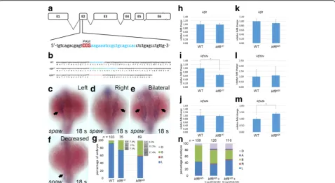
klf8d25 and klf8i17 mutants, we did not observe smaller eyes and abnormal cerebellar morphology that were detected in klf8 morphant embryos [35]. Next, we investigated whether spaw expression was affected in klf8 mutant embryos. Bilateral spaw expression was more frequently observed in embryos from the inter-cross of respective klf8d25 (27 out of 69, 39.1%) or
klf8i17 (9 out of 35, 25.7%) F2 heterozygous mutants as compared to wild type embryos (7%) at 18 s stage (Fig. 6c-g). In order to evaluate the genotype of F3 embryos from klf8i17 and klf8d25 mutants with bilat-eral spaw expression, we sequenced seven klf8i17 and 18 klf8d25embryos with the phenotype. From sequen-cing data, we obtained two (28.6%) wild type, three (42.9%) heterozygotes and two (28.5%) homozygotes from a total of seven klf8i17 F3 embryos, as well as three (16.7%) wild type, seven (38.9%) heterozygotes
and eight (44.4%) homozygotes from a total of 18
klf8d25 embryos. We then deduced that 7.3% of klf8i17
or 17.4% of klf8d25 F3 embryos were homozygous mu-tant embryos that also had bilateral spaw expression. This observation was based on the following calcula-tion [0.285 (% of sequenced embryos that were homo-zygous) × 9 (total number with bilateral spaw
expression) / 35 (total number of embryos) = 7.3% for klf8i17; 0.444 × 27 / 69 = 17.4% for klf8d25]. The other F3 embryos that exhibited bilateral spaw ex-pression were also deduced to be either heterozygous mutant embryos (11% in klf8i17, 15.2% in klf8d25) or sibling wild type (7.4% in klf8i17, 6.5% in klf8d25) based on similar calculations.
Because human KLF8, KLF3 and KLF12 form a sub-group in phylogenetic tree analysis due to the pres-ence of CtBP-binding sites [41], we wondered whether expression of zebrafish klf3, klf12a, or klf12b
may be upregulated to compensate for klf8deficiency. We discovered substantial upregulation of klf12b in
klf8d25 F5 homozygous mutant embryos and
Fig. 4Overexpression ofklf8mRNA caused bilateral expression of Nodal signalling component genes. Injection ofklf8mRNA induced bilateral
downregulation of klf12ain klf8i17 F5 homozygous mutant embryos at 10–12 s stages, while no alteration ofklf3 expres-sion was observed in either klf8d25 or klf8i17 mutant embryos (Fig. 6h-m). Subsequently, we knocked down
klf12b in klf8d25 F6 homozygous mutant embryos and
evaluated the spaw expression pattern at 18 s stage (Fig. 6n). In klf8d25 F6 homozygous mutant embryos, bilateral (50.3%) and decreased (6.3%) spaw expres-sion patterns were detected. In homozygous mutant embryos injected with 5 or 10 ng klf12b MO, we found a dose-dependent reduction that did not reach significance in the percentage (42.1% for 5 ng, 29.3% for 10 ng) of embryos with bilateral spaw expression, which was accompanied by the increased occurrence of right (1.6% for 5 ng, 1.7% for 10 ng), decreased (15.9% for 5 ng, 16.4% for 10 ng) or absent (4.0% for 5 ng, 3.4% for 10 ng) spaw expression patterns.
These results indicate that although klf8 mutant em-bryos display different spaw expression patterns than
morphant embryos, this effect may be partly attributed to a compensatory induction ofklf12bexpression.
Discussion
Establishing asymmetric spawexpression in the LPM is essential for left-right patterning in zebrafish embryos. Expression of spaw is first apparent in bilateral cells flanking KV between the 4–6 s stages, while asymmetric
spaw expression emerges in the posterior LPM during the 10–12 s stages, and extends to the anterior LPM by the 18 s stage [22]. Knockdown of spawabolishesspaw
expression in the LPM but not in peri-KV domains [21], suggesting that autoregulation of spaw occurs only in the left LPM. We used morpholino antisense oligomers to knockdown klf8, and found that spaw expression in the left LPM was reduced or eliminated in the majority of 18–22 s stage morphants (Fig. 2, Additional file 2: Figure S2). Spaw activates expression of itself, as well as
lft1, lft2, and pitx2 in the left LPM during the
Fig. 5Knockdown ofklf8affected dorsal forerunner cell number, KV cilia number and length. Cell number of dorsal forerunner cells (DFCs) was
affected inklf8-MO1atg- orklf8-MO2atg-injected embryos as compared toklf8-4 mm MO1-injectedTg(sox17:gfp)control embryos (4 mm) at 75% epiboly (a-d). Images of KV cilia stained with acetylated tubulin antibody inklf8morphants (g-i) and control embryos (e, f) at 10s stage (e-i). Images of acetylated tubulin stained KV cilia and GFP stained DFCs inTg(sox17:gfp)embryos injected with differentklf8MOs (l-n),klf8-4 mm MO1 (k) or wild type (j) embryos at 10s stage (j-n). DFC number (o), KV cilia number (q), length (r) but not lumen area (p) were affected inklf8
segmentation stage [22]. As such, the observed reduction or elimination oflft1,lft2, andpitx2in the left dienceph-alon, heart or LPM at 18–22 s stages in the majority of
klf8 morphants was not unexpected (Fig. 2, Additional file 2: Figure S2). Overall, defects in the expression of
spawand its downstream genes, and the subsequent de-fects in internal organ patterning observed inklf8 mor-phants are consistent with those detected in spaw
morphants [22]. Although spaw/sfw mutant embryos more frequently displayed a D-loop heart (68%), com-pared to klf8 morphants, the difference may be attrib-uted to heart specific actomyosin activity. Furthermore, insfwmutant embryos, expression of laterality genes in-cluding lft1, lft2, and pitx2 were lost, and spaw expres-sion did not propagate toward the anterior of the left LPM [42]. In our study, we saw that overexpression of
klf8 but not klf8△zf resulted in bilateral expression of
spaw, lft1, lft2, and pitx2 at 18 s or 19–22 s stages (Fig. 4, Additional file 3: Figure S3), demonstrating a requirement for the Klf8 zinc finger DNA binding
domain. Expression of ntl in the notochord was found to be unaltered in klf8-overexpressing embryos, sug-gesting that the midline structure is in intact. Overall, our results demonstrate that Klf8 may regulate asym-metric spaw expression in the left LPM, which in turn affects left-side specific expression of lft1, lft2,
and pitx2 in zebrafish embryos.
Proper morphogenesis of KV is also important for ini-tial asymmetric spaw expression in the posterior LPM. The development of KV involves the formation of DFCs from surface epithelial cells, ingression at the dorsal germ ring margin, DFC migration, formation of rosette-like epithelial structures, coalescence of epithelial ro-settes, and differentiation of ciliated KV with interior lumen [15, 43]. Both Tbx16 and Ntl were shown to regu-late a mesenchymal to epithelial transition that partici-pates in the formation of rosette-like epithelia [44]. Wnt11- and Prickle1a-mediated planar cell polarity sig-nalling, as well as Cnpy1-mediated FGF sigsig-nalling, were shown to regulate cell adhesion between adjacent dorsal
Fig. 6Generation ofklf8mutants by CRISPR-Cas9 gene editing and the effect onspawexpression.aklf8genomic structure withklf8sgRNA (blue
lettering) targeted to exon 2. Protospacer adjacent motif (PAM) sequence is shown inred.bNucleotide and predicted amino acid sequences of
klf8in wild type,klf8I17andklf8d25mutants are shown. Deleted nucleotides are shown by ared dashed line, while inserted nucleotides are shown ingreen lettering. Representative images of embryos with differentspawexpression patterns in the LPM at 18 s stage (arrow;c-f).gPercentage of embryos displayed left (L), right (R), decreased (D) or bilateral (B) expression ofspawin the LPM from intercross of respectiveklf8d25orklf8i17F2 heterozygous mutants. Deduced percentage of wild type (+/+), heterozygote (+/−) or homozygote (−/−) genotype of embryos from intercross of respectiveklf8d25orklf8i17F2 heterozygous mutants exhibited bilateralspawexpression pattern. Expression levels ofklf3(h, k),klf12a(i, l) orklf12b
(j, m) were compared between wild type and respectiveklf8d25andklf8i17F5 homozygous mutant embryos at 10–12 s stages (h-m). Knockdown ofklf12breduced the percentage of embryos with bilateralspawexpression in the LPM ofklf8d25F6 homozygous mutant embryos, but
forerunner cells to maintain cluster formation [45, 46]. Defects in these signalling events resulted in small KV lumen, with shortened and decreased number of KV cilia. In klf8 morphants, a similar smaller KV lumen, with decreased number and length of KV cilia was fre-quently observed at the 10s stage. These abnormal struc-tures may result from lower number of DFCs that was observed in 75% epiboly morphants (Fig. 5). Whether Klf8 may participate in Wnt11- and Prickle1a-mediated planar cell polarity signalling, or Cnpy1-mediated FGF signalling to modulate DFC cluster formation, remains to be determined.
Zebrafish KV architecture is asymmetric along the anterior-posterior axis, with more ciliated cells in the anter-ior region. Furthermore, the positioning of the basal body of motile cilia at the posterior end of the epithelial cells may result in cilia tilting [17, 47]. These motile cilia then use a vortical motion to generate swirling fluid flow consist-ing of a relatively stronger leftward flow across the anterior pole of KV and a weaker rightward flow at the posterior end [16, 18]. Based on experimental tracking of native par-ticles within the KV of wild type,did−/−mutant anddnah7
morphants, and simulated flow by mathematically model-ling, it was determined that a threshold of 30 cilia, with dorsal anterior clustering, is essential to generate proper swirling flow in the anti-clockwise direction [40]. In control embryos, with strong left-sided flow across the anterior pole of KV, asymmetric expression is established for charonin the right side of the KV, andspawin the left LPM. In em-bryos with non-directional flow, symmetriccharon expres-sion and a lack ofspawexpression may be found. Embryos without motile cilia, and therefore no KV flow, may exhibit symmetric and slightly weakercharonexpression and bilat-eral spaw expression in the posterior LPM [40]. In klf8
morphant embryos, injected with differentklf8MOs, a sig-nificantly reduced number of cilia (< 30), with random dis-tribution was detected (Fig. 5). KV with such a cellular architecture may exhibit a weak and homogenous fluid flow, resulting in the symmetriccharonexpression around KV that was detected in the majority of klf8 morphants, and leading to drastically reducedspaw expression at late somite stage (Additional file 4: Figure S4, Fig. 2). Overall, our results clearly indicate that Klf8 is required for normal KV morphogenesis, which is known to be critical for initiat-ing asymmetricspawexpression in the left LPM.
In mouse and chick, BMP signalling plays either a positive or negative role in regulating asymmetricNodal
expression. Moreover, the presence of BMP antagonists, such as Noggin, Chordin, or Caronte, can relieve BMP-inhibition to promote asymmetric Nodal expression in the left LPM [48–51]. In zebrafish embryos, heat– acti-vated BMP2bexpression inhibitsspawexpression, while heat-activatednoggin3induced bilateralspawexpression, indicating that BMP signalling is required to repress
spawexpression in the right LPM of early segmentation stage embryos, [25]. In addition, expression of Lft1 in the midline, and Lft2 in the left cardiac field, serve to generate a posterior or anterior barrier to restrict Spaw activity to the left LPM during segmentation stages in zebrafish embryos [27]. Dvr1, a zebrafish Vg1 ortholog, was also shown to facilitate the transfer ofspaw expres-sion from the peri-KV region to the left LPM. Thus, reduced or absent expression of spaw and downstream
lft1 and lft2 in the LPM, diencephalon, notochord, or heart were detected indvr1morphants [52]. In order to investigate whether Klf8 may regulate asymmetric
spaw expression via modulation of expressions of
bmp2b or dvr1, we then compared expression of
these two genes between klf8 morphants and control embryos (Additional file 5: Figure S5). Similar bmp2b
expression level around tailbud region was identified in 3 s stage wild type and embryos injected with klf8 -MO1atg, klf8-MO2atg or klf8- 4 mm MO1. Likewise, no alteration of dvr1 expression around the tailbud region was detected in wild type and embryos injected with klf8-MO1atg, klf8-MO2atg or klf8-4 mm MO1 at bud stage. Thus, Klf8 does not act via BMP2b or Dvr1 signalling pathway to regulate asym-metric spaw expression, and the underlying mechan-ism remains to be determined.
In addition to our studies with klf8 morphants, we generated klf8 mutants by a CRISPR-Cas9 system (Fig. 6). Intriguingly, obvious phenotypic differences were found between morphants and mutants. In 24 hpf homozygous F3 embryos of klf8d25 and klf8i17
mutants, we did not observe smaller eyes and abnor-mal cerebellar morphology that were detected in klf8
morphant embryos [35]. In addition, bilateral spaw
expression was detected in the LPM of klf8d25 and
klf8i17 mutants at the 18 s stage (Fig. 6). Discrepant phenotypes between mutants created by TALENs or CRISPR-Cas genome editing systems and antisense morpholino mediated-morphants have been frequently encountered. Previously, differences have been attrib-uted to off-target effects of morpholinos [53], or com-pensatory effects, which have been described in vasculature development [54], reproduction [55], or neurogenesis [56]. With regard to the two klf8 mutant alleles from our study, more klf8d25 mutant embryos showed bilateral spaw expression in the LPM, com-pared to klf8i17 mutants (Fig. 6g, n). This difference in outcome may correlate with aberrant upregulation of
klf12b in klf8d25 that was further confirmed by klf12b
compensate instmn4△5but notstmn4△4mutants [56]. In our study, we observed that in response toklf8deficiency,
klf12b, a member of a subgroup of KLF family with a CtBP
interaction site, was induced to compensate for the loss of
klf8. However aberrant upregulation ofklf12bfurther re-sulted in bilateral spaw expression. On the contrary, downregulation ofklf12awas detected inklf8i17mutants (Fig. 6). In these mutants, bilateral spaw expression was observed to a lesser degree, suggesting that klf12a may have undergone functional divergence withklf12b, and as such,klf12amay play a role in restrictingspawexpression to the left side of embryos. Overall,klf8mutant embryos showed bilateralspaw expression, which was quite differ-ent from klf8morphants that exhibited reduced or elimi-nated spaw expression in the LPM. This dissimilar phenotype may have been partly related to the compensa-tory induction of klf12b expression in the mutant embryos.
Conclusions
In this report, we have demonstrated a novel role for zebrafish Klf8 in left-right asymmetric patterning. Dur-ing gastrulation, Klf8 may regulate DFC cell number to control proper KV morphogenesis, which is essential to initiate asymmetric spaw expression in the left LPM. During somitogenesis, Klf8 may further modulate asym-metric spaw expression in the left LPM to ensue asym-metric organ positioning.
Additional files
Additional file 1: Figure S1.Knockdown ofklf8expression by splicing
morpholino oligomers resulted in embryos with reduced or absentspaw
expression in the left LPM.Aklf8genomic structure showing position of translational morpholino oligomers (klf8-MO1atg,klf8-MO2atg) and splicing morpholino oligomers (klf8DO2,klf8AC,klf8DO). Arrows indicate the positions of
forward and reverse primers.BRT-PCR showing the efficacy ofklf8splicing morpholino oligomers.CThe majority of embryos injected withklf8splicing MOs had decreased or absentspawexpression in the left LPM. (TIFF 153 kb)
Additional file 2: Figure S2.Heart looping and downregulated
expression ofspawand its downstream genes were not caused by induction ofp53expression inklf8morphants. Embryos co-injected with p53-MOspand
klf8-MO1atgorklf8-MO2atgdisplayed no-loop or L-loop heart defects at 72 hpf
(A). The majority of embryos co-injected with p53-MOspandklf8-MO1atgor
klf8-MO2atgexhibited decreased or absent expression ofspaw(B),lft1(C),lft2
(D), orpitx2(E) in the left LPM, diencephalon or heart at the 18–22 s stages. Expression levels ofgata5andoepwhich are known to be expressed in the LPM at the 22 s stage were unaffected byklf8knockdown (F-M). (TIFF 797 kb)
Additional file 3: Figure S3.Overexpression ofklf8but notklf8△zf
mRNA induced bilateral expression of Nodal signalling component genes. Percentages of embryos injected with either 100 pg ofklf8or
klf8△zfmRNA that exhibit left (L), right (R), decreased (D) or bilateral (B) expression ofspawin the LPM,lft1in the diencephalon and heart,lft2
in the heart, andpitx2in the LPM at 18 s or 19–22 s stages. (TIFF 74 kb)
Additional file 4: Figure S4.Symmetriccharonexpression around KV
was observed in the majority ofklf8morphants. Representative images of embryos showing strongercharonexpression on the right side (A) or left side (B) and symmetriccharonexpression on both sides (C) of KV are shown. Quantification of differentcharonexpression patterns in embryos
injected with differentklf8MOs, control MO or wild type embryo is shown (D). Statistical significance was determined by Student’st-test. *p< 0.05. Error bars indicate standard deviation. (TIFF 397 kb)
Additional file 5: Figure S5.Expression level ofbmp2bordvr1around tailbud region was not affected byklf8knockdown. Representative images show similar expression level ofbmp2b(A-D) ordvr1(E-H) around the tailbud region in the wild type or embryos injected withklf8-MO1atg,klf8-MO2atgor
klf8-4 mm MO1 at 3 s or bud stages. (TIFF 382 kb)
Abbreviations
ASE:Left-side specific enhancers; CRISPR-Cas: Clustered regularly interspaced short palindromic repeats- CRISPR-associated system; CtBP: C-terminal binding protein; DFCs: Dorsal forerunner cells; Hpf: Hours post fertilization; klf8: krüppel-like factor 8; KV: Kupffer’s vesicle; lft1: lefty1; lft2: lefty2; LPM: Lateral plate mesoderm; MO: Morpholino oligomer; Ndr: Nodal-related genes; RT: Room temperature; RT-PCR: Reverse transcription PCR; RT-qPCR: Reverse transcription quantitative real-time PCR; S: Somite
Acknowledgments
We thank the members of the Core Facility of the Institute of Cellular and Organismic Biology, Academia Sinica, for their assistance with DNA sequencing and confocal imaging. We thank the Taiwan Zebrafish Core Facility at Academia Sinica (TZCAS) for providing the ASAB wild type strain. We thank ZIRC and TZCAS for providingTg(sox17:gfp)s870/+fish line. We thank Dr. M. Rebagliati for providing
full-lengthspawcDNA. We thank Ms. Mei-Chen Chen for fish maintenance.
Funding
This work was supported by Academia Sinica Innovative Translational Agricultural Research Program [542300 to S.P.L.H.] and the Ministry of Science and Technology, Taiwan [MOST 105-2313-B-001-005-MY3 to S.P.L.H.].
Availability of data and materials
All materials are available by the corresponding author.
Authors’contributions
CYL, MYT, YCC, YRL, YHL, YFL, HCL, HWL, CHY performed experiments; CYL, MYT, CJH and SPLH analysed data; SPLH conceived the project and wrote the manuscript. All authors read and approved the final manuscript.
Ethics approval and consent to participate
All animal procedures were approved by the Institutional Animal Care and Use Committee of Academia Sinica (Protocol ID: 15-12-918).
Consent for publication
Not applicable.
Competing interests
The authors declare that they have no competing interests.
Publisher’s Note
Springer Nature remains neutral with regard to jurisdictional claims in published maps and institutional affiliations.
Author details
1
Department of Bioscience and Biotechnology, National Taiwan Ocean University, Keelung, Taiwan.2Graduate Institute of Life Sciences, National Defence Medical Center, National Defence University, Neihu, Taipei, Taiwan. 3Department of Life Science, National Taiwan University, Taipei, Taiwan.
4
Received: 5 April 2017 Accepted: 7 July 2017
References
1. Bisgrove BW, Morelli SH, Yost HJ. Genetics of human laterality disorders: insights from vertebrate model systems. Annu Rev Genomics Hum Genet. 2003;4:1–32.
2. Kosaki K, Casey B. Genetics of human left-right axis malformations. Semin Cell Dev Biol. 1998;9:89–99.
3. Hamada H, Meno C, Watanabe D, Saijoh Y. Establishment of vertebrate left-right asymmetry. Nat Rev Genet. 2002;3:103–13.
4. Shiratori H, Hamada H. The left-right axis in the mouse: from origin to morphology. Development. 2006;133:2095–104.
5. Hirokawa N, Tanaka Y, Okada Y. Left-right determination: involvement of molecular motor KIF3, cilia, and nodal flow. Cold Spring Harb Perspect Biol. 2009;1:a000802.
6. Nonaka S, Tanaka Y, Okada Y, Takeda S, Harada A, Kanai Y, Kido M, Hirokawa N. Randomization of left-right asymmetry due to loss of nodal cilia generating leftward flow of extraembryonic fluid in mice lacking KIF3B motor protein. Cell. 1998;95:829–37.
7. Neugebauer JM, Amack JD, Peterson AG, Bisgrove BW, Yost HJ. FGF signalling during embryo development regulates cilia length in diverse epithelia. Nature. 2009;458:651–4.
8. Thomas J, Morle L, Soulavie F, Laurencon A, Sagnol S, Durand B. Transcriptional control of genes involved in ciliogenesis: a first step in making cilia. Biol Cell. 2010;102:499–513.
9. Nakamura T, Mine N, Nakaguchi E, Mochizuki A, Yamamoto M, Yashiro K, Meno C, Hamada H. Generation of robust left-right asymmetry in the mouse embryo requires a self-enhancement and lateral-inhibition system. Dev Cell. 2006;11:495–504.
10. Schier AF. Nodal signaling in vertebrate development. Annu Rev Cell Dev Biol. 2003;19:589–621.
11. Saijoh Y, Adachi H, Sakuma R, Yeo CY, Yashiro K, Watanabe M, Hashiguchi H, Mochida K, Ohishi S, Kawabata M, Miyazono K, Whitman M, Hamada H. Left-right asymmetric expression of lefty2 and nodal is induced by a signaling pathway that includes the transcription factor FAST2. Mol Cell. 2000;5:35–47. 12. Shiratori H, Sakuma R, Watanabe M, Hashiguchi H, Mochida K, Sakai Y,
Nishino J, Saijoh Y, Whitman M, Hamada H. Two-step regulation of left-right asymmetric expression of Pitx2: initiation by nodal signaling and maintenance by Nkx2. Mol Cell. 2001;7:137–49.
13. Ryan AK, Blumberg B, Rodriguez-Esteban C, Yonei-Tamura S, Tamura K, Tsukui T, de la Pena J, Sabbagh W, Greenwald J, Choe S, Norris DP, Robertson EJ, Evans RM, Rosenfeld MG, Izpisua Belmonte JC. Pitx2 determines left-right asymmetry of internal organs in vertebrates. Nature. 1998;394:545–51.
14. Essner JJ, Amack JD, Nyholm MK, Harris EB, Yost HJ. Kupffer’s vesicle is a ciliated organ of asymmetry in the zebrafish embryo that initiates left-right development of the brain, heart and gut. Development. 2005;132:1247–60. 15. Oteiza P, Koppen M, Concha ML, Heisenberg CP. Origin and shaping of the
laterality organ in zebrafish. Development. 2008;135:2807–13. 16. Amack JD. Salient features of the ciliated organ of asymmetry.
BioArchitecture. 2014;4:6–15.
17. Wang G, Cadwallader AB, Jang DS, Tsang M, Yost HJ, Amack JD. The Rho kinase Rock2b establishes anteroposterior asymmetry of the ciliated Kupffer’s vesicle in zebrafish. Development. 2011;138:45–54.
18. Smith DJ, Montenegro-Johnson TD, Lopes SS. Organized chaos in Kupffer’s vesicle: how a heterogeneous structure achieves consistent left-right patterning. BioArchitecture. 2014;4:119–25.
19. Bisgrove BW, Snarr BS, Emrazian A, Yost HJ. Polaris and Polycystin-2 in dorsal forerunner cells and Kupffer’s vesicle are required for specification of the zebrafish left-right axis. Dev Biol. 2005;287:274–88.
20. Borovina A, Superina S, Voskas D, Ciruna B. Vangl2 directs the posterior tilting and asymmetric localization of motile primary cilia. Nat Cell Biol. 2010;12:407–12. 21. Wang X, Yost HJ. Initiation and propagation of posterior to anterior (PA)
waves in zebrafish left-right development. Dev Dyn. 2008;237:3640–7. 22. Long S, Ahmad N, Rebagliati M. The zebrafish nodal-related gene southpaw
is required for visceral and diencephalic left-right asymmetry. Development. 2003;130:2303–16.
23. Ahmad N, Long S, Rebagliati M. A southpaw joins the roster: the role of the zebrafish nodal-related gene southpaw in cardiac LR asymmetry. Trends Cardiovasc Med. 2004;14:43–9.
24. Hashimoto H, Rebagliati M, Ahmad N, Muraoka O, Kurokawa T, Hibi M, Suzuki T. The Cerberus/Dan-family protein Charon is a negative regulator of Nodal signaling during left-right patterning in zebrafish. Development. 2004; 131:1741–53.
25. Chocron S, Verhoeven MC, Rentzsch F, Hammerschmidt M, Bakkers J. Zebrafish Bmp4 regulates left-right asymmetry at two distinct developmental time points. Dev Biol. 2007;305:577–88.
26. Smith KA, Noel E, Thurlings I, Rehmann H, Chocron S, Bakkers J. Bmp and nodal independently regulate lefty1 expression to maintain unilateral nodal activity during left-right axis specification in zebrafish. PLoS Genet. 2011;7: e1002289.
27. Lenhart KF, Lin SY, Titus TA, Postlethwait JH, Burdine RD. Two additional midline barriers function with midline lefty1 expression to maintain asymmetric Nodal signaling during left-right axis specification in zebrafish. Development. 2011;138:4405–10.
28. Kaczynski J, Cook T, Urrutia R. Sp1- and Kruppel-like transcription factors. Genome Biol. 2003;4:206.
29. Pearson R, Fleetwood J, Eaton S, Crossley M, Bao S. Kruppel-like transcription factors: a functional family. Int J Biochem Cell Biol. 2008;40:1996–2001. 30. Lahiri SK, Zhao J. Kruppel-like factor 8 emerges as an important regulator of
cancer. Am J Transl Res. 2012;4:357–63.
31. van Vliet J, Turner J, Crossley M. Human Kruppel-like factor 8: a CACCC-box binding protein that associates with CtBP and represses transcription. Nucleic Acids Res. 2000;28:1955–62.
32. Zhang P, Basu P, Redmond LC, Morris PE, Rupon JW, Ginder GD, Lloyd JA. A functional screen for Kruppel-like factors that regulate the human gamma-globin gene through the CACCC promoter element. Blood Cells Mol Dis. 2005;35:227–35.
33. Funnell AP, Mak KS, Twine NA, Pelka GJ, Norton LJ, Radziewic T, Power M, Wilkins MR, Bell-Anderson KS, Fraser ST, Perkins AC, Tam PP, Pearson RC, Crossley M. Generation of mice deficient in both KLF3/BKLF and KLF8 reveals a genetic interaction and a role for these factors in embryonic globin gene silencing. Mol Cell Biol. 2013;33:2976–87.
34. Zhao J, Bian ZC, Yee K, Chen BPC, Chien S, Guan J-L. Identification of transcription factor KLF8 as a downstream target of focal adhesion kinase in its regulation of cyclin D1 and cell cycle progression. Mol Cell. 2003;11:1503–15.
35. Tsai MY, Lu YF, Liu YH, Lien HW, Huang CJ, Wu JL, Hwang SP. Modulation of p53 and met expression by Kruppel-like factor 8 regulates zebrafish cerebellar development. Dev Neurobiol. 2015;75:908–26.
36. Kimmel CB, Ballard WW, Kimmel SR, Ullmann B, Schilling TF. Stages of embryonic development of the zebrafish. Dev Dyn. 1995;203:253–310. 37. Chen J, Ruan H, Ng SM, Gao C, Soo HM, Wu W, Zhang Z, Wen Z, Lane DP,
Peng J. Loss of function of def selectively up-regulates Delta113p53 expression to arrest expansion growth of digestive organs in zebrafish. Genes Dev. 2005;19:2900–11.
38. Jao LE, Wente SR, Chen W. Efficient multiplex biallelic zebrafish genome editing using a CRISPR nuclease system. Proc Natl Acad Sci U S A. 2013;110: 13904–9.
39. Bustin SA, Benes V, Garson JA, Hellemans J, Huggett J, Kubista M, Mueller R, Nolan T, Pfaffl MW, Shipley GL, Vandesompele J, Wittwer CT. The MIQE guidelines: minimum information for publication of quantitative real-time PCR experiments. Clin Chem. 2009;55:611–22.
40. Sampaio P, Ferreira RR, Guerrero A, Pintado P, Tavares B, Amaro J, Smith AA, Montenegro-Johnson T, Smith DJ, Lopes SS. Left-right organizer flow dynamics: how much cilia activity reliably yields laterality? Dev Cell. 2014;29: 716–28.
41. McConnell BB, Yang VW. Mammalian Kruppel-like factors in health and diseases. Physiol Rev. 2010;90:1337–81.
42. Noel ES, Verhoeven M, Lagendijk AK, Tessadori F, Smith K, Choorapoikayil S, den Hertog J, Bakkers J. A Nodal-independent and tissue-intrinsic mechanism controls heart-looping chirality. Nat Commun. 2013;4:2754. 43. Matsui T, Ishikawa H, Bessho Y. Cell collectivity regulation within migrating
cell cluster during Kupffer’s vesicle formation in zebrafish. Front Cell Dev Biol. 2015;3:27.
44. Amack JD, Wang X, Yost HJ. Two T-box genes play independent and cooperative roles to regulate morphogenesis of ciliated Kupffer’s vesicle in zebrafish. Dev Biol. 2007;310:196–210.
46. Matsui T, Thitamadee S, Murata T, Kakinuma H, Nabetani T, Hirabayashi Y, Hirate Y, Okamoto H, Bessho Y. Canopy1, a positive feedback regulator of FGF signaling, controls progenitor cell clustering during Kupffer’s vesicle organogenesis. Proc Natl Acad Sci U S A. 2011;108:9881–6.
47. Wang G, Manning ML, Amack JD. Regional cell shape changes control form and function of Kupffer’s vesicle in the zebrafish embryo. Dev Biol. 2012;370:52–62. 48. Schlange T, Arnold HH, Brand T. BMP2 is a positive regulator of Nodal
signaling during left-right axis formation in the chicken embryo. Development. 2002;129:3421–9.
49. Mine N, Anderson RM, Klingensmith J. BMP antagonism is required in both the node and lateral plate mesoderm for mammalian left-right axis establishment. Development. 2008;135:2425–34.
50. Fujiwara T, Dehart DB, Sulik KK, Hogan BL. Distinct requirements for extra-embryonic and extra-embryonic bone morphogenetic protein 4 in the formation of the node and primitive streak and coordination of left-right asymmetry in the mouse. Development. 2002;129:4685–96.
51. Raya A, Izpisua Belmonte JC. Unveiling the establishment of left-right asymmetry in the chick embryo. Mech Dev. 2004;121:1043–54. 52. Peterson AG, Wang X, Yost HJ. Dvr1 transfers left-right asymmetric signals
from Kupffer's vesicle to lateral plate mesoderm in zebrafish. Dev Biol. 2013; 382:198–208.
53. Kok FO, Shin M, Ni CW, Gupta A, Grosse AS, van Impel A, Kirchmaier BC, Peterson-Maduro J, Kourkoulis G, Male I, DeSantis DF, Sheppard-Tindell S, Ebarasi L, Betsholtz C, Schulte-Merker S, Wolfe SA, Lawson ND. Reverse genetic screening reveals poor correlation between morpholino-induced and mutant phenotypes in zebrafish. Dev Cell. 2015;32:97–108. 54. Rossi A, Kontarakis Z, Gerri C, Nolte H, Holper S, Kruger M, Stainier DY.
Genetic compensation induced by deleterious mutations but not gene knockdowns. Nature. 2015;524:230–3.
55. Spicer OS, Wong TT, Zmora N, Zohar Y. Targeted mutagenesis of the hypophysiotropic Gnrh3 in Zebrafish (Danio rerio) reveals no effects on reproductive performance. PLoS One. 2016;11:e0158141.
56. Lin MJ, Lee SJ. Stathmin-like 4 is critical for the maintenance of neural progenitor cells in dorsal midbrain of zebrafish larvae. Sci Rep. 2016;6:36188.
• We accept pre-submission inquiries
• Our selector tool helps you to find the most relevant journal
• We provide round the clock customer support
• Convenient online submission
• Thorough peer review
• Inclusion in PubMed and all major indexing services
• Maximum visibility for your research
Submit your manuscript at www.biomedcentral.com/submit