R E S E A R C H
Open Access
Gap junction blockers: a potential approach
to attenuate morphine withdrawal symptoms
Sabah Moradi
1, Mohammad Charkhpour
1, Hamed Ghavimi
1,2, Rasoul Motahari
1, Majid Ghaderi
3and Kambiz Hassanzadeh
4,5*Abstract
Background:The exact mechanisms of morphine-induced dependence and withdrawal symptoms remain unclear.
In order to identify an agent that can prevent withdrawal syndrome, many studies have been performed. This study was aimed to evaluate the effect of gap junction blockers; carbenoxolone (CBX) or mefloquine (MFQ); on morphine withdrawal symptoms in male rat.
Adult male Wistar rats (225–275 g) were selected randomly and divided into 10 groups. All groups underwent stereotaxic surgery and in order to induce dependency, morphine was administered subcutaneously) Sc) at an interval of 12 hours for nine continuous days. On the ninth day of the experiment, animals received vehicle or CBX (100, 400, 600μg/10μl/rat, icv) or MFQ (50, 100 and 200μg/10μl/rat, icv) after the last saline or morphine (Sc) injection. Morphine withdrawal symptoms were precipitated by naloxone hydrochloride 10 min after the
treatments. The withdrawal signs including: jumping, rearing, genital grooming, abdomen writhing, wet dog shake and stool weight, were recorded for 60 minutes.
Results:Results showed that CBX and MFQ decreased all withdrawalsigns; and the analysis indicated that they could attenuate the total withdrawal scores significantly.
Conclusion:Taking together it is concluded that gap junction blockers prevented naloxone-precipitated withdrawal symptoms.
Keywords:Carbenoxolone, Mefloquine, Morphine, Withdrawal symptoms
Background
It is well known that repeated administration of opiates results in physical dependence. This major side effect of opiates administration, limits their clinical application [1]. Dependence is a behavioral state requiring continued drug administration to avoid a series of aversive with-drawal symptoms. Therefore, new drugs and strategies are under investigation for preventing of opiate depend-ence as well as withdrawal signs in a wide variety of ani-mal species. The neurotransmitter systems have been widely studied to find out the involved mechanisms of withdrawal symptoms. Several lines of evidence indicate the involvement of noradrenergic system in opiate
withdrawal symptoms [2-4]. Although the factors and the brain regions or nucleolus involved in opiate dependence and withdrawal symptoms have been heavily investigated during two past decades. However the exact mechanisms of these phenomena are not completely understood.
The locus coeruleus (LC) area has been found to be the most sensitive site for the elicitation of motor aspects of opiate withdrawal [5,6]. It is a bilateral nucleus in the brainstem consisting mostly of noradrenergic neurons. Through a widespread efferent projection system, the locus coeruleus–noradrenergic (LC-NE) system supplies norepin-ephrine (NE) throughout the central nervous system [7]. During withdrawal of the opiates, the LC neurons exhibit an augmented activation of their noradrenergic discharge activ-ity. Also there is growing evidence that gap junctions play an important role in the synchronization of neuronal oscilla-tory activity that has been implicated in many cognitive processes and in the generation of epileptic discharges [8]. * Correspondence:[email protected]
4
Cellular and Molecular Research Center, Kurdistan University of Medical Sciences, Sanandaj, Iran
5
Department of Physiology and Pharmacology, Faculty of Medicine, Kurdistan University of Medical Sciences, Sanandaj, Iran
Full list of author information is available at the end of the article
Gap junctions are the channel-forming structures be-tween the membranes of two abutting cells which allow direct electrical communication between cells [8]. Inter-cellular communication mediated by gap junction chan-nels plays an important role in a variety of tissues, including the nervous system, lens, and heart, by allowing the passage of ions and small molecules between adjacent cells [9]. To date, the most thoroughly studied problem has been the involvement of gap junctions in seizure activ-ity and the possibilactiv-ity of applying gap junction blockers to decrease epileptic discharges [10].
From the other side, carbenoxolone (CBX), a well-known gap junction inhibitor, could block the electrical coupling of neurons in LC therefore decreased synchronization of the spontaneous activity in this site [11].
CBX is a derivative of glycyrrhetinic acid, which has been used in the treatment of gastric and duodenal ul-cers [12], directly binds to and blocks a broad spectrum of the connexins (Cx) that make up gap junctions or he-michannels [13,14].
In addition CBX could block the voltage-gated Ca2+ channels [15] and NMDA-evoked currents [16]. Fur-thermore, CBX is known to enhance the effects of endogenous glucocorticoid hormones by inhibiting 11beta-hydroxysteroid dehydrogenase [17].
Mefloquine, another potent gap junction blocker has been found to be relatively selective for certain subtypes of gap junctions [18]. Mefloquine has been commonly used in the prophylaxis and treatment of malaria and it could inhibit the IP3- induced Ca2+ release [19], inhibition of acetylcholinesterase activity [20], blockade of adenosine A2A receptors [21] and inhibition of ATP-sensitive K channels [22].
Because of the similarity between withdrawal sings and the firings occur during the seizure and the role of gap junction inhibitors on preventing of epileptic dis-charges, in the present study we were interested to verify the effect of intracerebroventricular (icv) central admin-istration of carbenoxolone and mefloquine as a gap junc-tion blockers on morphine withdrawal symptoms.
Methods Animals
Male Wistar rats (225-275 g) were purchased from the Pasteur Institute of Iran. They were housed six rats per cage (40 × 40 × 20 cm) at laboratory temperature (20 ± 3°C) and humidity (60%) under a 12-h light–dark cycle (lights on at 07:00 A.M). Food (lab chow) and water were availablead libitum. All procedures for animals were ap-proved by the research committee of the Tabriz University of Medical Sciences and were performed according to the Guide for Care and Use of Laboratory Animals published by the United States National Institutes of Health (NIH Publication No. 85–23, revised 1985).
Experimental groups
Rats were randomly divided into 10 groups:
1) One chronically subcutaneously (sc) normal saline solution (1 mL/kg, sc) and normal saline solution (10μL/rat, icv) treated group (n = 8)
2) One chronically subcutaneously (sc) normal saline solution (1 mL/kg, sc) and DMSO 25% in saline (10μL/rat, icv) treated group (n = 8)
3) One chronically subcutaneously (sc) Morphine and normal saline solution (10μL/rat, icv) treated group (n = 8): Control for carbenoxolone
4) One chronically subcutaneously (sc) Morphine and DMSO 25% in saline (10μL/rat, icv) treated group (n = 8): Control for mefloquine
5) Three chronically subcutaneously (sc) Morphine and CBX (100, 400, 600μg/10ul/rat, icv) treated groups (n = 8)
6) Three chronically subcutaneously (sc) Morphine and MFQ (50, 100 and 200μg/10μl/rat, icv) treated groups (n = 8).
Surgery and icv cannula implantation
Rats were anesthetized with a mixture of ketamin (100 mg/Kg, ip) and xylazine (5 mg/Kg, ip) and then placed on a stereotaxic apparatus and secured using blunt rodent ear bars. Lidocaine (2% V/V) was used for local anesthesia then the skull was surgically exposed and a stainless-steel guide cannula (3.4 mm long, 0.65 mm outer diameter, 23 gauge needle) was unilaterally implanted in to the injection site (coordinates for site of in injection: −0.8 mm posterior, -1.3 mm midline to lateral and 3.4 mm ventral according to the Rat Brain Atlas) [23]. The guide cannula tips were placed 1 mm above the injection site to minimize cellular damage. The guide cannula was fixed to the skull using two stainless-steel screws anchored to the skull and dental acrylic cement. Animals were allowed to recover by about 7 days after surgery [24].
Drugs
The following drugs were used: Morphine sulfate and Nalox-one naloxNalox-one hydrochloride (Darou Pakhsah Co. Iran), carbenoxolone (Sigma-Aldrich, St. Louis, USA), mefloquine (Sigma-Aldrich, St. Louis, USA), ketamin (Trittau, Germany) and xylazine (Alfasan, Netherland). Morphine, carbenoxo-lone and naloxone were dissolved in normal saline (0.9%) and MFQ dissolve in 25% dimethyl sulfoxide (DMSO) in sa-line before injection. Naloxone was injected to the all groups.
Intracerebroventricular drug administration
1.0 mm beyond the implanted guide cannula tip. The in-jector cannula was connected to a 25-μl hamilton microsyringe by a polyethylene −20 (PE-20) tubing and 10 μl of CBX, MFQ or their vehicles were infused over 60s period. In order to minimize the drug back flow into the injection track, the cannula was gently withdrawn 60s after the injection and followed by replacement of the stylet. During the filling of injection system, a small air bubble was introduced into the PE-20 tubing to monitor the movement of the fluid during the injection.
Induction of morphine dependence
In order to induce the dependency, additive doses of morphine were administered subcutaneously for nine days as follows: Day 1: 5 mg/kg /12 h, Day 2,3: 10 mg/ kg/12 h, Day 4,5: 15 mg/kg /12 h, Day 6,7: 20 mg/kg/ 12 h, Day 8,9: 25 mg/kg/12 h. On the ninth day only the morning dose of morphine was injected then CBX or MFQ or their vehicles (saline or DMSO 25%, respect-ively) administered after 20 minutes. The method for additive doses of morphine was adapted from previous studies [25,26] which indicated that the dependency was established using this summing doses.
Induction of morphine withdrawal
In order to induce withdrawal symptoms, naloxone (4 mg/kg, ip) was injected on the ninth day, 30 minutes after the morning dose of morphine injection (10 minutes after drugs or their vehicles injection). The chosen dose for naloxone was based on previous studies [25].
Measurement of behavioral signs
Immediately after naloxone injection, the behavioral assess-ment of opioid withdrawal signs including jumping, rearing, genital grooming, abdomen writhing and wet-dog-shake was started for one hour. Animals were studied individually in a clear plexiglass chamber (50 × 25 × 15 cm) that was placed in other dark chamber to avoid environmental per-turbations. A digital camera connected to a recording com-puter was placed on the inner chamber to simultaneously show the rats behaviors. The reactions of each animal were evaluated by an observer who was not aware of the nature of the treatment received by that animal. The behaviors of all animals were evaluated by the same observer.
In present study the total withdrawal score was calcu-lated for each animal in addition to reporting the symp-toms alone because the individual withdrawal signs under the various conditions appear different patterns of activity and there were significant differences in the spe-cific behaviors exhibited by individual animals. Thus, the total withdrawal score measuring shows a general trend in the composite withdrawal scores which might be dif-fered under different conditions [27].
To do this, 5 assessed signs in an animal were scored on the basis of the division of absolute frequency of each be-havior into its respective weighting factor (Table 1). Then the scores of withdrawal signs were added to obtain a total withdrawal score (TWS) for each animal [24,27,28].
Locomotor activity test
Locomotor activity was assessed by direct observation using interval scale that measures the frequency of cross-ing the lines painted on the underside of floor of the Plexi-glas behavioral arena by each animal. Locomotor activity test was done before and after icv injection [29].
Histological verification of cannula placement
On completion of each experiment for evaluation the pre-cise injecting site , methylene blue solution (5μL/rat, icv) was injected to the cannula, and then animals were Table 1 Weighing factor of different withdrawal signs
Behavior signs Weighing factor
Jumping 4
Wet dog shake 5
Abdomen writhing 5
Genital grooming 5
Rearing 20
euthanized with pentobarbital followed by decapitation. The brain of each animal was dissected out and cut along the coronal plane to verify the placement of the guide can-nula and the distribution of the methylene blue in the ven-tricles after the behavioral tests. Data for only those animals that displayed a uniform distribution of methylene blue in the ventricles were considered for statistical ana-lysis. Overall, six animals were discarded because the placement of the guide cannula was incorrect.
Data analysis
Statistical comparisons among the experimental groups were made by a one way analysis of variance (ANOVA)
followed by Tukey’s post-hoc test for multiple comparisons. All results are shown as the mean ± SEM (standard error of the mean), with statistical significance set at p < 0.05.
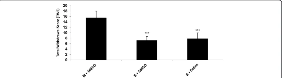
Results and discussion Naloxone-induced withdrawal
Animals received additive doses of morphine twice a day for nine days. In order to induce withdrawal symptoms, na-loxone (4 mg/kg, ip) was injected. The results in Figure 1 show that there was a significant (P < 0.001) difference in the TWS between saline + saline or DMSO 25% and con-trol groups (morphine + saline or DMSO 25%). Data ana-lysis indicated that the withdrawal signs including: jumping, Figure 2Effects of icv injection of carbenoxolone (100, 400, 600μg/rat) on the expression of naloxone (4 mg/kg)-induced withdrawal signs in morphine-dependent rats.Data are expressed as mean ± S.E.M.*p < 0.05,**p < 0.01,***p < 0.001 different from control (morphine-dependent DMSO 25% microinjected group). M = Morphine, S = Saline, CBX = carbenoxolone.
Figure 3Effects of icv injection of carbenoxolone (100, 400, 600μg/rat) on the expression of naloxone (4 mg/kg)-induced TWS in morphine-dependent rats.Data are expressed as mean ± S.E.M.***p < 0.001 different from control (morphine-dependent DMSO 25%
rearing, genital grooming, abdomen writhing and wet dog shake were significantly greater in the control groups which received morphine compared to saline or DMSO 25% treated animals which means that the animals in the con-trol group has already became dependent.
Effect of icv administration of carbenoxolone on morphine withdrawal signs
The results in Figure 2 revealed that icv injection of CBX (600 μg/10ul/rat, icv) significantly decreased all withdrawal signs except abdomen writhing [Figure 2] (P < 0.001 for all signs). In addition we found that carbenoxolone with all doses used in this study induced a significant decrease in TWS in comparison with the control group [Figure 3] (P < 0.001).
Effect of icv administration of mefloquine on morphine withdrawal signs
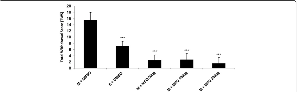
Our findings in this study have shown that icv administra-tion of mefloquine (50, 100 and 200 μg/10 μl/ rat)
decreased all withdrawal signs significantly [Figure 4] (P < 0.001). In addition the results indicated that icv injection of mefloquine resulted in a significant decrease in TWS compared to the control group [Figure 5] (P < 0.001).
The results of this study showed that stereotaxic sur-gery and icv injection of DMSO 25% had no effect on naloxone-induced opioid withdrawal symptoms in morphine-dependent rats while gap junction inhibitors (CBX and MFQ) significantly decreased the withdrawal signs. Locomotor activity test of rats with icv microin-jected by CBX or MFQ was done to rule out the occur-rence of motor performance hampering effect of drugs. The data showed that icv injection of CBX or MFQ had no significant effect on rats’motor performance. Thus, the alleviative effect of CBX or MFQ on opioid withdrawal is not ensued from suppression of motor activity.
Different drugs and strategies have been investigated to find an effective approach for preventing opioid de-pendence and withdrawal symptoms. Neurotransmitter systems, especially noradrenergic pathway has been Figure 4Effects of icv injection of mefloquine (50, 100, 200μg/rat) on the expression of naloxone (4 mg/kg)-induced withdrawal signs in morphine-dependent rats.Data are expressed as mean ± S.E.M.*p < 0.05,**p < 0.01,***p < 0.001 different from control (morphine-dependent
DMSO 25% microinjected group). M = Morphine, S = Saline, MFQ = Mefloquine.
Figure 5Effects of icv injection of mefloquine (50, 100, 200μg/rat) on the expression of naloxone (4 mg/kg)-induced TWS in morphine-dependent rats.Data are expressed as mean ± S.E.M.***p < 0.001 different from control (morphine-dependent DMSO 25%
found to play a key role in appearance of withdrawal symptoms. It has been reported that chronic morphine treatment may lead to a reduction in expression of in-hibitory opioid peptides. These processes with the con-currently increase in the activity of glutamatergic neurons may lead to the enhancement of activity of nor-adrenergic system and somatic signs during drug de-pendence and withdrawal [30].
Some brain nucleuses especially LC appears to mediate somatic signs of opiate withdrawal. It has been reported that acute opiates administration inhibit LC neurons and subsequently inhibit cAMP This effect may lead to de-crease in protein kinase A (PKA) activity and the phos-phorylation rate of cAMP response element binding protein (CREB). These selective effects of morphine on cAMP protein phosphorylation indirectly generate spe-cific behaviors commonly associated with addiction, tol-erance and withdrawal [31,32]. Indeed if morphine treatment persists and is then abruptly terminated, LC neurons will increase the frequencies of action poten-tials. Therefore, the symptoms of morphine withdrawal will be produced [33,34].
Also it has been suggested that neuronal connections outside the LC play an important role in the withdrawal-induced activation of these nucleus [35].
The results of the present study for the first demon-strate that central administration of gap junction blocker such as carbenoxolone could alleviate opioid withdrawal symptoms in morphine-dependent rats.
Previous studies reported that CBX directly binds to and blocks a broad spectrum of the connexins that make up gap junctions [13,14]. CBX microinjected bilaterally into the substantia nigra (pars reticulata) also produced a dose-dependent reduction in the duration and severity of seizures [10].
CBX is a broad-spectrum gap junction blocker believed to act on a range of connexins and pannexins, with add-itional anti-inflammatory and mineralocorticoid- like properties [36], and has been shown in vitro to reduce seizure-like after discharges and spontaneous activity in electrical stimulation [37,38].
Experimental and theoretical evidence suggests that direct electrotonic communication between neurons via gap junctions, in combination with synaptic and ionic mechanisms, might contribute to the generation or maintenance of seizures [39-41].
Furthermore in a previous study, it has been shown that CBX might enhance the anticonvulsant action of some antiepileptics, such as diazepam, gabapentin, phenobarbital, felbamate and valproate [42], suggesting its potential usefulness in the human therapy of some types of pharmacoresistant epilepsies.
In another part of study we showed that MFQ as a po-tent and selective Cx36 gap junction blocker [43] could
prevent opioid withdrawal signs and gathering the data from different symptoms according to Rasmussen et al. method, showed that mefloquine could attenuate the total withdrawal scores which is depicted in Figure 5.
Cx36 expression is found in almost all brain areas, in-cluding neocortex, brainstem, basal ganglia, hippocampus, and cerebellum [44,45]. Also MFQ is find to acts as an hibitor of P-glycoprotein [46] and, therefore, it can in-crease the concentrations of other drugs in the brain [47].
In addition it has been shown that MFQ significantly increased IPSCs frequency in brain slices isolated from mouse [48] and this property might be helpful in attenu-ating of firings during withdrawal sings exhibition.
Conclusion
Taking together we found that central administration of gap junction blockers; carbenoxolone or mefloquine prevented morphine-induced withdrawal symptom in rats.
Competing interests
The authors declare that they have competing interests.
Authors’contributions
SM: contribution in doing the experiments.MC: contribution in study design.HG: contribution in data analysis and manuscript preparation.RM: contribution in doing the experiments.MG: contribution in doing the experiments and manuscript preparation.KH: contribution in study design, data analysis and manuscript preparation. All authors read and approved the final manuscript.
Author details
1Department of Pharmacology and Toxicology, Faculty of Pharmacy, Tabriz
University of Medical Sciences, Tabriz, Iran.2Student Research Committee,
Tabriz University of Medical Sciences, Tabriz, Iran.3Department of Biology,
Faculty of Science, Islamic Azad University, Sanandaj Branch, Sanandaj, Iran.
4Cellular and Molecular Research Center, Kurdistan University of Medical
Sciences, Sanandaj, Iran.5Department of Physiology and Pharmacology,
Faculty of Medicine, Kurdistan University of Medical Sciences, Sanandaj, Iran.
Received: 1 May 2013 Accepted: 14 October 2013 Published: 21 October 2013
References
1. Bailey CP, Connor M:Opioids: cellular mechanisms of tolerance and physical dependence.Curr Opin Pharmacol2005,5(1):60.
2. MALDONADO R:Participation of noradrenergic pathways in the expression of opiate withdrawal: biochemical and pharmacological evidence.Neurosci Biobehav Rev1997,21(1):91–104.
3. Nestler EJ, Alreja M, Aghajanian GK:Molecular control of locus coeruleus neurotransmission.Biol Psychiatry1999,46(9):1131–1139.
4. Van Bockstaele EJ, Menko AS, Drolet G:Neuroadaptive responses in brainstem noradrenergic nucleic following chronic morphine exposure. Mol Neurobiol2001,23(2):155–171.
5. Maldonado R, Koob GF:Destruction of the locus coeruleus decreases physical signs of opiate withdrawal.Brain Res1993,605(1):128–138. 6. Koob GF, Maldonado R, Stinus L:Neural substrates of opiate withdrawal.
Trends Neurosci1992,15(5):186–191.
7. Akaoka H, Aston-Jones G:Indirect serotonergic agonists attenuate neuronal opiate withdrawal.Neuroscience1993,54(3):561–565. 8. McCracken CB, Roberts D:Neuronal gap junctions: expression, function,
and implications for behavior.Int Rev Neurobiol2006,73:125–151. 9. Willecke K, Eiberger J, Degen J, Eckardt D, Romualdi A, Güldenagel M,
10. Medina-Ceja L, Cordero-Romero A, Morales-Villagrán A:Antiepileptic effect of carbenoxolone on seizures induced by 4-aminopyridine: a study in the rat hippocampus and entorhinal cortex.Brain Res2008,1187:74–81. 11. Ballantyne D, Andrzejewski M, Mückenhoff K, Scheid P:Rhythms, synchrony
and electrical coupling in the locus coeruleus.Respir Physiol Neurobiol
2004,143(2):199–214.
12. Turpie A, Thomson T:Carbenoxolone sodium in the treatment of gastric ulcer with special reference to side-effects.Gut1965,6(6):591–594. 13. Gladwell SJ, Jefferys JG:Second messenger modulation of electrotonic
coupling between region CA3 pyramidal cell axons in the rat hippocampus.Neurosci Lett2001,300(1):1–4.
14. Bukauskas FF, Bennett MV:Connexin-based gap junction hemichannels: gating mechanisms.Biochim Biophys Acta2005,1711:215–224. 15. Vessey JP, Lalonde MR, Mizan HA, Welch NC, Kelly ME, Barnes S:
Carbenoxolone inhibition of voltage-gated Ca channels and synaptic transmission in the retina.J Neurophysiol2004,92(2):1252–1256. 16. Crawley JN C, Aisa N, Sergeeva OA, Haas HL:Carbenoxolone impairs LTP
and blocks NMDA receptors in murine hippocampus.Neuropharmacology
2008,55(2):139–147.
17. Bujalska ISM, Howie A, Stewart PM:Human 11 beta-hydroxysteroid dehydrogenase: studies on the stably transfected isoforms and localization of the type 2 isozyme within renal tissue.Steroids1997,62:77–82. 18. Cruikshank SJ, Hopperstad M, Younger M, Connors BW, Spray DC, Srinivas
M:Potent block of Cx36 and Cx50 gap junction channels by mefloquine. Proc Natl Acad Sci U S A2004,101(33):12364–12369.
19. Lee H, Go M:Effects of mefloquine on Ca2+ uptake and release by dog brain microsomes.Arch Int Pharmacodyn Ther1996,331(3):221. 20. Lim L, Go M:The anticholinesterase activity of mefloquine.Clin Exp
Pharmacol Physiol1985,12(5):527–531.
21. Weiss S, Benwell K, Cliffe I, Gillespie R, Knight A, Lerpiniere J, Misra A, Pratt R, Revell D, Upton R:Discovery of nonxanthine adenosine A2A receptor antagonists for the treatment of Parkinson’s disease.Neurology2003,
61(11 suppl 6):S101–S106.
22. Gribble FM, Davis TM, Higham CE, Clark A, Ashcroft FM:The antimalarial agent mefloquine inhibits ATP‐sensitive K‐channels.Br J Pharmacol2000,
131(4):756–760.
23. Paxinos G, Watson C:The Rat Brain in Stereotaxic Coordinates. Hard Cover Edition.Academic press; 2007.
24. Dizgah IM, Karimian SM, Zarrindast MR, Sohanaki H:Attenuation of morphine withdrawal signs by a D1 receptor agonist in the locus coeruleus of rats.Neuroreport2005,16(15):1683.
25. Parvizpour A, Charkhpour M, Habibi-asl B, Shakhsi M, Ghaderi M, Hassanzadeh K:Repeated central administration of selegiline attenuated morphine physical dependence in rat.Pharmacol Rep2013,65(593):593–599. 26. Habibi Asl B, Sa O, Charkhpour M, Hassanzadeh K:A novel pharmacological
role for minocycline: attenuating the withdrawal syndrome of morphine in rat.Pharm Sci (journal of Faculty of Pharmacy)2009,15(1):67–74. 27. Rasmussen K, Beitner-Johnson DB, Krystal JH, Aghajanian GK, Nestler EJ:
Opiate withdrawal and the rat locus coeruleus: behavioral, electrophysiological, and biochemical correlates.J Neurosci1990,
10(7):2308–2317.
28. Riahi E, Mirzaii-Dizgah I, Karimian SM, Sadeghipour HR, Dehpour AR:
Attenuation of morphine withdrawal signs by a GABAB receptor agonist in the locus coeruleus of rats.Behav Brain Res2009,196(1):11–14. 29. Crawley JN, GG R, MA S, DR S, Wray S:Short Protocols in Neuroscience.New
Jersey: John Wiley & Sons Inc; 2007.
30. Van Bockstaele EJ, Peoples J, Menko AS, McHugh K, Drolet G:Decreases in endogenous opioid peptides in the rat medullo-coerulear pathway after chronic morphine treatment.J Neurosci2000,20(23):8659–8666. 31. Nestler EJ:Molecular basis of long-term plasticity underlying addiction.
Nat Rev Neurosci2001,2(2):119–128.
32. Hyman SE, Malenka RC:Addiction and the brain: the neurobiology of compulsion and its persistence.Nat Rev Neurosci2001,2(10):695–703. 33. Saiepour MH, Semnanian S, Fathollahi Y:Occurrence of morphine
tolerance and dependence in the nucleus paragigantocellularis neurons. Eur J Pharmacol2001,411(1):85–92.
34. Robbe D, Bockaert J, Manzoni OJ:Metabotropic glutamate receptor 2/3‐ dependent long‐term depression in the nucleus accumbens is blocked in morphine withdrawn mice.Eur J Neurosci2002,16(11):2231–2235. 35. Ivanov A, Aston-Jones G:Local opiate withdrawal in locus coeruleus
neurons in vitro.J Neurophysiol2001,85(6):2388–2397.
36. Nilsen KE, Cock HR:Focal treatment for refractory epilepsy: hope for the future?Brain Res Rev2004,44(2):141–153.
37. Juszczak GR, Swiergiel AH:Properties of gap junction blockers and their behavioural, cognitive and electrophysiological effects: animal and human studies.Prog Neuro-Psychopharmacol Biol Psychiatry2009,33(2):181–198. 38. Wang PP, Frazier J, Brem H:Local drug delivery to the brain.Adv Drug
Deliv Rev2002,54(7):987–1013.
39. Carlen PL, Skinner F, Zhang L, Naus C, Kushnir M, Perez Velazquez JL:The role of gap junctions in seizures.Brain Res Rev2000,32(1):235–241. 40. Perez Velazquez JL, Carlen PL:Gap junctions, synchrony and seizures.
Trends Neurosci2000,23(2):68–74.
41. Traub RD, Whittington MA, Buhl EH, LeBeau FE, Bibbig A, Boyd S, Cross H, Baldeweg T:A possible role for gap junctions in generation of very fast EEG oscillations preceding the onset of, and perhaps initiating, seizures. Epilepsia2008,42(2):153–170.
42. Gareri P, Condorelli D, Belluardo N, Gratteri S, Ferreri G, Donato Di Paola E, De Sarro A, De Sarro G:Influence of carbenoxolone on the anticonvulsant efficacy of conventional antiepileptic drugs against audiogenic seizures in DBA/2 mice.Eur J Pharmacol2004,484(1):49–56.
43. Allison DW, Ohran AJ, Stobbs SH, Mameli M, Valenzuela CF, Sudweeks SN, Ray AP, Henriksen SJ, Steffensen SC:Connexin‐36 gap junctions mediate electrical coupling between ventral tegmental area GABA neurons. Synapse2006,60(1):20–31.
44. Belluardo N, Mudò G, Trovato-Salinaro A, Le Gurun S, Charollais A, Serre-Beinier V, Amato G, Haefliger J-A, Meda P, Condorelli DF:Expression of connexin36 in the adult and developing rat brain.Brain Res2000,
865(1):121–138.
45. Condorelli DF, Belluardo N, Trovato-Salinaro A, Mudò G:Expression of Cx36 in mammalian neurons.Brain Res Rev2000,32(1):72–85.
46. Pham YT, Régina A, Farinotti R, Couraud PO, Wainer IW, Roux F, Gimenez F:
Interactions of racemic mefloquine and its enantiomers with P-glycoprotein in an immortalised rat brain capillary endothelial cell line, GPNT.Biochimica et Biophysica Acta (BBA)-General Subjects2000,1524(2):212–219.
47. Pussard E, Merzouk M, Barennes H:Increased uptake of quinine into the brain by inhibition of P-glycoprotein.Eur J Pharm Sci2007,32(2):123–127. 48. Allison DW, Wilcox RS, Ellefsen KL, Askew CE, Hansen DM, Wilcox JD, Sandoval
SS, Eggett DL, Yanagawa Y, Steffensen SC:Mefloquine effects on ventral tegmental area dopamine and GABA neuron inhibition: A physiologic role for connexin‐36 GAP junctions.Synapse2011,65(8):804–813.
doi:10.1186/1423-0127-20-77
Cite this article as:Moradiet al.:Gap junction blockers: a potential approach to attenuate morphine withdrawal symptoms.Journal of Biomedical Science201320:77.
Submit your next manuscript to BioMed Central and take full advantage of:
• Convenient online submission
• Thorough peer review
• No space constraints or color figure charges
• Immediate publication on acceptance
• Inclusion in PubMed, CAS, Scopus and Google Scholar
• Research which is freely available for redistribution