Open Access
Research
Action potential repolarization enabled by Ca
++
channel
deactivation in PSpice simulation of smooth muscle propagation
Lakshminarayanan Ramasamy
1and Nicholas Sperelakis*
2,3Address: 1Dept. of Electrical Computer Engineering and Computer Science University of Cincinnati College of Engineering Cincinnati, OH 45219
USA, 2Dept. of Molecular & Cellular Physiology University of Cincinnati College of Medicine Cincinnati, OH 45267-0576 USA and 3Dept. of
Molecular & Cellular Physiology University of Cincinnati College of Medicine Cincinnati, OH 45267-0576 USA
Email: Lakshminarayanan Ramasamy - [email protected]; Nicholas Sperelakis* - [email protected] * Corresponding author
Abstract
Background: Previously, only the rising phase of the action potential (AP) in cardiac muscle and smooth muscle could be simulated due to the instability of PSpice upon insertion of a second black box (BB) into the K+ leg of the basic membrane unit. This restriction was acceptable because only the transmission of excitation from one cell to the next was investigated.
Methods: In the current work, the repolarization of the AP was accomplished by inserting a second BB into the Ca++ leg of the basic membrane unit. Repolarization of the AP was produced, not through an activation of the K+ channel conductance, but rather through a mimicking of the deactivation of the Ca++ channel conductance. Propagation of complete APs was studied in a chain (strand) of 10 smooth muscle cells, in which various numbers of gap-junction (gj) channels (assumed to be 100 pS each) were inserted across the cell junctions.
Results: The shunt resistance across the junctions produced by the gj-channels (Rgj) was varied from 100,000 MΩ (0 gj-channels) to 10,000 MΩ (1 gj-channel), to 1,000 MΩ (10 channels), to 100 MΩ (100 channels), to 10 MΩ (1000 channels), and to 1.0 MΩ (10,000 channels). Velocity of propagation (θ, in cm/sec) was calculated from the measured total propagation time (TPT, the time difference between when the AP rising phase of the first cell and the last cell crossed -20 mV), assuming a constant cell length of 200 µm. When there were no gj-channels, or only one, the transmission of excitation between cells was produced by the electric field (EF), i.e., the negative junctional cleft potential, that is generated in the narrow junctional clefts (e.g., 100 A) when the prejunctional membrane fires an AP (a fraction of a millisecond before the adjacent surface membrane). There were significant end-effects at the termination of the strand, such that the last cell (cell #10) failed to fire, or fired after a prolonged delay. This end-effect was abolished when the strand termination resistance (Rbt) was increased from 1.0 KΩ to 600 MΩ. When there were 1000 or 10,000 gj-channels, the transmission of excitation was produced by local-circuit current flow from one cell to the next through the gj-channels.
Discussion: In summary, it is now possible to simulate complete APs in smooth muscle cells that could propagate along a single chain of 10 cells, even when there were no gj-channels between the cells.
Published: 30 December 2005
BioMedical Engineering OnLine 2005, 4:71 doi:10.1186/1475-925X-4-71
Received: 21 October 2005 Accepted: 30 December 2005
This article is available from: http://www.biomedical-engineering-online.com/content/4/1/71
© 2005 Ramasamy and Sperelakis; licensee BioMed Central Ltd.
Introduction
There are no low-resistance connections between the cells in several different cardiac muscle and smooth muscle preparations [1-3]. In a computer simulation study of propagation in cardiac muscle, firing of the pre-junctional membrane generated an electric field (EF) in the narrow junctional clefts, which depolarized the postjunctional membrane to its threshold [4-6]. This causes excitation of the postjunctional cell, after a brief junctional delay. The total propagation time consists primarily of the summed junctional delays. This results in a staircase-shaped prop-agation, the surface sarcolemma of each cell firing almost simultaneously [5]. Propagation has been demonstrated to be discontinuous (or saltatory) in cardiac muscle [7-10]. A high density of fast Na+ channels are localized in
the junctional membranes of the intercalated disks of car-diac muscle [6,11,12], consistent with the requirement of the junctional membrane being more excitable than the surface membrane (for the EF mechanism to work [3-6]). In connexin-43 and Cx40 knockout mice, propagation in the heart still occurs, but it is slowed [13-16], as predicted by the previous PSpice simulation studies. Propagation by mechanisms not requiring low-resistance connections have also been proposed by others [6,8,9].
Propagation of APs in cardiac muscle and smooth muscle were previously modeled using the PSpice program for circuit design and analysis. The studies corroborated the earlier reports that the EF developed in the junctional cleft is sufficiently large to allow the transfer of excitation
with-out the requirement for a gap junction [17-19]. In those studies, a second black-box (BB-2) could not be inserted into the K+ leg of the Hodgkin-Huxley circuit of the basic
membrane units because that caused the PSpice program to become very erratic and unstable. Therefore, only the rising phase of the APs was simulated. This defect in the program was acceptable, since only the mechanism(s) for the transfer of excitation from one cell to the next was under investigation. For this purpose, only the fast rising phase of the AP was necessary. PSpice has recently used by others to model conduction in nerve fibers [20].
The purpose of the present study was to determine whether another method for repolarization could be devised that would circumvent the instability problem. So it was decided to try inserting a BB-2 into the same leg as the first BB (BB-1), namely into the Ca++ leg, to mimic the
deactivation of the Ca++ channel, and thereby produce
repolarization of the AP. This method worked. Thus, repo-larization was produced, not by activation of the K+
con-ductance, but instead by deactivation of the Ca++conductance. Similar results were found for cardiac
muscle simulations [21].
Methods
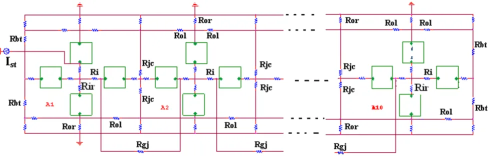
As shown in Figure 1, there were two surface membrane units in each cell (one facing upwards and one inverted) and one unit for each of the junctional membranes. The values for the circuit parameters used (standard condi-tions) are listed in Table 1 for both the surface units and
Circuit diagram for the single chain of ten smooth muscle cells bathed in a bath of Ringer solution Figure 1
junctional units, and are consistent with those used previ-ously [3,17,18].
The smooth muscle cell was assumed to be a cylinder 200
µm long and 6 µm in diameter. The cell capacitance was assumed to be 30 pF, and the input resistance to be 20 MΩ. But to increase +dV/dt max to a value appropriate for visceral smooth muscle, the total cell capacitance was reduced 11 pF. A junctional tortuosity (interdigitation) factor of 4 was assumed for the cell junction [3]. The junc-tional cleft potential (Vjc) is produced across Rjc, the radial resistance of the narrow and tortuous junctional cleft. The junctional cleft contained two radial resistances (Rjc) of 600 MΩ each in parallel (i.e., Rjc = 300 MΩ). The value assigned to Rjc reflects the thickness of the junc-tional gap (end-to-end) and the tortuosity factor.
The circuit used for each unit was kept as simple as possi-ble, using only those ion channels that set the resting potential (RP) and predominate during the rising phase of the AP. The RP was -56 mV, and the overshoot potential was +9 mV (AP amplitude of 65 mV). Because the PSpice program does not have a V-dependent resistance to repre-sent the increase in conductance for Ca++ ions in smooth
muscle cells during depolarization and excitation, this function was simulated by a V-controlled current source ("black-box", BB) in each of the basic circuit units. The current output of the BB, at various membrane voltages,
was calculated assuming a sigmoidal relationship between membrane voltage and resistance between -50 mV and -20 mV. The appropriate values were inserted into the GTABLEs for each BB [3].
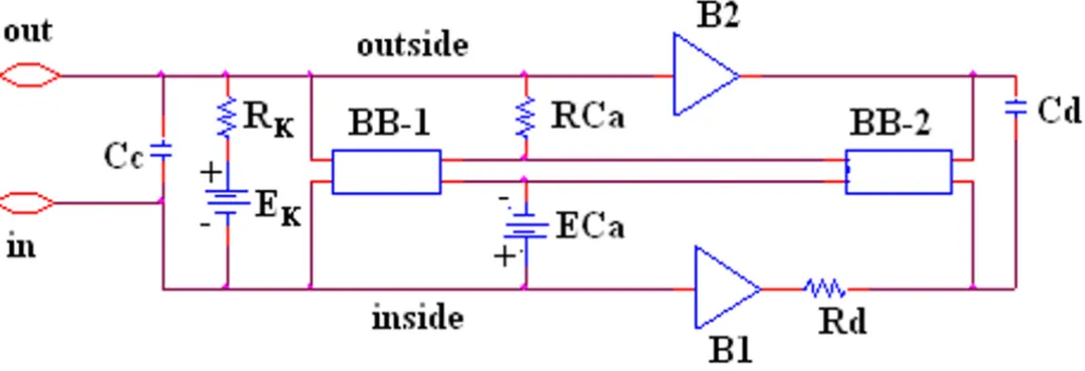
Our novel approach to simulation of the entire action potential (AP) waveform was achieved by inserting a sec-ond BB into the Ca++ leg of the basic unit. Thus, the first
BB mimics Ca++ activation, and the second BB mimics
deactivation of the Ca++-channel conductance. The latter
allowed repolarization of the AP to occur. BB-2 is con-nected between the outside and inside of the membrane unit, with reversed polarity compared to BB-1, as shown in Figure 2. The outputs of BB-1 and BB-2 were tied together in such a way that BB-2's output current nullifies BB-1's output current. BB-2 was activated with a delay time corresponding to the physiological delay value (i.e., to give an appropriate action potential duration (APD50)). The required delay time was generated using a delay ele-ment Rd Cd (RC time constant) (Fig. 2). At the resting potential, BB-2's output current was set to 0 nA. Once the cell has fired using BB-1's current, the potential across the input of BB-2 starts increasing with a rate corresponding to the RC time constant of the delay element. Thereby, BB-2 starts responding to the input rising voltage. Two buffer elements (unity gain operational amplifiers) were added to isolate the input terminal of BB-2 from BB-1, thus avoiding any hindrance between the two BBs.
The chain of 10 cells was assumed to be bathed in a large volume of Ringer solution connected to ground. The external resistance (Ro) of this fluid was divided into two components: a radial resistance (Ror) and a longitudinal
Table 2: Black-box values for surface membraneunit and junctional membrane unit
A. Surface membrane unit
Black-box #1 Black-box #2
Voltage (mV) Current (nA) Voltage (mV) Current (nA)
-55 -0.57 55 0
-50 -0.71 -
-3 -1.65 -
-10 -1.67 -10 -1.1
B. Junctional membrane unit
Black-box #1 Black-box #2
-55 -0.057 55 0
-50 -0.013 -
-3 -0.165 -
-10 -0.167 -10 -0.11
Table 1: Parameter values used understandard conditions.
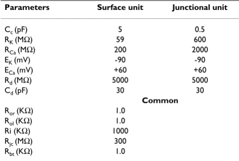
Parameters Surface unit Junctional unit
Cc (pF) 5 0.5
RK (MΩ) 59 600
RCa (MΩ) 200 2000
EK (mV) -90 -90
ECa (mV) +60 +60
Rd (MΩ) 5000 5000
Cd (pF) 30 30
Common
Ror (KΩ) 1.0 Rol (KΩ) 1.0 Ri (KΩ) 1000 Rjc (MΩ) 300 Rbt (KΩ) 1.0
Cc = Total cell capacitance RK = Potassium resistance RCa = Calcium resistance
resistance (Rol). The cells in the chain were either con-nected by low-resistance pathways (gap-junction chan-nels) or not interconnected, so that transmission of excitation from one cell to the next had to rely on the elec-tric field (EF) developed in the junctional cleft. The ends of the chains had a bundle termination resistance (Rbt) of 1.0 KΩ to mimic the physiological condition, but was increased to 600 MΩ in some experiments to counter end-effects. Rbt was increased to equal Rjc. Electrical stimulation (rectangular current pulses of 0.25 nA and 0.50 ms duration) was applied to the inside of the first cell of the chain (cell # 1). To minimize confusion, the voltage was recorded from only one surface unit
(upward-facing) in each cell. Propagation velocity was cal-culated from the measured total propagation time (TPT) (measured as the difference between when the APs (rising phase) of the first cell and last cell crossed -20 mV) and the assumed cell length of 200 µm.
Results
The AP waveform and propagation down the chain of 10 cells, when the strand termination resistance (Rbt) was 1.0 KΩ, is illustrated in Figure 3. Propagation was studied with 0, 1, 10, 100, 1000 and 10,000 gj-channels traversing the cell junctions. Assuming each gj-channel has a con-ductance of 100 pS, these channels corresponded to a shunt resistance across each junction (Rgj) of 100,000
Circuit diagram for one of the basic units Figure 2
Circuit diagram for one of the basic units. The unit circuitry was the same for both the surface units and junctional units, but the values of the various components were adjusted to reflect the long surface membrane and short junctional membrane. These values are listed in Table 1. The GTABLE values for the two types of membrane were also different, and these are listed in Table 2. Note that there are two black-boxes (BB) in the basic unit, and both are in the same Ca++ conductance leg of the Hodgkin-Huxley circuit. The first BB produced activation of the Ca++conductance, and the second BB produced deactivation of the Ca++channel conductance. It was necessary to produce a time delay (RC time constant) before the second BB came into play to cause deactivation. The two operational amplifiers (unity gain) depicted acted as buffers. In the K+leg, the K+ conduct-ance is in series with EK, the K+equilibrium potential.
Table 3: Summary of TPT and θ values as a function of number of gap-junction channels.
Rbt = 1.0 KΩ Rbt = 600 MΩ
Number of gap-junction channels #
Rgj (MΩ) TPT (ms) Θ (cm/s) TPT (ms) Θ (cm/s)
0 100,000 9.0* 17.8* 9.3 19.4
1 10,000 8.8* 18.2* 8.0 22.6
10 1,000 8.0 22.5 6.3 28.7
100 100 2.8 64.3 2.0 90.0
1,000 10 0.1 1800 0.04 5143
10,000 1 0 8 0 8
Propagation of APs simulated by PSpice along a chain of ten smooth muscle cells Figure 3
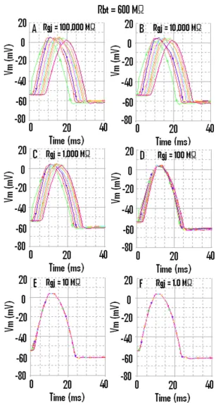
When the strand termination resistance was increased to 600 MΩ, then the last cell (cell #10) did not fail when there were no gj-channels (A) or only one gj-channel (B)
Figure 4
MΩ, 10,000 MΩ, 1,000 MΩ, 100 MΩ, 10 MΩ, and 1.0 MΩ, respectively. The corresponding records are shown in Figure 3, panels A through F.
When there were no gj-channels (Fig. 3A), or only one channel (Fig. 3B), fast propagation still occurred, which was initiated by the EF mechanism. As can be seen, the last cell (cell #10) failed to fire (Fig. 3A). The reason for the failure of cell #10 will be evident when the next figure is discussed. In Figure 3B (1 gj-channel), cell #10 fired after a prolonged delay; this delay was reduced in Fig. 3C (10 gj-channels). When there are 1000 (Fig. 3E) or 10,000 (Fig. 3F) gj-channels, then the APs of all 10 cells are super-imposed. Thus, the TPT is nearly 0 ms in these panels.
Propagation down the chain when the strand termination resistance Rbt was 600 MΩ is illustrated in Figure 4. As can be seen, all 10 cells fired an AP, even with no gj-channels (Fig. 4A) or only 1 gj-channel (Fig. 4B). Also evident in Figure 4 is the fact that the overshoot of the APs in cells #10 and #1 are smaller than in cells 2–9. When there were 1000 or 10,000 gj-channels, the APs of all 10 cells were
superimposed (Fig. 4E, F). The TPT is nearly 0 msec in Fig. 4E and 4F. The APD50 is slightly lower at the very high Rbt value. The peak of the AP was also more smooth (or rounded) at the high Rbt value (e.g., compare Fig. 4F with Fig. 3F). The hyperpolarizing after-potential was about the same in the two cases.
Figure 5 gives the graphic plots of TPT and θ as a function of Rgj for both Rbt of 1.0 KΩ (Fig. 5A, B) and Rbt of 600 MΩ (Fig. 5C, D). As shown, as Rgj decreases (reflecting more and more gj-channels), the TPT decreases and θ increases, for both low (1.0 KΩ) and high (600 MΩ) values of Rbt. These data are also summarized in Table 3, to facilitate comparison of the quantitative numbers.
Discussion
The present results demonstrate that it is now possible to simulate the entire AP, both the rising phase and the fall-ing phase, in PSpice simulations. Since a second BB (BB-2) could not be inserted into the K+leg of the basic
Hodg-kin-Huxley membrane units because of instability of the PSpice program, the problem was solved by inserting the
Graphic summary of the total propagation time (TPT) (A, C) and propagation velocity (θ, theta) (B, D) as a function of the shunt resistance across the 9 cell junctions (Rgj)
Figure 5
BB-2 into the Ca++ leg to produce deactivation of the Ca++
conductance turned on by the first BB (BB-1). BB-2 sup-plied current in the opposite polarity of that supsup-plied by BB-1, giving a net current of zero. BB-2 supplied its current after a time delay provided by an RC time constant. Thus, BB-1 produced the rising phase of the AP, and BB-2 pro-duced the falling (repolarizing) phase. Therefore, it is now possible to use the PSpice program to study propagation of complete APs in single strands and in two-dimensional sheets (parallel strands).
The after-hyperpolarization that was produced following the "spike" of the AP, (e.g., see Fig. 3A) is equivalent to the physiological hyperpolarizing after-potential seen in smooth muscle cells. It is due to a "feed-forward" mecha-nism. When BB-2 is supplying its opposing current and producing partial repolarization of the AP, the current supplied by BB-1 starts to decrease because the GTABLE is reversible [22]. Therefore, BB-2 supplies an excess of cur-rent, causing the after-hyperpolarization.
The data showed that, when Rbt was 1.0 KΩ, cell #10 failed to fire when the transmission of excitation from cell to cell depended on the EF mechanism (Fig. 3A). It was previ-ously shown that, with the EF mechanism, the distal por-tion of the cell reached threshold and fired before the proximal portion [3,5]. Increasing Rbt to 600 MΩ enabled the firing of cell #10 when the EF mechanism was domi-nant (Fig. 4A).
When cell #10 failed to fire, there was no passive partial depolarization produced due to electrotonic spread of current (Fig. 3A). This underscores the fact that there were no low-resistance connections or gj-channels between cells 9 and 10 or between the other cells. Yet propagation occurred at a fast velocity, enabled entirely by the EF mechanism. When there were 1000 or 10,000 gj-chan-nels, the propagation velocity became extremely fast, i.e., non-physiological.
There were substantial end (edge) effects at both ends of the strand. It can be seen in Figure 3A–C, that the over-shoot of the APs (hence that AP amplitude) was greater in cell #1 and cell #10 compared to those in cells 2 – 9. In contrast, when Rbt was 600 MΩ, the overshoot of the APs in cell #1 and cell #10 was smaller than in cells 2 – 9 (Fig. 4 A-B). Thus, the edge effect caused the AP amplitude in the end two cells to be either greater or smaller, depending on the value of Rbt . We are unable, at this time, to com-pare the end-effects observed in the PSpice simulations with those reported for biological tissues.
In summary, it was now possible to produce complete APs in PSpice simulation by placing a second BB (BB-2) into the Ca++ leg of the basic membrane units which supplied
an opposing current after a short time delay. This oppos-ing current essentially deactivated the inward Ca++ current
that was supplied by BB-1. The single strand of 10 cells exhibited prominent edge effects at the two ends of the strand. As previously reported, propagation in smooth muscle occurred at physiological velocities when no gj-channels, one gj-channel or 10 gj-channels was present. The propagation velocity became very fast and non-phys-iological when 1000 or 10,000 gj-channels were present. A large Rbt value enabled the firing of cell #10 by the EF mechanism.
References
1. Sperelakis N, Editorial: An electric field mechanism for trans-mission of excitation between myocardial cells. Circ Res 2002,
91:985-987.
2. Sperelakis N, McConnell K: Electric field interactions between closely abutting excitable cells. IEEE-Eng Med Biol 2002,
21:77-89.
3. Sperelakis N, Ramasamy L: Propagation in cardiac muscle and smooth muscle based on electric field transmission at cell junctions: An analysis by PSpice. IEEE-Eng Med Biol 2002,
21:130-143.
4. Sperelakis N, Mann JE Jr: Evaluation of electric field changes in the cleft between excitable cells. J Theor Biol 1977, 64:71-96. 5. Picone JB, Sperelakis N, Mann JE Jr: Expanded model of the
elec-tric field: Hypothesis for propagation in cardiac muscle. Math and computer modeling 1991, 15:17-35.
6. Kucera JP, Rohr S, Rudy Y: Localization of sodium channels in intercalated disks modulates cardiac conduction. Circ Res 2002, 91:1176-1182.
7. Spach MS, Miller WT, Geselowitz DB, Barr R, Kootsey JM, Johnson EA: The discontinuous nature of propagation in normal canine cardiac muscle. Evidence for recurrent discontinuity of intracellular resistance that affects the membrane cur-rents. 1981, 48:39-54.
8. Diaz PJ, Rudy Y, Plonsey R: Intercalated discs as a cause for dis-continuous propagation in cardiac muscle. A theoretical simulation. Ann Biomed Eng 1983, 11:177-189.
9. Shaw RM, Y Rudy: Ionic mechanisms of propagation in cardiac tissue. Roles of the sodium and L-type calcium currents dur-ing reduced excitability and decreased gap junction coupldur-ing.
Circ Res 1997, 81:727-741.
10. Henriquez AP, R Vogel, BJ Muller-Borer, CS Henriquez, R Weingart, WE Cascio: Influence of dynamic gap junction resistance on impulse propagation in ventricular myocardium: a computer simulation study. Biophys J 2001, 81:2112-2121.
11. Cohen SA: Immunocytochemical localization of rH1 sodium channel in adult rat heart atria and ventricle. Presence in terminal intercalated disks. Circulation 1994, 74:1071-1096. 12. Sperelakis N: Cable properties and propagation of action
potentials. CH 18. In Cell Physiology Source Book 1st edition. Aca-demic PressPublishers. San Diego; 1995:245-254.
13. Morley GE, Vaidya D, Samie FH, Lo CW, Taffet SM, Delmar M, Jalife J: Characterization of conduction in the ventricles of normal and heterozygous Cx43 knockout mice using optical map-ping. J Cardiovasc Electrophysiol 1999, 10:1361-1375.
14. Tamaddon HS, Vaidya D, Simon AM, Paul DL, Jalife J, Morley GE:
High resolution optical mapping of the right bundle branch in connexin40 knockout mice reveals low conduction in the specialized conduction system. Circ Res 2000, 87:929-936. 15. Gutstein DE, Morley GE, Tamaddon H, Vaidya D, Schneider MD,
Chen J, Chien KR, Stuhlmann H, Fishman GI: Conduction slowing and sudden arrythmic death in mice with cardiac restricted inactivation of connexin43. Circ Res 2001, 88:333-339. 16. Vaidya D, Tamaddon HS, Lo CW, Taffet SM, Delmar M, Morley GE:
Nullmutation of connexin43 causes slow propagation of ven-tricular activation in the late stages of mouse embryonic development. Circ Res 2001, 88:1196-1202.
Publish with BioMed Central and every scientist can read your work free of charge "BioMed Central will be the most significant development for disseminating the results of biomedical researc h in our lifetime."
Sir Paul Nurse, Cancer Research UK
Your research papers will be:
available free of charge to the entire biomedical community
peer reviewed and published immediately upon acceptance
cited in PubMed and archived on PubMed Central
yours — you keep the copyright
Submit your manuscript here:
http://www.biomedcentral.com/info/publishing_adv.asp
BioMedcentral
and smooth muscle in PSpice simulation. J Electrocardiol 2003,
36(4):279-293.
18. Sperelakis N, Kalloor B: Effect of variation in membrane excit-ability on propagation velocity of simulated action potentials for cardiac muscle and smooth muscle in the electric field model for cell to cell transmission of excitation. IEEE-Eng Med Biol 2004, 51:2216-2219.
19. Sperelakis N, Kalloor B, Ramasamy L: Boundary effects influence velocity of transverse propagation of simulated cardiac action potentials. Theor Biol Med Modeling 2005, 2:36.
20. Peasgood W, Dissado AL, Lam KC, Armstrong A, Wood W: A novel electrical model of nerve and muscle using PSpice. J Phys D: Appl Phys 2003, 36:311-329.