Zahra Hemati
1, B–D, Reza Ghanbarpour
1, A, E, F, Hesam Alizade
2, C–FThe Distribution of Beta Lactamase Genes
in
Escherichia Coli
Phylotypes Isolated from Diarrhea
and UTI Cases in Northwest Iran
1 Molecular Microbiology Department, Faculty of Veterinary Medicine, Shahid Bahonar University, Kerman, Iran 2 Department of Microbiology, International Branch, Shahid Beheshti University of Medical Science, Tehran, Iran
A – research concept and design; B – collection and/or assembly of data; C – data analysis and interpretation; D – writing the article; E – critical revision of the article; F – final approval of article; G – other
Abstract
Background. Pathogenic Escherichia coli strains are a common cause of intestinal and extra-intestinal infections, especially in developing countries. Extended spectrum beta-lactamases (ESBLS), a heterogeneous group of plasmid-encoded beta-lactamases, are common throughout the world.
Objectives. The aim of the present study was to determine the phenotypic and genotypic characteristics of ESBLS produced by E. coli isolates taken from patients with diarrhea and urinary tract infections (UTI) in northwest Iran.
Material and Methods. A total of 132 E. coli isolates (92 isolates from UTI and 40 isolates from diarrheic cases) were recovered and confirmed by biochemical tests. The isolates were examined for blaTEM and blaSHV genes and
phylogenetic background by two multiplex PCR assays. The isolates were tested for antibiotic susceptibility against nine antibiotic agents by the disk diffusion method.
Results. Thephylogenetic analysis showed that the UTI isolates mostly fell into phylo-group B2, followed by D, while the diarrheic isolates belonged to phylo-groups D and A. Out of 92 UTI isolates, 29.3% and 17.4% pos-sessed blaTEM and blaSHV genes, respectively. Ten diarrheic isolates were positive for blaTEM, two isolates possessed the blaSHV gene, and one isolate was positive for both genes. The UTI isolates that were positive for blaTEM and
blaSHV genes mostly belonged to phylo-groups D and B2, whereas the diarrhea isolates were in phylo-groups D and
A. Phylogenetic group D isolates have an accumulation of ESBLS genes in the diarrheic and UTI isolates. In both the UTI and diarrhea isolates, the maximum rate of resistance was against cefazolin, and the minimum rate of resistance was against nitrofurantoin. Twenty-four antibiotic resistance patterns were observed among the isolates. The amikacin, ciprofloxacin, cefotaxime, cefuroxime, cefazolin, gentamicin, nalidixic acid and trimethoprim/sulfa-methoxazole resistance pattern was the most prevalent in the isolates that belonged to phylo-group D.
Conclusions. The correct choice of effective antibiotic policy is needed to limit the spread of antibiotic resistance in bacteria (Adv Clin Exp Med 2014, 23, 4, 523–529).
Key words: extended spectrum beta-lactamases (ESBLs), Escherichia coli, diarrhea, UTI, phylogeny.
Adv Clin Exp Med 2014, 23, 4, 523–529 ISSN 1899–5276
ORIGINAL PAPERS
© Copyright by Wroclaw Medical University
Escherichia coli (E. coli) strains are versatile bacteria, comprising harmless commensal bacte-ria as well as different pathogenic vabacte-riants with the ability to cause both intestinal and extra-intesti-nal diseases in humans and other hosts [1]. Some strains have specific virulence attributes, which confer the ability to adapt to new niches and cause a broad spectrum of diseases [2]. Several vari-ants have been described, and the strains capable
of causing infection of the gastrointestinal sys-tem are called intestinal pathogenic E. coli, while those that cause infections outside the gastrointes-tinal system are called extra-intesgastrointes-tinal pathogenic
and infectious arthritis [4, 5]. Diarrheagenic E. coli
strains (DEC) are among the most important bac-terial enteric pathogens, and are classified into six categories, including enteropathogenic E. coli
(EPEC), enterohemorrhagic E. coli (EHEC), en-terotoxigenic E. coli (ETEC), enteroinvasive E. coli
(EIEC), enteroaggregative E. coli (EAEC), and dif-fusely adhering E. coli (DAEC) [6]. Pathogenic
E. coli isolates have a relatively high potential for developing resistance, so the treatment of E. coli
infections is becoming increasingly difficult [7]. There are several mechanisms of drug resistance; one of the most important resistant mechanisms is the production of beta-lactamases [8]. Beta-lac-tamase enzymes hydrolyze the four-membered be-ta-lactam ring common to a wide range of beta-lactam type antibiotics [9].
Among the clinically bacterial isolates, blaTEM,
blaSHV and blaCTX-M are the most frequently reported
extended-spectrum beta-lactamase (ESBL) genes [10]. E. coli and Klebsiella pneumoniae (K. pneu-moniae) are common species of Enterobacteriacae
that both have pathogenic potential and frequently incorporate ESBL-encoding genes [11].
There are four well-recognized phylogroups of
E. coli strains and these have been designated A, B1, B2 and D. These groups can be divided into six phylogenetic subgroups including (A0, A1, B22, B23,
D1, and D2), according to the combination of the
three genetic markers chuA, yjaA and DNA frag-ment TspE4.C2 [12]. Phylogenetic group distri-bution may be related to antimicrobial resistance prevalence in isolates of E. coli [13].
The aim of this study was to determine the
blaTEM and blaSHV genes, phylogenetic background
and antibiotic resistance profile of E. coli isolates from diarrhea and UTI samples in the Hamadan province in northwest Iran.
Material and Methods
Sampling and Bacteriological
Examinations
The study was carried out on 92 UTI samples and 40 diarrhea specimens from patients referred to lab-oratories in Hamadan, in northwest Iran. The sam-ples were obtained from patients aged between one and 65 years old. They were collected between April and November 2012. The specimens were cultivated on 5% sheep blood agar and MacConkey agar plates (Biolife Laboratories, Milano, Italy). The isolates were confirmed to be E. coli based on colony morphology, gram stain and standard biochemical methods. A sin-gle colony was obtained from each sample. Cultures of each isolate confirmed as E. coli were maintained in Luria-Bertani broth (Invitrogen, Paisley, Scotland) with 30% sterile glycerol at –80°C.
Polymerase Chain
Reaction (PCR)
and Reference Strains
DNA was extracted from the E. coli isolates and reference strains by the lysis method. The E. coli ref-erence strain ECOR62 (chuA+, yjaA+ and Tspe4. C2+), ATCC E. coli 35218 strain (blaTEM+) and
K. pneumoniae 700603 strain (blaSHV+) were used
as positive controls. E. coli strain ATCC 25922 was used as a negative control. The PCR primers spe-cific for blaTEM and blaSHV were described by Sharma
et al. [14]; phylogenetic groups (A, B1, B2 and D) were described by Clermont et al. [15]. Classifica-tion of the isolates in to phylogenetic subgroups (A0, A1, B22, B23, D1 and D2) was undertaken as
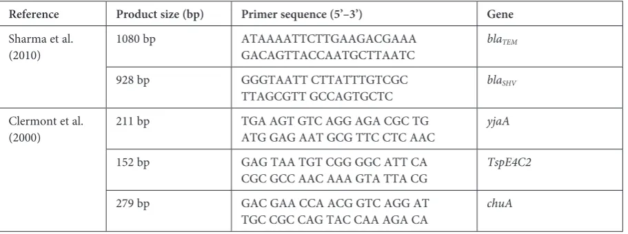
described by Escobar-Paramo et al. [12]. The se-quences and sizes of the PCR products are shown in Table 1.
Table 1. Oligonucleotide primers used in this study
Reference Product size (bp) Primer sequence (5’–3’) Gene
Sharma et al.
(2010) 1080 bp ATAAAATTCTTGAAGACGAAAGACAGTTACCAATGCTTAATC
blaTEM
928 bp GGGTAATT CTTATTTGTCGC
TTAGCGTT GCCAGTGCTC blaSHV Clermont et al.
(2000) 211 bp TGA AGT GTC AGG AGA CGC TGATG GAG AAT GCG TTC CTC AAC yjaA 152 bp GAG TAA TGT CGG GGC ATT CA
CGC GCC AAC AAA GTA TTA CG TspE4C2 279 bp GAC GAA CCA ACG GTC AGG AT
Antimicrobial Susceptibility Test
The antibiotic resistance of all the isolates to nine antibacterial agents was assessed by the disk diffusion method according to the Clinical and Lab-oratory Standards Institute’s guidelines [16]. The antibiotic disks (Mast, England) used in this study were amikacin (30 μg), cefazolin (30 μg), cefotaxime (30 μg), cefuroxime (30 μg), ciprofloxacin (5 μg), gentamicin (10 μg), nalidixic acid (30 μg), nitrofu-rantoin (300 μg) and trimethoprim/sulfamethoxa-zole (1.25/23.75 μg). E. coli ATCC 25922 was used as the positive control for the disk diffusion test.
Statistical Analysis
The statistical analysis for the descriptive data was carried out using SPSS software, version 17.
Results
Following the bacteriological tests, 132 confirmed
E. coli isolates (92 urine samples and 40 diarrhea sam-ples) were chosen for antibiogram and molecular ex-aminations. The isolates were recovered from both female (88) and male (44) patient samples.
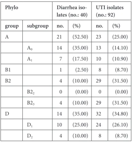
The phylogenetic analysis showed that the 132 E. coli isolates segregated into phylogenetic groups A (33.3%), B1 (6.8%), B2 (25%) and D (34.9%). The PCR assays for phylotyping of the 92 UTI E. coli isolates indicated that those isolates fell into 4 phylogenetic groups: 23 of them belonged to A (25%), 8 belonged to B1 (8.7%), 29 were B2 (31.5%) and 32 isolates belonged to group D (34.8%). The PCR assays of the 40 diarrheic E. coli isolates showed that 21 isolates (52.5%) belonged to phylo-genetic group A, one isolate (2.5%) to B1, 4 isolates (10%) to B2 and 14 isolates (35%) to group D.
Further analysis of the PCR phylotyping of UTI
E. coli isolates showed that the isolates fell into five phylogenetic subgroups: A0, A1, B23, D1 and D2;
most of them were in subgroups B23 (31.5%) and
D1 (26.1%). The 40 diarrheic E. coli isolates also fell into five phylogenetic subgroups: A0, A1, B23, D1 and
D2; the A0 35% (14 isolates) and D1 25% (10 isolates)
were the most prevalent subgroups (Table 2). The genotyping of the E. coli isolates revealed that out of 92 UTI isolates and 40 diarrheic isolates, 56 (42.4%) isolates were positive for the blaTEM and blaSHV
genes. Specifically, 37 isolates (28.03%) were positive for blaTEM; 18 (13.6%) were positive for blaSHV genes;
and 1 isolate (0.76%) was positive for both genes. Thirteen diarrheic isolates (32.5%) and 43 UTI isolates (47%) contained blaTEM and/or blaSHV genes (Table 3).
Out of 13 (32.5%) ESBL positive isolates from diarrheic cases, 10 (25%) isolates carried blaTEM genes and 2 (5%)
had blaSHV genes, while 1 (2.5%) isolate included both
genes. Out of the 43 (47%) ESBL positive isolates from UTI cases, 27 isolates (29.3%) possessed blaTEM genes
and 16 (17.4%) had blaSHV genes (Table 3).
Table 2. Distribution of diarrhea and UTI isolates in phy-logenetic groups/subgroups
Phylo Diarrhea
iso-lates (no.: 40) UTI isolates (no.: 92) group subgroup no. (%) no. (%)
A 21 (52.50) 23 (25.00) A0 14 (35.00) 13 (14.10)
A1 7 (17.50) 10 (10.90)
B1 1 (2.50) 8 (8.70) B2 4 (10.00) 29 (31.50) B22 0 (0.00) 0 (0.00)
B23 4 (10.00) 29 (31.50)
D 14 (35.00) 32 (34.80) D1 10 (25.00) 24 (26.10)
D2 4 (10.00) 8 (8.70)
Table 3. Combination of beta-lactamase genes in 40 diarrheic and 92 UTI isolates in relation to phylogenetic groups/subgroups
Gene Diarrhea isolates UTI isolates
A B1 B2 D Total A B1 D Total A0 A1 B1 B22 B23 D1 D2 A0 A1 B1 B22 B23 D1 D2
blaTEM 1 2 1 – 2 3 1 10 3 2 1 – 11 7 3 27
blaSHV – – – – – 1 1 2 3 2 2 – 3 6 – 16
blaTEM,blaSHV – – – – – 1 – 1 – – – – – – – –
The phylotyping of 56 diarrhea and UTI E. coli
isolates possessing beta-lactamase genes indicat-ed that these isolates belongindicat-ed mainly to phylo-genetic groups D and B2. The 10 blaTEM positive
isolates from diarrheic cases fell into phylogenetic groups A (3 isolates), B1 (1), B2 (2) and D (4); the 27 UTI isolates belonged to phylogenetic groups A (5 isolates), B1 (1), B2 (11) and D (10). Both of the 2 blaSHV positive isolates from diarrheic cases
fell into phylogenetic group D; the 16 blaSHV
posi-tive UTI isolates belonged to groups A (5 isolates), B1 (2), B2 (3) and D (6). The 1 isolate (0.76%) that was positive for both genes fell into phylogenetic group D (Table 3).
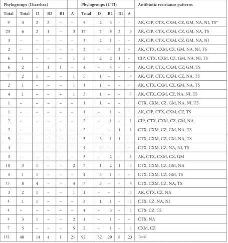
According to the results of the antibiotic sus-ceptibility test, in the diarrheic isolates the maxi-mum rate of resistance was against cefazolin 97.5% (39 isolates) and the minimum resistance rate was against nitrofurantoin 10% (4 isolates). The rates of resistance to other antibiotics were as follows: cefotaxim 34 (85%), nalidixic acid 25 (62.5%), ce-furoxime 24 (60%), cotrimoxazole 20 (50%), ami-kacin 19 (47.5%), ciprofloxacin 13 (32.5%), and gentamicin 14 (35%). In the UTI isolates the maxi-mum rate of resistance was against cefazolin 95.7% (88 isolates), followed by cefotaxim 87% (80), co-trimoxazole 66% (61), cefuroxime 65.2% (60), na-lidixic acid 59% (54), gentamicin 53.3% (49), ami-kacin 39.1% (36), ciprofloxacin 39.1% (36), and nitrofurantoin 23.9% (22). The results showed that 24 antibiotic resistance patterns were found among the E. coli isolates. Fourteen antibiotic re-sistance patterns were detected in the diarrhe-ic isolates. The cefotaxime, cefuroxime, cefazolin, nalidixic acid and trimethoprim/sulfamethoxazole resistance pattern was the most prevalent pattern in the diarrheic isolates that belonged to phylo-genetic groups A and D. Among the UTI isolates, 24 antibiotic resistance patterns were detected. The most frequent antibiotic resistance pattern among these isolates was against amikacin, ciprofloxa-cin, cefotaxime, cefuroxime, cefazolin, gentami-cin, nalidixic acid and trimethoprim/sulfamethox-azole, which was observed in phylogenetic group D. Twenty-four antibiotic resistance patterns were determined, distributed among the isolates in the 4 phylogenetic groups (Table 4).
Discussion
E. coli is one of the principal bacterial patho-gen causes of UTIs, enteric infections and sys-temic infections in humans and other hosts [3]. Diarrheagenic E. coli strains (DECs) are an im-portant cause of diarrhea, especially in develop-ing countries and constitute an important public
health problem [6, 17]. Extensive use of antibiotics to treat E. coli infections caused by resistant iso-lates will increase the problem of resistance [18, 19]. blaTEM and blaSHV genes, which are commonly
found in E. coli and K. pneumoniae, confer resis-tance to a broad spectrum of beta lactam antibi-otics and are a significant therapeutic concern for infections caused by many species of gram-neg-ative bacteria [20–23]. At the same time, the ep-idemiology of ESBL-producing E coli is compli-cated and varies among hospitals and countries [24]. There are alarming trends of associated re-sistance to other classes of antimicrobial drugs among ESBL-producing organisms isolated from the community [25].
In the current study, antibiotic resistance pat-terns were studied for E. coli isolates. Resistance was observed to antibiotics in widespread use, such as cefazolin, ciprofloxacin, cotrimoxazole, cefotaxi-me, gentamicin, amikacin and nalidixic acid. Most of the isolates in the present study were resistant to multiple antibiotic groups. High resistance to com-mon antibiotics has also been reported in previous studies [7, 11, 26]. The present study offers the first characterization of TEM-type and SHV-type ESBL variants produced by E. coli circulating in hospi-tals in Hamadan (northwest Iran). The progres-sive spread of ESBL-producing E. coli seems to be caused mainly by the extensive use of broad-spec-trum beta-lactam antibiotics in empiric therapy and rapid plasmid mediated distribution of resistance genes between bacterial species [23]. In this study, the blaTEM gene is the most prevalent ESBL, followed
by the blaSHV gene. Therefore, it seems that these
two genes are appropriate candidates for molecular screening of ESBL positive samples [8].
Enterobacteriaceae harboring transferable
blaTEM, blaCTX-M, blaSHV and blaOXA genes have been
reported worldwide in several studies of clinical isolates. Isendahl et al. [11] reported that 32.6% fe-cal samples from children were carriers of ESBL-producing E. coli or K. pneumonia. Shahid et al. [22] reported findings similar to those of the pres-ent study: 38.4% of the isolates in their study were positive for bla genes, and the blaTEM gene was
more prevalent than the blaSHV gene. In another
study, the prevalence of blaTEM, blaCTX-M and blaSHV
were carrying blaSHV, 75 isolates (68.8%) were
car-rying blaCTX-M, and 95 isolates (87.1%) were
carry-ing blaTEM genes, while 40 isolates (36.6%) isolates
had all three of these genes [8].
Phylogenetic analyses have shown that ex-tra-intestinal and intestinal E. coli are segregated in four main phylogenetic groups: A, B1, B2, and D [15]. In the present study, UTI isolates fell most-ly into phylogenetic group B2, followed by D; and diarrheic isolates belong to groups D and A. Simi-larly, previous reports indicated that most ExPEC strains belonged to group B2, although some fell into group D [3, 28]. In this study, UTI isolates that
were positive for blaTEM and blaSHV genes mostly
belonged to phylogenetic groups D and B2, while diarrhea isolates that were positive for these genes were in groups D and A. A study of community-ac-quired fecal carriage of ESBL-producing E. coli iso-lates in children showed that phylo-group A pre-dominated and that the most frequent ESBL was
CTX-M-1. The CTX-M producing isolates main-ly carried one other beta-lactamase-encoding gene (TEM-1 and SHV). Most of the ESBL-producing
E. coli isolates belonged to groups A and B1, possi-bly because of greater antibiotic exposure of group A/B1 strains in the fecal flora [28].
Table 4. Antibiotic resistance patterns in relation to phylogenetic groups
Phylogroups (Diarrhea) Phylogroups (UTI) Antibiotic resistance patterns Total Total D B2 B1 A Total D B2 B1 A
9 4 2 2 – – 5 2 3 – – AK, CIP, CTX, CXM, CZ, GM, NA, NI, TS*
23 6 2 1 – 3 17 7 5 2 3 AK, CIP, CTX, CXM, CZ, GM, NA, TS 3 – – – – – 3 2 1 – – AK, CIP, CTX, CXM, CZ, GM, NA, NI 2 – – – – – 2 – – 2 – AK, CTX, CXM, CZ, GM, NA, NI, TS 6 1 – – – 1 5 2 2 1 – CIP, CTX, CXM, CZ, GM, NA, NI, TS 6 2 – 1 1 – 4 – 4 – – AK, CIP, CTX, CXM, CZ, GM, TS
7 2 1 – – 1 5 1 – – 4 AK, CIP, CTX, CXM, CZ, NA, TS 2 1 – – – 1 1 1 – – – AK, CTX, CXM, CZ, GM, NA, TS 4 1 – – – 1 3 1 – – 2 AK, CTX, CXM, CZ, NA, NI, TS 1 – – – – – 1 1 – – – CTX, CXM, CZ, GM, NA, NI, TS 1 – – – – – 1 – 1 – – AK, CIP, CTX, CXM, CZ, TS
2 – – – – – 2 – 1 – 1 CIP, CTX, CXM, CZ, GM, NA 2 – – – – – 2 – – 1 1 CTX, CXM, CZ, GM, NA, TS
5 – – – – – 5 3 1 1 – CTX, CXM, CZ, GM, NA, TS 4 – – – – – 4 4 – – – CTX, CXM, CZ, NA, NI, TS 3 – – – – – 3 – 2 – 1 AK, CTX, CXM, CZ, GM 10 3 1 – – 2 7 1 2 1 3 CTX, CXM, CZ, GM, NA 5 1 1 – – – 4 3 1 – – CTX, CXM, CZ, GM, TS
15 8 4 – – 4 7 3 – – 4 CTX, CXM, CZ, NA, TS 3 2 1 – – 1 1 – – – 1 AK, CTX, CZ, NA
4 1 1 – – – 3 1 1 – 1 CTX, CZ, NA, NI
4 – – – – – 4 – 3 – 1 CTX, CZ, TS
4 3 1 – – 2 1 – 1 – – CTX, NA
7 5 – – – 5 2 – 1 – 1 CXM, CZ
132 40 14 4 1 21 92 32 29 8 23 Total
In conclusion, as Abdou et al. wrote: “Resis-tance in gram negative bacteria (such as E. coli) presents a major challenge for the antimicrobi-al therapy and significantly narrows the treatment options of human infections” [29]. Pathogenic iso-lates of E. coli have relatively high potentials for developing resistance and can make the treatment of ESBL-producing E. coli infections difficult [7]. Therefore, correctly detecting drug-resistant E. coli
is important to prevent the further spread of beta-lactamase-positive resistance in humans and other hosts [20]. The correct choice of safe and effective
antibiotic policy is needed to limit the emergence and spread of antibiotic resistance in bacteria [7]. The present study examined the genes codifying for beta-lactamases, but this does not necessarily reflect the expression of these genes during infec-tion; and the results might not be applicable to areas with a different epidemiology of ESBL-producing strains of E. coli. Discrepancies in the distribution of genotypes of ESBLs may be due to differences in the patterns of antibiotic use in various geograph-ical areas. In addition, frequent surveillance of re-sistance to antimicrobial agents is necessary.
References
[1] Leimbach A, Hacker J, Dobrindt U:E. coli as an all-rounder: The thin line between commensalism and pathoge-nicity. Curr Top Microbiol Immunol 2013, 358, 3–32.
[2] Oliveira FA, Paludo KS, Arend LNV, Farah SMSS, Pedrosa FO, Souza EM, Surek M, Picheth G, Fadel-Picheth CMT: Virulence characteristics and antimicrobial susceptibility of uropathogenic Escherichia coli strains. Genet Mol Res 2011, 10, 4114–4125.
[3] Pitout JD: Extraintestinal pathogenic Escherichia coli: A combination of virulence with antibiotic resistance. Front Microbiol 2012, 3.
[4] Katouli M: Population structure of gut Escherichia coli and its role in development of extra-intestinal infections. Iran J Microbiol 2010, 2, 59–72.
[5] Carvalho VM, Osugui L, Setzer AP, Lopez RPG, Pestana de Castro AF, Irino K, Catão-Dias JL: Characterization of extraintestinal pathogenic Escherichia coli isolated from captive wild felids with bacteremia. J Vet Diagn Invest 2012, 24, 1014–1016.
[6] Moreno AC, Filho AF, Gomes TAT, Ramosc STS, Montemora LPG, Tavaresa VC, Filho LS, Irinoe K, Martineza MB: Etiology of childhood diarrhea in the northeast of Brazil: significant emergent diarrheal pathogens. Diagn Microbiol Infect Dis 2010, 66, 50–57.
[7] Banu A, Kabbin J, Anand M: Extraintestinal infections due to Escherichia coli: an emerging issue. J Clin Diag Res 2011, 5, 486–490.
[8] Yazdi M, Nazemi A, Mirinargasi M, Jafarpour M, Sharifi SH: Genotypic versus phenotypic methods to detect extended-spectrum beta-lactamases (ESBLs) in Uropathogenic Escherichia coli. Ann Biol Res 2012, 3, 2454–2458. [9] Brown NG, Pennington JM, Huang W, Ayvaz T, Palzkill T: Multiple global suppressors of protein stability
defects facilitate the evolution of extended-spectrum TEM β-lactamases. J Mol Biol 2010, 404, 832–846.
[10] Ojer-Usoz E, Gonzalez D, Vitas AI, Leiva J, García-Jalón I, Febles-Casquero A, Escolano Mde L: Prevalence of extended-spectrum β-lactamase-producing Enterobacteriaceae in meat products sold in Navarra, Spain. Meat Sci 2013, 93, 316–321.
[11] Isendahl J, Turlej-Rogacka A, Manjuba C, Rodrigues A, Giske CG, Naucler P: Fecal carriage of ESBL-producing
E. coli and K. pneumoniae in children in Guinea-Bissau: a hospital-based cross-sectional study. PLoS One 2012, 7, 51981.
[12] Escobar-Paramo P, Grenet K, Le Menac’h A, Rode L, Salgado E, Amorin C, Gouriou S, Picard B, Rahimy MC, Andremont A, Denamur E, Ruimy R: Large-scale population structure of human commensal Escherichia coli
isolates. Appl Environ Microbiol 2004, 70, 5698–5700.
[13] Asai T, Masani K, Sato C, Hiki M, Usui M, Baba K, Ozawa M, Harada K, Aoki H, Sawada T: Phylogenetic groups and cephalosporin resistance genes of Escherichia coli from diseased food-producing animals in Japan. Acta Vet Scand 2011, 53.
[14] Sharma J, Sharma M, Ray P: Detection of TEM & SHV genes in Escherichia coli & Klebsiella pneumoniae isolates in a tertiary care hospital from India. Indian J Med Res 2010, 132, 332–336.
[15] Clermont O, Bonacorsi S, Bingen E: Rapid and simple determination of the Escherichia coli phylogenetic group. Appl Environ Microbiol 2000, 66, 4555–4558.
[16] Clinical and Laboratory Standards Institute: Performance standards for antimicrobial susceptibility testing: twen-ty-second. Wayne, PA, USA 2012, 22th informational supplement, M100–S22.
[17] Franiczek R, Sobieszczanska B, Turniak M, Kasprzykowska U, Krzyzanowska B, Jermakow K, Mokracka- -Latajka G: ESBL-producing Escherichia coli isolated from children with acute diarrhea-antimicrobial susceptibil-ity, adherence patterns and phylogenetic background. Adv Clin Exp Med 2012, 21, 187–192.
[18] Gayathri D, Eramma NK, Devaraja TN: New Delhi metallo beta-Lactamase-1; Incidence and threats. Int J Biol Med Res 2012, 3, 1870–1874.
[19] Nasehi N, Shahcheraghi F, Nikbin VS, Nematzadeh S:PER, CTX-M, TEM and SHV beta-lactamases in clinical isolates of Klebsiella pneumonia isolated from Tehran, Iran. Iran J Basic Med Sci 2010, 13.
[21] Lahlaoui H, Anis BH, Mohamed K, Mohamed BM: Emergence of SHV-12 extended spectrum beta-lactamase among clinical isolates of Enterobacter cloacae in Tunisia. Microb Pathog 2012, 53, 64–65.
[22] Shahid M, Singh A, Sobia F Rashid M, Malik A, Shukla I, Khan HM: blaCTX-M, blaTEM, and blaSHV in
Enterobacteriaceae from North-Indian tertiary hospital: high occurrence of combination genes. Asian Pac J Trop Med 2011, 4, 101–105.
[23] Mirsalehian A, Akbari-Nakhjavani F, Peymani A, Kazemi B, Jabal Ameli F, Mirafshar SM: Prevalence of extended spectrum β-lactamase-producing Enterobacteriaceae by phenotypic and genotypic methods in intensive care units in Tehran, Iran. DARU 2008, 16.
[24] Akpaka PE, Legall B, Padman J: Molecular detection and epidemiology of extended-spectrum beta-lactamase genes prevalent in clinical isolates of Klebsiella pneumoniae and E. coli from Trinidad and Tobago. West Indian Med J 2010, 59, 591–596.
[25] Herindrainy P, Randrianirina F, Ratovoson R, Ratsima Hariniana E, Buisson Y, Genel N, Decré D, Arlet G, Talarmin A, Richard V: Rectal carriage of extended-spectrum beta-lactamase-producing gram-negative bacilli in community settings in Madagascar. PLoS One 2011, 6, e22738.
[26] Wu JH, Chiou YH, Chang JT, Wang HP, Chen YY, Hsieh KS: Urinary tract infection in infants: a single-center clinical analysis in southern Taiwan. Pediatr Neonatol 2012, 53, 283–288.
[27] Hawser SP, Badal RE, Bouchillon SK, Hoban DJ, Biedenbach DJ, Cantón R, Paterson DL: Monitoring the global in vitro activity of ertapenem against Escherichia coli from intra-abdominal infections: SMART 2002–2010. Int J Antimicrob Agents 2013, 41, 224–228.
[28] Birgy A, Cohen R, Levy C, Bidet P, Courroux C, Benani M, Thollot F, Bingen E: Community faecal carriage of extended-spectrum beta-lactamase-producing Enterobacteriaceae in French children. BMC Infect Dis 2012, 12. [29] Abdou E, Daoud Z, Roula AM: Leaf and branch extracts of Eriobotrya japonica extracts antibacterial activity
against ESBL-producing Escherichia coli and Klebsiella pneumoniae. Int J Phytomedicine 2011, 3, 120–128. [30] Shanthi M, Sekar U: Extended spectrum beta lactamase producing Escherichia coli and Klebsiella pneumoniae: risk
factors for infection and impact of resistance on outcomes. J Assoc Physicians India 2010, 58, 41–44.
Address for correspondence:
Hesam AlizadeDepartment of Microbiology, International Branch, Shahid Beheshti University of Medical Science Tehran
Iran
Tel.: +98 913 245 65 62
E-mail: [email protected]
Conflict of interest: None declared