Anna Górska
1, A, C, D, Wojciech Przystupa
1, B–D, Lidia Rutkowska-Sak
3, B, F,
Małgorzata Kwiatkowska
3, B, Sławomir Chlabicz
1, E, Izabela Szarmach
2, E, FTemporomandibular Joint Dysfunction and Disorders
in the Development of the Mandible in Patients
with Juvenile Idiopathic Arthritis – Preliminary Study
1 Department of Family Medicine and Community Nursing, Medical University of Białystok, Poland 2 Department of Orthodontics, Medical University of Białystok, Poland3 Pediatric Clinic, Institute of Rheumatology, Warszawa, Poland
A – research concept and design; B – collection and/or assembly of data; C – data analysis and interpretation;
D – writing the article; E – critical revision of the article; F – final approval of article; G – other
Abstract
Background. The temporomandibular joint (TMJ) may be affected unilaterally or bilaterally in the course of juve-nile idiopathic arthritis (JIA). Permanent complications involve joint damage or stiffness and disorders in the development of the mandible, such as micrognathia, posterior rotation of the mandible, crowding and protrusion of the front teeth and malocclusion.
Objectives. The aim of the study was the clinical and radiological assessment of TMJ dysfunctions and disorders in the development of the mandible in patients suffering from JIA, depending on the duration of the disease.
Material and Methods. The research involved 46 patients with JIA, recognized according to the criteria estab-lished by the International League of Associations for Rheumatology (ILAR). Among the patients, 20 suffered from polyarticular JIA and 26 from pauciarticular JIA. The clinical assessment included determination of the facial profile according to Ricketts, intraoral assessment according to Angle’s classification and canine class. There were 15 patients (9 with polyarticular and 6 with pauciarticular JIA) qualified for radiological examination. The loca-tion of the mandible was determined with the use of a lateral cephalometric image on the basis of a compilaloca-tion of various analyses.
Results. Out of the 46 patients, 15 individuals (32.6%) displayed clinical features of TMJ dysfunction (pain, click-ing, crepitus). Of these, 6 patients (40.0%) reported disorders in mandibular development typical of JIA in the form of retrognathia and posterior rotation of the mandible. According to the grading system developed by Rohlin and Petersson, articular surface damage was considerably higher in the patients with a longer history of the disease (p < 0.01) and positively correlated with the deficit in mandible growth (r = 0.66, p < 0.008).
Conclusions. Early detection of temporomandibular joint damage, even in the case of juvenile idiopathic arthri-tis with a low level of inflammation, may prevent permanent and significant facial deformities when combined with orthodontic treatment of disorders in the development of the mandible (Adv Clin Exp Med 2014, 23, 5, 797–804).
Key words: juvenile idiopathic arthritis, temporomandibular joint, malocclusion.
Adv Clin Exp Med 2014, 23, 5, 797–804
ISSN 1899–5276
ORIGINAL PAPERS
© Copyright by Wroclaw Medical University
Juvenile idiopathic arthritis (JIA) is a system-ic connective tissue disease characterized by a self- -sustaining immunological inflammatory pro-cess which can affect 1 or multiple joints, includ-ing temporomandibular joints (TMJ) [1]. Synovial inflammation and hyperplasia form the patholog-ic basis of inflammatory changes in the TMJ and in peripheral joints. Pannus, which is formed
The clinical symptoms of damage to the TMJ in the course of JIA may appear both during exac-erbations and remissions of the disease. They usu-ally involve transient pain in the TMJ and acoustic symptoms such as clicking and crepitus during the movement of the joint, which are often unconnect-ed with the rheumatoid process [6, 7].
A dangerous clinical consequence of inflamma-tion of the TMJ is restricinflamma-tion in opening the mouth, leading to difficulty in eating solid food [6, 8].
The aim of this study was the clinical and ra-diological assessment of TMJ dysfunctions and disorders in the development of the mandible, such as retrognathia and posterior rotation, in pa-tients suffering from juvenile idiopathic arthritis, depending on the clinical form of the disease and its duration.
Material and Methods
The study involved 46 patients (27 girls and 19 boys), aged 6–19, whose JIA had been diag-nosed according to the ILAR classification criteria of 2001; 20 of them suffered from polyarticular JIA and 26 from pauciarticular JIA. The patients were under medical supervision at the Pediatric Clinic of the Warsaw Institute of Rheumatology.
The following aspects were evaluated in the ex-amined patients:
– the existence of pain and acoustic symptoms in the area of TMJ during the medical inquiry and clinical examination (during palpation and during maximal mouth opening);
– occlusion according to Angle’s class and ca-nine class, determining the relations between the first molars and canines of the jaw and mandible:
– Angle’s Class I: the first anterior molar cusp of the superior first molar is located in the anterior molar intercuspal sulcus of the inferior first molar;
– Angle’s Class II: the first anterior molar cusp of superior first molar is located in the space between the second premolar tooth and the first molar of the mandible;
– Angle’s Class III: the first anterior molar cusp of the superior first molar is located in the posterior molar intercuspal sulcus of the inferior first molar or in the space between the first and second molars of the mandible;
– Canine Class I: the cusp of the superior ca-nine impinges on the distal plane of the inferior canine;
– Canine Class II: the cusp of the superior ca-nine impinges on the proximal plane of the inferi-or canine tooth;
tooth;
– facial profile according to Ricketts analysis: – straight = normal profile: the lower lip is lo-cated 2–3 mm behind the so-called “esthetic line” E
from the tip of the nose to the most forward pro-jecting point on the anterior surface of the chin (the pogonion); the lower lip is located farther be-hind line E;
– Convex profile: the lower lip is located over 1mm in front of line E;
– Concave profile: the lower lip is located over 5 mm behind line E;
– maximal mouth opening: measurement of the vertical aperture between the incisal edge of the maxillary central incisor and the labio-inci-sal edge of the opposing mandibular incisor with maximum mandibular abduction.
No previous cases of malocclusion in the fam-ily eliminated the possibility of a genetic back-ground of the malocclusion.
From among the examined patients, 15 were qualified for radiological examination and statis-tical analysis (8 with polyarticular JIA and 7 with pauciarticular JIA).
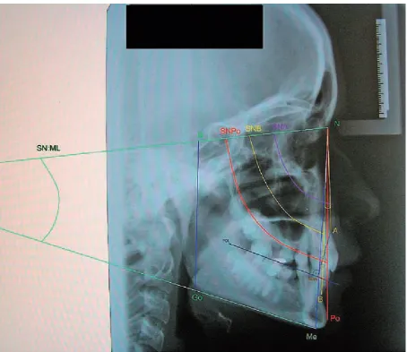
The location of the mandible was determined with the use of a lateral cephalometric image on the basis of a compilation of various analyses (Fig. 1). All lateral cephalometric images were tak-en in maximal intercuspidation using an OP 100 orthopantomograph with Kodak T-Mat E film.
The articular surface of the TMJ was described according to the grading system developed by Roh-lin and Petersson [9]:
Grade 0 – normal conditions: marked border of the cortical bone of articular surface;
Grade 1 – slight abnormality: single, mild os-teophytes, sclerosis and flattening of the articular surface;
Grades 2 and 3 – early and moderate destruc-tive abnormality, such as erosion and cysts;
Grade 4 – severe destructive abnormality: ex-tensive erosions of the lateral and medial parts of the condyle and the temporal joint;
Grade 5 – mutilating abnormality: temporo-mandibular joint anchylosis.
The assessment of the TMJ surface and peri-apical changes in the teeth was possible on the ba-sis of pantomographic images taken with an Or-thoceph OC 100 using Kodak T-Mat G/RA film.
Ethical Issues
Statistical Analysis
The results obtained were subjected to statis-tical analysis in which the arithmetic mean and standard deviation were calculated for the mea-surable features, and the distribution in quantity- -percentage terms was calculated for the qualitative features. The Shapiro-Wilk test was used to check for normal distribution of the data. The parametric Student t-test or Mann-Whitney U-tests were ap-plied as appropriate.
The relationships between selected parameters were determined using the Spearman rank correla-tion coefficient. P < 0.05 was assumed as statistical-ly significant. The statistical power of the tests was also calculated. The calculations were carried out using the Statistica 10 software package (StatSoft).
Results
The age range in the group of patients in-volved in the study was 6–19 years (mean age 13.8 ± 4.52 years), and the range of duration of the disease was 2–13 years (mean 6.7 ± 4.25 years). The disease activity was determined using the Juvenile Arthritis Disease Activity Score (JADAS) [10]. The JADAS score consists of a global disease activity assessment by a doctor, a global assessment of the child’s well-being by the patient/parent, the num-ber of joints involved, and the erythrocyte sedi-mentation rate (ESR) value.Three grades of dis-ease activity are distinguished: low, medium and high. According to this scale the study population was divided into 2 groups: 35 patients (76%) were classified as having low disease activity, while the
Fig. 1. A compilation of various analyses of a lateral cephalogram of one of the examined patients
disease activity in 11 patients (24%) was moder-ate. Clinical symptoms in the temporomandibular joints mainly included pain, crepitus and restrict-ed mouth opening. The highest value of maximal mouth opening in patients with these symptoms ranged from 44 mm to 35 mm.
Symptoms of joint dysfunction or changes within the facial skeleton were observed in 15 out of the 46 patients, which constituted 32.6% of all the participants of the study; 7 of those 15 (46.7%) had pauciarticular JIA and 8 (53.3%) had polyarticular JIA. Inflammatory disease activity in these 15 pa-tients during the examination was low, according to JADAS. Table 1 shows the frequency of dys-functions within the facial skeleton observed dur-ing clinical examinations. It should be emphasized that only 2 patients with TMJ dysfunction had no changes in the mandibular condyle and articular
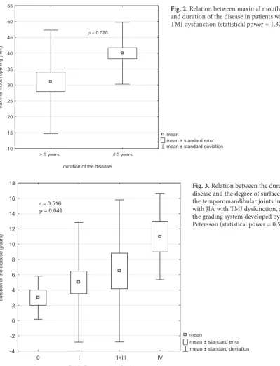
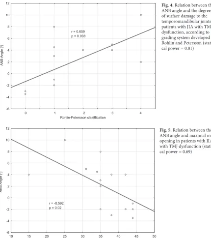
surface, and 6 demonstrated significant damage, including 2 patients with very serious changes. In addition, periapical tissues of teeth were evaluated in pantomographic images, and changes were not-ed in 12 patients (Table 2a).Theresults presented in Table 2b show a higher tendency toward retrog-nation and posteriorotation of the mandible in pa-tients with JIA and TMJ dysfunction in compari-son to healthy children. No significant differences were identified between patients with pauciarticu-lar JIA and patients with polyarticupauciarticu-lar JIA in terms of temporomandibular joint destruction (85.7% to 75%). Dysfunction of these joints, indicated in maximal mouth opening restriction, increases with the duration of the disease (Fig. 2; statistical pow-er = 1.37), as does damage to the temporoman-dibular bones observed in radiographic imaging (Fig. 3; statistical power = 0.54). A significant pos-itive correlation between the degree of mandibular condylar damage and the suppression of mandible development was observed (Fig. 4; statistical pow-er = 0.81). A significant negative correlation be-tween the ANB angle (retrognathia) and maximal mouth opening was also ascertained (Fig. 5; statis-tical power = 0.69).
Discussion
The chronic inflammatory process in the course of JIA can affect the temporomandibular joints as well as the surfaces of the bones which form them, including the temporal bones and mandibular condyloid processes. Symptoms of TMJ inflam-mation in JIA may occur at the beginning of the disease, during its course, or may even precede the disease itself [11, 12]. In the study group, clinical symptoms of TMJ inflammation such as transient pain, clicking and difficulty in opening the mouth were observed in 15 out of 46 patients (over 1/3 of the patients). Among those 15, the maximal mouth opening during the examination was 38 mm, and dysfunction
Cephalometric measurments: Mean SD ±
SGo:NMe (norm 58º ± 3.6º) 64.14 6.36
ANB (norm 2 ± 3 mm) 3.0 5.42
SN:ML (norm 33º ± 6º) 35.47 9.10
SNA (norm 82º ± 3.5º) 80.13 4.96
SNB (norm 80º ± 3.5º) 76.30 5.70
SNPo (norm 81º ± 3.5º) 76.80 6.50
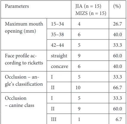
Parameters JIA (n = 15)
MIZS (n = 15) (%)
Maximum mouth
opening (mm) 15–3435–38 4 6 26.740.0
42–44 5 33.3
Face profile
ac-cording to ricketts straightconcave 9 6 60.040.0
Occlusion –
an-gle’s classification III 510 33.366.7
Occlusion
– canine class III 5 9 33.360.0
III 1 6.7
Table 2a. Pantomographic analysis in patients with
juve-nile idiopathic arthritiswith TMJ dysfunction
Parameters JIA (n = 15)
MIZS (n = 15) (%)
Rohlin and Petersson grad-ing system*
0o 2 13.3
I o 7 46.7
IIo and IIIo 4 26.7
IV o 2 13.3
Periapical chang-es in the teeth (pantomograph-ic imaging)
no
changes 3 20
changes
present 12 80
the minimal value was 15 mm. The differences de-pended on the severity of the symptoms and the degree of articular surface damage. Due to the considerable discrepancies in the ages of the ex-amined patients, these values should be compared cautiously with the results of other studies, which indicate a maximal mouth opening of 35 mm [6], 29.2 mm [13] and 49.5 mm [14]. It should be em-phasized that persistent pain in TMJ may lead to a weakening of the muscles or even to masseter muscle atrophy, which affects the functioning and development of the mandible [15]. The current study confirms this, because an increased ANB
angle reduced maximal mouth opening (negative correlation – Fig. 5).
Furthermore, in the developmental years the mandible is especially prone to inflammatory fac-tors, partly because the mandibular growth cen-ter is located just below the articular surface of the mandibular condyloid process. Therefore, it can be easily damaged, which can lead to mandibu-lar underdevelopment, abnormality in facial skele-ton morphology or (ultimately) to “bird face” fea-tures [7, 8, 16, 17].
According to the available literature, the fre-quency of inflammatory changes in the TMJ ranges mean
mean ± standard error mean ± standard deviation > 5 years ≤ 5 years
duration of the disease 10
15 20 25 30 35 40 45 50 55
maximal mouth opening (mm)
p = 0.020
mean
mean ± standard error mean ± standard deviation
0 I II+III IV
Rohlin-Petersson classification -4
-2 0 2 4 6 8 10 12 14 16 18
duration of the disease (years)
r = 0.516 p = 0.049
Fig. 2.Relation between maximal mouth opening and duration of the disease in patients with JIA with TMJ dysfunction (statistical power = 1.37)
from 17% to 87%. These differences may result ei-ther from the degree of severity of the symptoms and articular surface damage, or from the different populations examined the use of different meth-ods of assessing the affected area of the joint [2, 7, 18–23].
The present study was conducted with the use of equipment allowing for cephalometric and pan-tomographic imaging. The accuracy of assessment of bone changes with the use of these methods has been evaluated as being from 71% to 84% [24, 25]. Arguably, MRI or CT examination are far more accurate methods [26]. However, they are much more expensive, and in the case of CT, the pa-tient is exposed to a considerably higher dose of
radiation than in the case of cephalometric or pan-tomographic imaging [27]. Furthermore, the re-sults of a preliminary study by Abramowicz et al. indicate that in children with JIA, the combination of abnormal condyle morphology and accentuat-ed antegonial notching on a panoramic radiograph correlates with TMJ synovitis on an MRI [28].
The findings of this study may be compared to a study by Twilt et al., which also made use of the Rohlin and Petersson grading system [12]. Twilt et al. found the occurrence of changes in the TMJ to be from 37% to 46%. In the present study, clear ab-normalities in the cephalometric examination (above Grade 2 according to the Rohlin and Petersson grad-ing system) were observed in 39.9% of the patients. 0 1 2 3 4
Rohlin-Petersson clasiffication -6
-4 -2 0 2 4 6 8 10
ANB
Angle (
)
o
r = 0.659 p = 0.008
ANB angle and the degree of surface damage to the temporomandibular joints in patients with JIA with TMJ dysfunction, according to grading system developed by Rohlin and Petersson (statisti-cal power = 0.81)
10 15 20 25 30 35 40 45 50
maximal mounth opening (mm) -6
-4 -2 0 2 4 6 8 10 12
ANB
Angle (
)
o
r = -0.592 p = 0.02
The findings of the present study confirm the findings of other authors [2, 29] that relate the in-flammatory process in TMJ to bigger changes, which may include a retrognathic mandible and impaired maximal mouth opening; the range of maximal mouth opening was significantly low-er in patients who had sufflow-ered from JIA for ovlow-er 5 years (p < 0.02, Fig. 2). Similarly, damage to the TMJ according to the grading system developed by Rohlin and Petersson was significantly higher in patients with a longer duration of the disease (p < 0.049) and positively correlated with an in-creased ANB angle expressing a retrognathic posi-tion of the mandible (r = 0.66, p < 0.008, Fig. 4).
It is a common conviction that polyarticular JIA may have a greater influence (3 : 1) on damage to the articular surface than pauciarticular JIA [2]; however, according to the current study, the pro-portions are almost equal (7 : 6).
According to the available literature, bone ero-sions in JIA occur later than in rheumatoid arthri-tis, which is a result of the wider articular cartilage, especially in the case of younger children. How-ever, the resulting impairment of the mandible function in the developmental years can be con-siderable, to such extent that juvenile idiopathic arthritis which affects the TMJ is considered a ma-licious localization of JIA [30].
To sum up, the use of the presented meth-ods for the early detection of TMJ and
mandibu-lar damagemay prevent permanent and significant
facial deformities that can have a serious impact on the quality of life of young patients, regardless of the disease activity or remission. A lack of standards for the diagnosis and treatment of patients with TMJ arthritis is undoubtedly still a problem [31], but the authors believe that this study will contribute to the development and elaboration of such standards.
The authors concluded that clinical and radio-logical assessment of the mandible and temporo-mandibular joints indicated posterior rotation and Class II malocclusion in over 1/3 of the study pop-ulation of children and teenagers suffering from juvenile idiopathic arthritis. This suggests that it is important to include the clinical and radiolog-ical assessment of these joints during each rheu-matological control visit. Cephalometric analysis confirms the degree of articular surface damage of the temporomandibular joint in approximate-ly 40% of patients with clinical signs of dysfunc-tion, which indicates the need for early diagnostic imaging. The treatment of patients suffering from juvenile idiopathic arthritis with the symptoms of temporomandibular joint dysfunction should be multidisciplinary, and an orthodontist should be included in the treatment team.
References
[1] Fjeld M, Arvidsson L, Stabrun A, Birkeland K, Larheim T, Oggard B: Average craniofacial development from 6 to 35 years of age in a mixed group of patents with juvenile idiopathic arthrtitis. Acta Odontol Scand 2009, 67, 153–160.
[2] Sidiropoulou-Chatzigianni S, Papadopoulos MA, Kolokithas G: Mandibular condyle lesions in children with juvenile idiopathic arthritis. Cleft Palate Craniofac J 2008, 45, 57–62.
[3] Walton AG, Welbury RR, Thomason JM, Foster HE: Oral health and juvenile idiopathic arthritis; a review. Rheumatology 2000, 39, 550–555.
[4] Huntjens E, Kiss G, Wouters C, Carels C: Condylar asymmetry in children with juvenile idiopathic arthritis assessed by cone-beam computed tomography. Eur J Orthod 2008, 30, 545–551.
[5] Ronning O, Barnes SAR, Pearson MH, Pledger DM: Juvenile chronic arthritis: a cephalometric study of the facial skeleton. Eur J Orthod 1994, 16, 53–62.
[6] Mandall NA, Gray R, O’Brien KD, Baildam E, Macfarlane TV, Davidson J, Sills J, Foster H, Gardner-Medwin J, Garrahy A, Millett D, Mattick R, Walsh T, Ward S: Juvenile idiopathic arthritis (JIA): a screening study to mea-sure class II skeletal pattern, TMJ PDS and use of systemic corticosteroids. J Orthod 2010, 37, 6–15.
[7] Ronchezel MV, Hilário MO, Goldenberg J, Lederman HM, Faltin K Jr, de Azevedo MF, Naspitz CK:
Temporomandibular joint and mandibular growth alterations in patients with juvenile rheumatoid arthritis. J Rheumatol 1995, 22, 1956–1961.
[8] Larheim TA, Haanes HR, Ruud AF: Mandibular growth, temporomandibular joint changes, and dental occlusion in juvenile rheumatoid arthritis. Scand J Rheumatol 1981, 10, 225–233.
[9] Rohlin M, Petersson A: Rheumatoid arthritis of the temporomandibular joint: Radiologic evaluation based on standard reference films. Oral Surg Oral Med Oral Pathol 1989, 67, 594–599.
[10] Twilt, Schulten AJM, Nicolaas P, Dülger A, van Suijlekom-Smit LWA: Facioskeletal changes in children with juvenile idiopathic arthritis. Ann Rheum Dis 2006, 65, 823–825.
[11] Twilt M, Schulten AJM, Prahl-Andersen B, van Suijlekom-Smit LWA: Long-term Follow-up of Craniofacial Alternations in Juvenile Idiopathic Arthritis. Angle Orthod 2009, 79, 1057–1062.
[12] Larheim TA, Haanaes HR: Micrognathia, temporomandibular joint changes and dental occlusion in juvenile rheumatoid arthritis of adolescents and adults. Scand J Dent Res 1981, 89, 329–338.
[15] Twilt M, van der Giesen E, Mobers SMLM, ten Cate R, van Suijlekom-Smit LWA: Abrupt condylar destruction of the mandibula in juvenile idiopathic arthritis. Ann Rheum Dis 2003, 62, 366–367.
[16] Kjellberg H, Fasth A, Kiliardis S, Wenneberg B, Thilander B: Craniofacialstructure in children with JCA com-pared with healthy children with ideal or postnormal occlusion. Am J Orthod Dentofacial Orthop 1995, 107, 67–78.
[17] Twilt M, Mobers SMLM, Arends LR, ten Cate R, van Suijlekom-Smit L: Temporomandibular involvement in juvenile idiopathic arthritis. J Rheumatol 2004, 31, 1418–1422.
[18] Twilt M, Arends LR, ten Cate R, Van Suijlekom-Smit LWA: Incidence of temporomandibular involvement in juvenile idiopathic arthritis. Scand J Rheumatol 2007, 36, 184–188.
[19] Twilt M, Schulten AJM, Verschure F, Wisse L, Prahl-Andersen B, van Suijlekom-Smit LW: Long-term follow-up of temporomandibular joint involvement in juvenile idiopathic arthritis. Arthritis Rheum 2008, 59, 546–552.
[20] Kjellberg H: Craniofacial growth in juvenile chronic arthritis. Acta Odontol Scand 1998, 56, 360–365.
[21] Mayne JG, Hatch GS: Arthritis of the temporomandibular joint. J Am Dent Assoc 1969, 79, 125–130.
[22] Mericle PM, Wilson VK, Moore TL, Hanna VE, Osborn TG, Rotskoff KS, Johnston LE Jr: Effects of polyarticu-lar and pauciarticupolyarticu-lar onset juvenile rheumatoid arthritis on facial and mandibupolyarticu-lar growth. J Rheumatol 1996, 23, 159–165.
[23] Kobayashi K, Kondoh T, Sawai K, Yamamoto A: Image diagnosis for internal derangements of the temporoman-dibular joint: the adventages and limitations of imaging techniques. Oral Radiol 1991, 7, 13–24.
[24] Kakudo K: The significance and problems of the rotational panoramic radiography as routinescreeningtests for osteoarthritis of the temporomandibular joint. J Jpn Assoc Dent Sci 1995, 14, 43–47.
[25] Brooks SL, Brand JW, Gibbs SJ, Hollender L, Lurie AG, Omnell KA: Imaging of the temporomandibular joint: a position paper of the American Academy of Oral and Maxillofacial Radiology. Oral Surg Oral Med Oral Pathol Oral Radio Endod 1997, 83, 609–618.
[26] Matsumoto R, Ioi H, Nishioka M: TMJ osteoarthritis/osteoarthrosis and dentofacial morphology in Japanese females. Orthod Waves 2006, 65, 101–106.
[27] Ronning O, Valiaho ML: Progress of mandibular condyle lesions in juvenile rheumatoid arthritis. Proc Finn Dent Soc 1981, 77, 151–157.
[28] Rutkowska-Sak L: Młodzieńcze idiopatyczne zapalenie stawów – problemy z klasyfikacją. Reumatologia 2011, 49, 5, 351–353.
Address for correspondence:
Anna Górska Jurowiecka 28/4 15-101 Białystok Poland
Tel.: +48 501 367 775 E-mail: [email protected]
Conflict of interest: None declared