Adrian Reśliński
1, Agnieszka Mikucka
2, Jakub Szmytkowski
1,
Katarzyna Głowacka
3, Wojciech Szczęsny
1, Eugenia Gospodarek
2,
Stanisław Dąbrowiecki
1Biofilm Detection on the Surface
of Hernia Mesh Implants*
Wykrywanie biofilmu na powierzchni siatek przepuklinowych
1 Department of General and Endocrine Surgery, Nicolaus Copernicus University of Torun, Collegium Medicum in Bydgoszcz, Poland
2 Department of Microbiology, Nicolaus Copernicus University of Torun, Collegium Medicum in Bydgoszcz, Poland
3 Department of Plant Physiology and Biotechnology, Warmia-Mazury University, Olsztyn, Poland
Abstract
Background. One serious complication of tension-free mesh hernioplasty is deep surgical site infection (SSI) involv-ing the implant (mesh infection). As evidenced by in vitro studies, bacterial colonization of a synthetic implant may lead to the development of a biofilm on its surface, which is a serious therapeutic problem.
Objectives. The goal of this study was to evaluate the usefulness of a biofilm detection method based on 2,3,5-tri-phenyltetrazolium chloride (TTC) for screening for the presence of bacterial biofilm on the surface of biomaterials utilized in hernia surgery.
Material and Methods. The study group included samples of 12 synthetic implants harvested from 12 patients treated for deep SSIs after abdominal and inguinal hernia repair. The presence of biofilm was evaluated using the TTC reduction method, scanning electron microscopy and a quantitative method.
Results. Bacterial biofilm was found on the surfaces of all of the implants. The presence of a biofilm detected by TTC reduction was confirmed in each case by scanning electron microscopy and a quantitative method. The quan-titative analysis yielded between3.25 × 105 and 2.50 × 10⁸ CFU/ml in the biofilm present on individual biomaterial samples.
Conclusion. The TTC reduction method is useful for initial screening for the presence of a bacterial biofilm on the surface of implants utilized in hernia surgery (Adv Clin Exp Med 2010, 19, 6, 685–690).
Key words: hernia, surgical mesh, biofilm, TTC.
Streszczenie
Wprowadzenie. Groźnym powikłaniem plastyki przepukliny z wszczepieniem biomateriału jest głębokie zakaże-nie miejsca operowanego (z.m.o.) obejmujące implantat. Konsekwencją bakteryjnej kolonizacji biomateriału jest powstanie biofilmu na jego powierzchni, co jest istotnym problemem terapeutycznym.
Cel pracy. Ocena przydatności metody redukcji chlorku 2,3,5-trójfenylotetrazoliowego (TTC) do wstępnej oceny występowania biofilmu bakteryjnego na powierzchni biomateriałów stosowanych w chirurgii przepuklin.
Materiał i metody. Badaniem objęto fragmenty 12 implantatów pochodzących od 12 pacjentów po plastyce prze-puklin brzusznych i pachwinowych, leczonych z powodu głębokiego z.m.o. Obecność biofilmu na powierzchni usuniętych biomateriałów badano metodą redukcji TTC, z użyciem skaningowego mikroskopu elektronowego oraz metodą ilościową.
Wyniki. Biofilm bakteryjny stwierdzono na powierzchni wszystkich badanych implantatów. Obecność biofilmu wykrytego za pomocą metody opartej na redukcji soli tetrazoliowych w każdym przypadku potwierdzono w bada-niu z użyciem skaningowego mikroskopu elektronowego i metody ilościowej. W badabada-niu ilościowym stwierdzono od3,25 × 105 i 2,50 × 10⁸ j.t.k./ml w biofilmie obecnym na jednej próbce biomateriału.
Adv Clin Exp Med 2010, 19, 6, 685–690 ISSN 1230-025X
ORIGINAl PAPERS
© Copyright by Wroclaw Medical University
Deep surgical site infection (SSI) is a serious complication of hernioplasty using a biomaterial implant. It has been documented that the inci-dence of SSI involving a mesh implant varies de-pending on the surgical technique and the type of biomaterial used. The incidence of polypropylene mesh infection ranges from 0.9 to 6.0% for open surgery [1, 2] and 0.0 to 4.0% for laparoscopic re-pair [3, 4]. With polyester implants, these values range from 2.0 to 28.5% [5, 6] for open surgery and 0.0 to 1.4% for laparoscopic repair [7, 8]. For hernioplasties using ePTFE patches, the infection rates are 0.0–22.1% [2, 9] for open and 0.0–1.0% for laparoscopic techniques [10, 11].
As evidenced by in vitro [12] studies and the authors’ clinical observations [13], bacterial colo-nization of a synthetic hernia implant may lead to the development of a biofilm on its surface. A biofilm is a heterogeneous structure composed of microorganisms which are bound to a surface, surrounded by an extracellular matrix and which display phenotypic traits not observed in their planktonic forms. Biofilm protects the bacteria within against the host’s immune mechanisms and antimicrobial agents. It can also become fragment-ed and detachfragment-ed, giving rise to secondary infection foci [14].
Many methods for detecting biofilm on the surface of surgical meshes have been described, in-cluding classic culture techniques, scanning elec-tron microscopy or evaluation by a confocal laser scanning microscope [12, 15]. In this study, col-orless 2,3,5-triphenyltetrazolium chloride (TTC) reduction to red formazan was used for the initial evaluation of biofilm presence on the surfaces of synthetic hernia implants.
Material and Methods
Biomaterials
The study group included samples of 12 syn-thetic implants harvested from 12 patients who had undergone abdominal and inguinal hernia re-pair performed at the authors’ center in the years 2009–2010, in whom deep SSI was diagnosed ac-cording to CDC criteria [16]. The biomaterials in-cluded: polypropylene meshes (three mono- and two multifilament), four multifilament polyester mesh implants, two expanded
polytetrafluoroeth-ylene (ePTFE) patches and one composite mesh (multifilament polyester mesh coated with a hy-drophilic layer of collagen, polyethylene glycol and glycerol). The biomaterials were harvested during surgery under general anesthesia. Each implant sample was derived from a different patient.
Microorganisms
The initial identification of the cultures was based on colony morphology on Columbia Agar with 5% sheep blood (Becton Dickinson, Sparks, USA) and selective differential media; specific tests were also performed, including coagulase production for Staphylococcus spp., esculin hy-drolysis for Enterococcus spp., oxidase production for Pseudomonas spp. and indole production for
Enterobacteriaceae. Species identification was per-formed using the ID32 Staph kit (bioMérieux S.A. RCS lyon, France) for staphylococci and ID32 E (bioMérieux S.A. RCS lyon, France) for rods of the Enterobacteriaceae family. The test results were read and interpreted using ATB Expression software (bioMérieux S.A. RCS lyon, France) with version 2.8.8 of the database.
Biofilm Detection
A qualitative method utilizing the property of metabolically active microorganisms to reduce col-orless 2,3,5-triphenyltetrazolium chloride (TTC) to red formazan [17] was used to detect biofilm on the biomaterial samples. Fragments (1 × 1 cm) of the implants were incubated in 4 ml of tryptic soy broth (TSB, Becton Dickinson, Sparks, USA) con-taining 50 µl of 1% TTC solution (POCH, Gliwice, Poland). The samples were then incubated at 37oC
for 24 hours and the appearance of red formazan was observed. The degree of TTC reduction was graded on the following scale: 0 – no TTC reduc-tion; +1 – slight pink dotting of the mesh surface, +2 – solid pink coloring of the entire surface, +3 – red coloring of the mesh surface, clouding and red coloring of the substrate. For each bioma-terial sample the experiment was done three times and the representative data were presented.
The presence of biofilm on the implant sur-faces was also evaluated by a scanning electron mi-croscope (SEM). The implant fragments were fixed in a 2.5% glutaraldehyde solution (POCH, Gliwice, Poland) in a 0.1 M phosphate buffer at a pH of 7.4
Wnioski. Metoda redukcji TTC jest przydatna do wstępnej oceny występowania biofilmu bakteryjnego na powierzchni implantatów stosowanych w chirurgii przepuklin (Adv Clin Exp Med 2010, 19, 6, 685–690).
for 24–48 hours at 4ºC. After fixation, the material was rinsed twice for 20 minutes in phosphate buffer at room temperature. The samples were dehydrated in a graded series of ethanol concentrations – 30%, 50%, 70%, 80% and 96% – for ten minutes in each solution and twice for 30 minutes in 99.8% ethanol (POCH, Gliwice, Poland) at room temperature. After dehydration, the samples were transferred to a dryer chamber (Critical Point Dryer CPD 030, Bal-Tec, Balzers, liechtenstein) filled with amyl acetate (Sig-ma-Aldrich, Steinheim, Germany) and dried at the critical point of CO2. The dried material was placed
on copper tables and sputter-coated with gold in an argon atmosphere in an ionic coater (Fine Coater, JCF-1200, JEOl, Tokyo, Japan). The sputter-coated material was placed in a SEM column (JSM-5310lV, JEOl, Tokyo, Japan) and analyzed at 25 kV. The re-sults were recorded on black and white IlFORD FP4 PlUS 125 photographic film.
A quantitative analysis was also performed. The biofilms were detached from the surfaces of the biomaterial samples (1 × 1 cm) by shaking in 0.5% saponin (Fluka, Steinheim, Germany). Serial 10-fold dilutions of the suspension thus obtained were performed with subsequent inoculation on trypticase soy agar (Tryptic Soy Agar, TSA, Bec-ton Dickinson, Sparks, USA). After a 24-hour in-cubation at 37ºC, the result (the average of three measurements) was recorded as the number of colony-forming units (CFUs) per one milliliter of suspension (CFU/ml).
In all the experiments a sterile sample (1 × 1 cm) of monofilament polypropylene mesh was used as a control. Polypropylene mesh is the most com-monly used implant type in hernia surgery [18].
Results
Fifteen bacterial strains were isolated from the implant samples. Among the most common etio-logical factors in implant infections were S. aureus
(seven strains) and E. coli (three strains). The mi-croorganisms isolated from the investigated im-plants are reviewed in Table 1.
Bacterial biofilm was confirmed on the surfaces of all of the synthetic materials. After one hour of incubation on a substrate containing 2,3,5-triphe-nyltetrazolium chloride, a pink hue corresponding to the first degree of TTC reduction was observed in eight samples. After four hours of incubation, slight pink dotting was observed on the surfaces of four implant samples, and in the remaining sam-ples the entire surface was uniformly pink. After 24 hours, nine implants exhibited a uniform red coloring on their entire surfaces, while three sam-ples showed coloring corresponding to the second degree of TTC reduction.
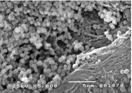
Scanning electron microscopy revealed the presence of bacteria adhering to the surfaces of the implants (Figures 1, 2 and 3), mainly in niches between individual filaments (in multifilament
Table 1. Evaluation of the bacterial biofilm on the surfaces of the implants (* – hour, ** – average of three measurements; m – monofilament mesh; mm – multifilament mesh; polypropylene – pp, polyester – pe, 0 – no TTC reduction, +1 – slight pink dotting of the mesh surface, +2 – solid pink coloring of the entire surface, +3 – red coloring of the mesh surface, cloud-ing and red colorcloud-ing of the substrate)
Tabela 1. Ocena biofilmu bakteryjnego na powierzchni implantatów (* – godzina, ** – średnia z trzech pomiarów, siatka mono-, multifilamentowa – odpowiednio: m, mm; polipropylen – pp, poliester – pe, 0 – brak redukcji TTC, +1 – lekkie punktowe zaróżowienie powierzchni implantatu, +2 – zaróżowienie całej powierzchni implantatu, +3 – zaczerwienienie całej powierzchni implantatu, zmętnienie oraz czerwona barwa podłoża)
No.
(lp.) Biomaterial (Biomateriał) Etiological factor of infection (Czynnik etiologiczny zakażenia) TTC reduction (Redukcja TTC) CFU/ml** (j.t.k./ml**) 1h* 4h 24h
1. 2. 3. 4. 5. 6. 7. 8. 9. 10. 11. 12. mm pp mm pp m pp m pp m pp mm pe mm pe mm pe mm pe ePTFE ePTFE
mm pe + collagen + polyethylene glycol + glycerol
Staphylococcus aureus Pseudomonas aeruginosa Staphylococcus aureus Staphylococcus aureus Staphylococcus epidermidis Staphylococcus aureus Staphylococcus aureus Staphylococcus aureus Escherichia coli
Escherichia coli, Enterococcus faecalis, Enterobacter cloaceae
Staphylococcus aureus
Escherichia coli, Enterococcus faecium
+1 0 +1 +1 0 +1 0 +1 +1 +1 +1 0 +2 +1 +2 +2 +1 +2 +1 +2 +2 +2 +2 +1 +3 +3 +2 +3 +2 +3 +2 +3 +3 +3 +3 +3
1.90 × 10⁸
6.50 × 10⁶
1.80 × 10⁶
1.85 × 10⁸
3.25 × 10⁵
2.50 × 10⁸
3.60 × 10⁶
1.55 × 10⁸
1.80 × 10⁷
4.50 × 10⁷
2.00 × 10⁷
implants) or at fiber cross-junctions (in monofila-ment implants).
The quantitative analysis yielded between 3.25 × 10⁵ and 2.50 × 10⁸ CFU/ml in the biofilm
present on individual biomaterial samples. The re-sults of biofilm assessment on the surfaces of the implants are shown in Table 1.
Discussion
This study evaluated the presence of bacterial biofilms on the surfaces of biomaterials obtained from patients with deep SSI following anterior ab-dominal wall hernioplasty. The presence of a bac-terial biofilm was detected on the surfaces of all of the investigated samples by a method utilizing the reduction of colorless TTC to red formazan, and was verified by scanning electron microscopy and a quantitative method. The presence of a biofilm was confirmed in each case.
In the material investigated, the etiological fac-tors of the implant infections included both Gram- -positive and Gram-negative bacteria (reviewed in Table 1). Reports have also been published on im-plant infections caused by Streptococcus pyogenes,
Acinetobacter baumannii, Acinetobacter lwoffii,
Citrobacter koseri, Proteus spp., Mycobacterium
spp., Peptostreptococcus spp. and fungi of the ge-nus Candida [1, 19–22]. The species colonizing the implant is influenced by the mechanism of im-plant contamination. The formation of a biofilm may result from intraoperative contamination, bacterial translocation from the large bowel [6] or a transient bacteremia.
The factors playing crucial roles in the cre-ation of a biofilm on the surface of a hernia im-plant have been identified. One of them is imim-plant hydrophobicity, which has been shown to facilitate biofilm formation on the surface of a biomaterial [12, 23]. Hydrophobic materials – i.e. polypropyl-ene or Teflon – had been used in seven patients in the current study. Another factor is the struc-ture of the surgical mesh. Multifilament implants facilitate biofilm formation due to the presence of interfilament niches, which provide favorable conditions for the bacteria to thrive and increase the overall area of the implant, providing a better opportunity for bacterial adhesion [12, 23]. In the case of monofilament implants, the bacteria usu-ally colonize the filament junctions [24]. Similar observations were made in the current study.
The current study entailed the use of a bio-chemical method based on the assessment of TTC reduction. This reaction is caused by metabolically active microorganisms present within the biofilm, utilizing dehydrogenase [25, 26]. In the material
Fig. 1. Biofilm on the surface of a multifilament poly-ester mesh (S. aureus); scanning electron microscopy (magnification 5000×)
Ryc. 1. Biofilm na powierzchni multifilamentowej siat-ki poliestrowej (S. aureus); skaningowa mikroskopia elektronowa (powiększenie 5000×)
Fig. 2. Biofilm on the surface of an ePTFE patch (polymicrobial biofilm); scanning electron microscopy (magnification 5000×)
Ryc. 2. Biofilm na powierzchni łaty z teflonu (biofilm wielogatunkowy); skaningowa mikroskopia elektrono-wa (powiększenie 5000×)
Fig. 3. A sample of sterile monofilament polypropylene mesh (control); scanning electron microscopy (magni-fication 5000×)
studied, alteration of the coloring of the implant sur-faces could already be observed after one hour, and its intensity increased over the period of incubation. Metabolism of TTC to formazan by the bacteria ad-hering to the implant surface took place regardless of the shape, type or color of the biomaterial.
The TTC method allows for biofilm detection without detaching it from the implant surface, which is a prerequisite of culture techniques. In our series, a quantitative result was obtained in each case. In vitro studies indicate that the sensitiv-ity of the TTC reduction method may exceed that of the classic culture techniques, as it allows for the detection of bacteria on the surface of the im-plant even if the number of the bacteria colonizing the biomaterial is below the detection threshold of culture techniques [27]. The sensitivity of the TTC
reduction method may reduce the number of false negative results, which delay the introduction of appropriate treatment of deep SSI [28].
Using a scanning electron microscope or con-focal laser scanning microscope [12, 15] enables the researcher to evaluate the morphology of film-forming bacteria and the structure of the bio-material itself; it also makes it possible to observe the three-dimensional structure of biofilm. These methods are, however, expensive and labor-inten-sive; and the limited availability of such equipment is another drawback of these methods. The TTC reduction method, on the other hand, is easy to perform and allows biofilm to be detected within a short time, making it a good diagnostic tool for the initial diagnosis of bacterial biofilm on the sur-face of hernia implants.
References
[1] Jezupors A, Mihelsons M: The analysis of infection after polypropylene mesh repair of abdominal wall hernia. World J Sur 2006, 30, 2270–2278.
[2] Leber GE, Garb JL, Alexander AI, Reed WP: long-term complications associated with prosthetic repair of inci-sional hernias. Arch Surg 1998, 133, 378–382.
[3] Andersson B, Hallen M, Leveau P, Bergenfelz A, Westerdahl J: laparoscopic extraperitoneal inguinal hernia repair versus open mesh repair: a prospective randomized controlled trial. Surgery 2003, 133, 464–472.
[4] Lal P, Kajla RK, Chander J, Saha R, Ramteke VK: Randomized controlled study of laparoscopic total extraperi-toneal vs. open lichtenstein inguinal hernia repair. Surg Endosc 2003, 17, 850–856.
[5] Hamy A, Pessaux P, Mucci-Hennekinne S, Radriamananjo S, Regenet N, Arnaud JP: Surgical treatment of large incisional hernias by an intraperitoneal Dacron mesh and aponeurotic graft. J Am Coll Surg 2003, 196, 531–534.
[6] Petersen S, Henke G, Freitag M, Faulhaber A, Ludwig K: Deep prosthesis infection in incisional hernia repair: predictive factors and clinical outcome. Eur J Surg 2001, 167, 453–457.
[7] Morrison JE, Jacobs VR: laparoscopic preperitoneal inguinal hernia repair using performed polyester mesh with-out fixation: prospective study with 1-year follow-up results in a rural setting. Surg laparosc Endosc Percutan Tech 2008, 18, 33–39.
[8] Rosen MJ: Polyester-based mesh for ventral hernia repair: is it safe? Am J Surg 2009, 197, 353–359.
[9] Bueno Lledó J, Sosa Quesada Y, Gómez I, Gavara I, Vaqué Urbaneja J, Carbonell Tatay F, Bonafé Diana S, García Pastor P, Baquero Valdelomar R, Mir Pallardó J: Prosthetic infection after hernioplasty. Five years expe-rience. Cir Esp 2009, 85, 158–164.
[10] Varghese TK, Denham DW, Dawes LG, Murayama KM, Prystowsky JB, Joehl RJ: laparoscopic ventral hernia repair: an initial institutional experience. J Surg Res 2002, 105, 115–118.
[11] Heniford BT, Park A, Ramshaw BJ, Voeller G: laparoscopic ventral and incisional hernia repair in 407 patients. J Am Coll Surg 2000, 190, 645–650.
[12] Engelsman AF, van der Mei HC, Francis KP, Busscher HJ, Ploeg RJ: Morphological aspects of surgical meshes as a risk factor for bacterial colonization. Br J Surg 2008, 95, 1051–1059.
[13] Reśliński A, Mikucka A, Szczęsny W, Szmytkowski J, Gospodarek E, Dąbrowiecki S:In vivo detection of biofilm on the surface of a surgical mesh implant – a case report. Chir Pol 2008, 3–4, 181–188.
[14] Bryers JD: Medical biofilms. Biotechnol Bioeng 2008, 100, 1–18.
[15] Saygun O, Agalar C, Aydinuraz K, Agalar F, Dephan C, Saygun M, Ceken S, Akkus A, Denkbas EB: Gold and gold-palladium coated polypropylene grafts in a Staphylococcus epidermidis wound infection model. J Surg Res 2006, 131, 73–79.
[16] Horan TC, Gaynes RP, Martone WJ, Jarvis WR, Emori G: CDC definitions of nosocomial surgical site infections, 1992: a modification of CDC definitions of surgical wound infections. Infect Control Hosp Epidemiol 1992, 13, 606–608.
[17] Gallimore B, Gagnon RF, Subang R, Richards GK: Natural history of chronic Staphylococcus epidermidis foreign body infection in a mouse model. J Infect Dis 1991, 164, 1220–1223.
[18] Robinson TN, Clarke JH, Schoen J, Walsh MD: Major mesh-related complications following hernia repair. Events reported to the Food and Drug Administration. Surg Endosc 2005, 19, 1556–1560.
[19] Rios A, Rodriguez JM, Munitiz V, Alcatraz P, Perez Flores D, Parrilla P: Antibiotic prophylaxis in incisional hernia repair using a prosthesis. Hernia 2001, 5, 148–152.
[21] Sohail MR, Smilack JD: Hernia repair mesh-associated Mycobacterium goodii infection. J Clin Microbiol 2004, 42, 2858–2860.
[22] Nolla-Salas J, Torres-Rodriguez JM, Grau S, Isbert F, Torrella T, Riveiro M, Sitges-Serra A: Successful treat-ment with liposomal amphotericin B of an intraabdominal abscess due to Candida norvegensis associated with a Gore-Tex mesh infection. Scand J Infect Dis 2000, 32, 560–562.
[23] Engelsman AF, van Dam GM, van der Mei HC, Ploeg RJ:In vivo evaluation of bacterial infection involving morphologically different surgical meshes. Ann Surg 2010, 251, 133–137.
[24] Bellón JM, N G-Honduvilla, Jurado F, G-Carranza A, Buján J:In vitro interaction of bacteria with polypropyl-ene/ePTFE prostheses. Biomaterials 2001, 22, 2021–2024.
[25] Umemoto K: Reduction mechanism of 2,3,5-triphenyltetrazolium chloride and 1,3,5-triphenylformazan. Bull Chem Soc Jpn 1985, 58, 2051–2055.
[26] Praveen-Kumar, Tarafdar JC: 2,3,5-triphenyltetrazolium chloride (TTC) as electron acceptor of culturable soil bacteria, fungi and actinomycetes. Biol Fertil Soils 2003, 38, 186–189.
[27] Jałoza D, Juda M, Malm A, Urban M, Bar K: The qualitative and quantitative detection of biofilm formation
in vitro on the biomaterials. Sepsis 2009, 2, 143–146.
[28] Rasnake MS, Dooley DP: Culture-negative surgical site infections. Surg Infect 2006, 7, 555–565.
Address for correspondence:
Adrian ReślińskiDepartment of General and Endocrine Surgery Nicolaus Copernicus University of Torun Collegium Medicum in Bydgoszcz M. Skłodowskiej-Curie 9
85-094 Bydgoszcz Poland
Tel.: +48 52 585 47 30 E-mail: [email protected]
Conflict of interest: None declared