© 2017 Amal S. Hathout et al. This is an open access article distributed under the terms of the Creative Commons Attribution License -NonCommercial-ShareAlikeUnported License (http://creativecommons.org/licenses/by-nc-sa/3.0/).
Journal of Applied Pharmaceutical Science Vol. 7 (01), pp. 086-092, January, 2017 Available online at http://www.japsonline.com
DOI: 10.7324/JAPS.2017.70111 ISSN 2231-3354
Synthesis and characterization of cobalt ferrites nanoparticles with
cytotoxic and antimicrobial properties
Amal S. Hathout
1, Abdulhadi Aljawish
2,3, Bassem A. Sabry
1, Aziza A. El-Nekeety
1, Mohamed H. Roby
4,
Nasseralla M. Deraz
5, Soher E. Aly
1, Mosaad A. Abdel-Wahhab
1*1
Food Toxicology & Contaminants Departmnt, National Research Center, Dokki, Cairo, Egypt. 2Nutrition Physiology and Toxicology Laboratory (NUTOX), INSERM U866, Bourgogne University, 1 Esplanade Erasme, F-21000 Dijon, France. 3ProBioGem Laboratory, EA 1026, Polytech'Lille-IUT "A" Bd Paul Langevin, Villeneuve d'Ascq cedex, Université Lille Nord de France, France. 4Food Science and Technology, Faculty of Agriculture, Fayoum University, Fayoum, Egypt. 5Physical Chemistry Department, National Research Center, Dokki, Cairo, Egypt.
ARTICLE INFO ABSTRACT
Article history:
Received on: 21/11/2016 Revised on: 02/12/2016 Accepted on: 28/12/2016 Available online: 31/01/2017
Recently, the application of nanotechnology in food sector and the agriculture attract the attention compared to its biomedical application. The aims of the current study was to synthesize and characterize cobalt ferrites nanoparticles [(CoFe2O4) NPs] by combustion method employing glycine as fuels and to evaluate their
antimicrobial against pathogenic bacteria and fungi and anti cancer properties against MCF-7 breast cancer cells line. The results indicated that the particles size of the synthesized (CoFe2O4) NPs was 40 nm. These (CoFe2O4)
NPs showed potential antibacterial properties against Gram-negative bacteria (Escherichia coli, Salmonella typhi) and Gram-positive bacteria (Staphylococcus aureus, Bacillus cereus) as well as the pathogenic fungi (Aspergillus flavus and Aspergillus ochraceus) in a dose dependent manner with maximum concentration of 1.8 mg/ml. (CoFe2O4) NPs also showed weak antiradical but have cytotoxic effects against MCF-7 breast cancer
cells line and succeeded to decrease the cell viability at a concentration of 2 mg/ml. It could be concluded that (CoFe2O4) NPs is a promise candidate as antimicrobial and anticancer agent for food sector and medical
application.
Key words:
Food safety; antimicrobial; anticancer; cobalt ferrites nanoparticles;
nanotechnology.
INTRODUCTION
Food safety is a major concern worldwide which has serious reflection on public health. Food contamination with pathogens can occur at different steps throughout the food production, processing, distribution, retail marketing, handling and preparation. Several epidemiological studies have implicated foods of animal origin as the major vehicles caused illnesses caused through food-borne pathogens (Petersen and James, 1998). Several attempts should be considered to limit the hazardous of these pathogens on human wellbeing. However, control constitutes the most significant component of overall
* Corresponding Author
Mosaad A Abdel-Wahhab, Food Toxicology & Contaminants Departmnt, National Research Center, Dokki, Cairo, Egypt.
E-mail: mosaad_abdelwahhab @ yahoo.com
management of food-borne pathogens. Recently, the unique properties of nanomaterials have given rise to enormous research activity towards nanoparticles fabrication and characterization as well as applications. Nanomaterials are rapidly expanding and used in various areas of research, such as food, feed, health management and environmental aspects (Bouwmeester et al. 2007; Kiruba Daniel et al. 2013). Metal-oxide nanoparticles draw attention due to their unique magnetic properties and other characteristics such as excellent chemical stability and mechanical hardness (Meng et
al., 2009; Phua et al., 2009; Sanpo et al., 2013). Cobalt ferrite
(CoFe2O4) nanoparticles offer potential applications in several
Yamamoto (2001) enumerated the influence of particle size on the antibacterial activity and reported that the activity was increased with decreasing particle size. The antimicrobial application of magnetic nanoparticles is gaining attention because they can be manipulated through an external magnetic field. Moreover, several nonmagnetic metals nanoparticles or metal oxides were found to be antimicrobials against pathogenic bacteria (Gajjar et al., 2009). The antimicrobial activities of magnetic nanoparticles are well established by Chifiriuc et al. (2011). Cobalt ferrites nanoparticles were synthesized using different techniques such as solid state reaction (Rafferty et al., 2008) microemulsion (Choi, 2003), combustion (Yan, 1999) the redox process (Rajendran, 2001), chemical co-precipitation (Pannaparayil and Komarneni, 1989), the hydrothermal method (Daou, 2006), and microwave synthesis (Bensebaa, 2004). Generally, the evaluation of magnetic nanoparticles for drug delivery, and sensing applications are widely studied. However, the use of cobalt ferrite nanoparticles as cytotoxicity and antimicrobials against pathogenic and drug resistant microbes are not well investigated. Therefore, the aim of the current work was to study the characteristics and antimicrobial activity of prepared cobalt ferrite nanoparticle against several pathogenic microorganisms and evaluation their cytotoxic effect against MCF-7 breast cancer cells.
MATERIALS AND METHODS
Preparation of cobalt ferrite nanoparticles [(CoFe2O4) NPs]
Cobalt ferrite nanoparticles [(CoFe2O4) NPs] were
prepared using the method described by Deraz (2010, 2012). In brief, the calculated proportions of iron and cobalt nitrates with a known amount of glycine, with molar ratio of 2:1:6, respectively, were mixed. The mixed precursors were concentrated in a porcelain crucible on a hot plate at 300 oC for 5 min. The crystal water was gradually vaporized during heating and when a crucible temperature was reached, a great deal of foams produced and spark appeared at one spot which spread through the mass, yielding a brown voluminous and fluffy product in the container. The product was washed several times with water and then dried in oven at 70 oC for 2h.
Characterization of nanoparticle
X-ray diffraction data collection of mixed oxide solids was carried out using a BRUKER D8 advance diffractometer (Germany). Data were collected with Cu Kα radiation at 40 KV and 40 mA with scanning speed in 2θ of 2o
/min. Scanning electron micrographs (SEM) were recorded on JEOL JAX-840A and JEOL JEM- 1230 electron micro-analyzers, respectively. The samples were dispersed in ethanol and then treated ultrasonically in order disperse individual particles over a gold grids.
Antifungal activity
The antifungal activity of (CoFe2O4) NPs was tested
against fungal cultures (Aspergillus flavus and Aspergillus
ochraceus). The fungal cultures were obtained from the Plant Pathology Department, National Research Centre, Cairo, Egypt. The qualitative assessment of the antifungal effect was done using well agar diffusion test. The sterilized Potato Dextrose Agar (PDA, BD Difco™, MD 21152, USA) medium was poured into petri plates and allowed to dry. Test cultures were inoculated over the dried surface of PDA plate. The appropriate number of wells was bored into the plates using sterile cork borer of 9-mm in diameter. Different concentrations of (CoFe2O4) NPs were added
to these wells and incubated for 7 days at 25 ± 2°C. The contact biocidal property was determined by measuring the diameter of the zone of inhibition around the well.
Antibacterial activity
The antibacterial activity of (CoFe2O4) NPs was tested
against bacterial cultures of Gram-negative bacteria (Escherichia
coli, Salmonella typhi) and Gram-positive bacteria
(Staphylococcus aureus, Bacillus cereus). The pathogenic bacteria were obtained from the Department of Microbial Chemistry, National Research Centre, Cairo, Egypt. The bacteria were pre-grown on Nutrient agar (NA, Sigma-Aldrich, St. Louis, MO63103, USA) for 16 h at 37.0 ± 0.1°C. The qualitative assessment of the antibacterial effect was done using two approaches (Disk Diffusion test and Bacterial Linear growth). Disc diffusion assay was determined using the protocol of Jorgensen and Turnidge (2007). Overnight grown bacterial cultures in nutrient broth were individually lawn cultured on nutrient agar plates. Three (CoFe2O4) NPs concentrations (0.6, 0.8 and 1.8 mg/mL) were
prepared in sterile water and dispensed by sonication. Sterile filter paper discs (5.0-mm) were saturated by the (CoFe2O4) NPs
solution and placed above the culture and incubated at 37 ± 0.1°C for 24 h after which the zone of inhibition were recorded. Sterile filter paper was saturated with sterile water and used as control. The same concentrations for bacterial linear growth were examined whereas each concentration was individually set in three petri dishes. The NA medium was poured in the plates and rotated to ensure an even distribution of the nanoparticle. Control Petri dishes were prepared containing no (CoFe2O4) NPs. Petri dishes
were individually inoculated at the center with equal disks (5.0-mm) of tested bacterial cultures. The average linear growth of each bacterium was measured after 24 h of incubation at 37.0 ± 0.1°C.
DPPH and ABTS free radical scavenging assay of (CoFe2O4)
NPs
(CoFe2O4) NPs at different concentrations was mixed
with 2 ml of methanol solution containing DPPH• radicals resulting in a final concentration of 6.10-5 M DPPH•. The mixture was shaken vigorously for 15 s and then left to stand in the dark at room temperature for 30 min. The absorbance (Abs) of the resulting solution was measured at 517 nm using a UV-visible spectrophotometer (Shimadzu UV-1605) against methanol as a blank according to the method described by Shimada et al. (1992). The scavenging ability was calculated by the following equation:
DPPH• radical scavenging activity was expressed as the half-maximal inhibition concentrations (IC50). IC50 value, which
expressed as the antioxidant concentration to reduce the radicals by 50%, was calculated for each compound by linear regression analysis (Gulcin, 2010).
The ABTS radical scavenging activity of (CoFe2O4) NPs
was determined according to the method described by Re et al. (1999). The reaction between 7 mM ABTS and 2.45 mM potassium persulfate in H2O made the ABTS
.+
radical cation. The last solution was left to stand in the dark, at room temperature, for 12–16h before use. Before analysis, the ABTS.+ solution was diluted with ethanol at 30°C, in order to obtain an absorbance of 0.700 ± 0.025 at 734 nm. Then, different concentrations of (CoFe2O4) NPs in solid form were mixed with 1 ml of diluted
ABTS.+ solution. The mixture was shaken vigorously for 15 s and then left to stand in the dark at room temperature for 20 min. The absorbance of ABTS.+ was measured at 734 nm against ethanol as a blanck. The extent of decolorization is calculated as the percentage reduction of ABTS absorbance by following equation: ABTS.+ radical scavenging activity (%) = (1 – Abs sample /Abs control)
x100
ABTS.+ radical scavenging activity was expressed as the half-maximal inhibition concentrations (IC50). All analyses were
carried out in triplicate and results represented the mean values with standard deviation.
Anti-proliferative activity Cell culture
MCF-7 (ECACC, United Kingdom) breast cancer cells line were cultured in monolayers at in Dulbecco’s modified eagle medium (DMEM) medium (Sigma, Germany), with 10% fetal bovine serum (FBS) (EuroBio, France) and 1% of antibiotic penicillin-streptomycin (GIBCO, USA). The cells were usually split when reaching confluence (2-3 days). They were first rinsed with Dulbecco’s phosphate-buffered saline without calcium (D-PBS) (Sigma, Germany) and then trypsinised with a solution containing 0.25% trypsin and 1 mM EDTA (GIBCO, USA). MCF-7 breast cancer cells lines were seeded into 96-well microplates at 5 x 10⁴ cells/well in 200µl of appropriate culture medium. After 24 h, the cells were exposed to 0.2 mg (final concentration 1 mg/ml) of (CoFe2O4) NPs and then incubated for 48 h at 37 °C, under 5%
CO2 atmosphere. A control was used without (CoFe2O4) NPs with
the same conditions.
Cell viability
The number of living cells was determined using methyl thiazolyldiphenyl-tetrazolium bromide (MTT) assay (Sigma, Germany) based on the reduction of the tetrazolium salt, methyl thiazolyldiphenyl-tetrazolium bromide into a crystalline blue formazan product by the cellular oxidoreductases of viable cells (Maher and McClean, 2006). The resultant formazan crystal formation is proportional to the number of living cells. After the incubation for 2 days, 250 µl of MTT (2 mg/ml in PBS, pH 7.4)
.
were added to all wells and the plate was then incubated at 37 °C under 5% CO2 atmosphere for 4 h. Medium with MTT was then
gently removed and 1 ml of isopropanol (60 %) was added to all wells for dissolving of the formazan crystals. The plates were then shaken at room temperature for 10 min and the absorbance was read at 540 nm in a microplate reader. The relative cell viability was calculated according to the following equation:
Relative cell viability (%) = [(1- (Abs treated cell/Abs control)] x 100
Where Abs treated cells and Abs control are the absorbance values at 540
nm of sample with treated cells and control, respectively. Each test was carried out in six copies, and each experiment was repeated triplicate. For analysis of cell morphology and spreading, images of MCF-7 breast cancer cells were taken after 2 days incubation. The cell images were taken at 10x magnification using an inverted optical microscope (Olympus, Tokyo, Japan) equipped with a digital camera (DXM1200F, Nikon).
Statistical analysis
All data were statistically analyzed using the General Linear Models Procedure of the Statistical Analysis System SAS (1982). The significance of the differences among treatment groups was determined by Waller-Duncan k-ratio. All statements of significance were based on probability of P ≤ 0.05.
RESULTS AND DISCUSSION
RD investigation of the synthesized sample showed that all the characteristic peaks of spinel CoFe2O4 phase with crystallite
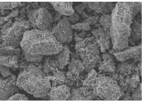
size of their particles 40 nm (Deraz, 2010, 2012). The microstructure of (CoFe2O4) NPs was analyzed using SEM
technique and the micrograph showed the formation of spongy and fragile network structure (Fig. 1).
Antimicrobial activity
The antifungal activity of (CoFe2O4) NPs was tested
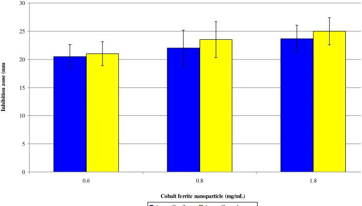
against fungal cultures (Aspergillus flavus and Aspergillus ochraceus). The qualitative assessment of the antifungal effect was done using well agar diffusion test and the current results showed that the highest antifungal activity of (CoFe2O4) NPs was recorded
at concentration 1.8 mg/mL against Aspergillus flavus and
Aspergillus ochraceus although no significant difference was
observed between the three tested concentrations of (CoFe2O4)
NPs. The zone inhibition of A. ochraceus was absent at concentration of 0.8 mg/mL (CoFe2O4) NPs (Fig. 2).
Although there is limited data about the use of (CoFe2O4) NPs
against pathogenic fungi, Chifiriuca et al. (2011) reported that (CoFe2O4) NPs produce a strong inhibitory effect on the adherence
to the cellular substrate of C. albicans strains. On the other hand, the use of other metals nanoparticles such as ZnO showed antifungal activity against Aspergillus, Fusarium, Penicillium
etc. (He et al., 2011; Lipovsky et al., 2011; Chitra and Annadurai 2013; Dimkpa et al., 2013). The antifungal activity of metals nanoparticle may be due to suppression of extracellular enzymes and metabolites that helps its survival when exposed to stress as reported by Bhainsa and D’ Souza (2006) and Vahabi et al. (2011).
The antibacterial activity of (CoFe2O4) NPs was
qualitatively measured by performing disk diffusion test against pathogenic microorganisms (Bacillus cereus, Staphylococcus
aureus, Salmonella typhi and Escherichia coli). The absence of
growth around the nanoparticle is an indirect measure of the ability of the material to inhibit the bacteria growth. The zone inhibition produced by the nanoparticle against the gram negative and the gram positive bacteria is shown in Fig. (3). These results revealed that (CoFe2O4) NPs efficiently inhibited the growth of the
microbes, whereas B. cereus was the most sensitive microorganism to the nanoparticles. It was also noticed that the
Fig. 2: Antifungal activity of (CoFe2O4) NPs using well agar diffusion test. Bars represent SD of three replicates.
Fig. 3: Antimicrobial activity of cobalt ferrite nanoparticles using disk diffusion test. Bars represent SD of two replicates.
0 5 10 15 20 25 30
0.6 0.8 1.8
Cobalt ferrite nanoparticle (mg/mL)
In
h
ib
it
ion
z
on
e (
m
m
)
Aspergillus flavus Aspergillus ochraceus
0 10 20 30 40 50 60 70 80 90 100
0.6 0.8 1.8
Cobalt ferrite nanoparticle (mg/mL)
In
h
ib
it
ion
z
on
e (
m
m
)
zone inhibition was increased by increasing the concentration from 0.6 to 1.8 mg/mL of (CoFe2O4) NPs. The effect of (CoFe2O4) NPs
on bacterial linear growth (Fig. 4) revealed that the bacterial growth was decreased by increasing the concentrations of the nanoparticles. Similar results were reported by Sanpo et al. (2013a) who reported an antibacterial activity of (CoFe2O4) NPs
against E. coli and S. aureus. In the same trend the rate of inhibition of E.coli was higher than that of S. aureus. The current results are in good harmony with several authors who reported that the nanocomposites of CoFe2O4 (Sanpo et al., 2013),polyaniline
and nanosilver (Kooti et al., 2014), and CoFe2O4/SiO2/Ag
composite (Kooti et al., 2015) showed high antibacterial activity. Moreover, Antoniea et al. (2012) found that the magnetite and (CoFe2O4) NPs induced the most significant effects in S. aureus
and B. subtilis. Grumezescu et al. (2010) studied the effect of
(CoFe2O4)/oleic acid nanoparticles and found that they induced
inhibitory effects on clinical isolates of bacterial and fungal strains at concentrations between 1000 and 1.9 µg/mL. Another study conducted by Sanpo et al. (2013b), the authors focused on the influence of chelating agents, namely polyvinyl alcohol and citric acid, on the microstructure and antibacterial property of cobalt ferrite nanopowders, and demonstrated that both types of cobalt
ferrite nanopowders showed an effective antibacterial activity against E. coli and S. aureus Several mechanisms have been suggested for the antibacterial action of nanoparticles. However, the actual inhibitory mechanism of the nanoparticles may require further studies (Moritz and Moritz, 2013). The antimicrobial activity of the nanoparticles is known to be the function of surface area which is adhesion to the microorganisms. The small size and the high surface to volume ratio i.e., large surface area enhances the interaction between the nanoparticles and the microbes to carry out a broad range of probable antimicrobial activities (Ravishankar and Jamuna, 2011).
Moreover, Vidya and Venkatesan (2015) suggested that nanoparticle bind to the membranes of microorganisms which can prolong the lag phase of the growth cycle and increase the generation time of the organisms. Other mechanisms contributing to the occurrence of antibacterial activity of nanoparticles is the release of reactive oxygen species (ROS) such as hydrogen peroxide (H2O2) and super-oxide (O
2–
) generated from the surface of nanoparticles (Sawai, 2003; Hashim et al., 2013). Furthermore, the penetration rate of an active oxide through the bacteria cell wall may play a part in the bacterial killing rate by nanoparticles (Xavier et al., 2014).
Fig. 4: Linear growth of pathogenic bacteria after treatment with different concentrations of cobalt ferrite nanoparticle. Bars represent SD of three replicates.
A B
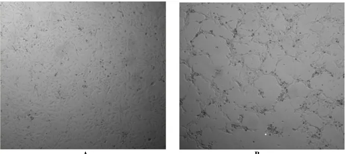
Fig. 5: Photos of MCF-7 breast cancer cells cultivated for 2 days (A) and after incubation with 0.1 mg/mL (CoFe2O4) NPs (B) by optical microscope (10x magnifications).
0 10 20 30 40 50 60 70
Control 0.6 0.8 1.8
Li
n
e
a
r
g
r
o
w
th
(m
m
)
Antiradical properties by DPPH free radical scavenging and ABTS radical cation decolorization assays
The results of DPPH and ABTS radical scavenging activities of (CoFe2O4) NPs were expressed as the half-maximal
inhibition concentrations (IC50). These results showed that IC50
values in DPPH and ABTS scavenging of (CoFe2O4) NPs were
determined as 6.8 ± 0.20 mg/ml and 0.25 ± 0.02 mg/ml, respectively. However, natural standard antioxidant Trolox as vitamin E analogue and ascorbic acid as vitamin C have IC50
values at 2.19 ± 0.02 µg/ml and 5.3 ± 0.3 µg/ml, respectively in ABTS scavenging (Aljawish et al., 2014). By comparing with this standard antioxidant, (CoFe2O4) NPs are considered as weak
antioxidant because the release of reactive oxygen species (ROS) generated from the surface of nanoparticles (Hashim et al., 2013).
Anti-proliferative activity
The effect of (CoFe2O4) NPs on MCF-7 breast cancer
cells lines (Figs. 5A, B) indicated that exposure of the breast cell lines to 1 mg/mL of CoFe2O4 NPs resulted in a significant
decrease in the cell viability and the relative cell viability reached 63 ± 3.6 %. It is well documented that the effect of a compound on tumor cells is mainly due to their oxidative stress effects. Tumor cells may, however, be dependent on a certain amount of hydrogen peroxide and the deficiency of hydrogen peroxide may resulted in cell cycle arrest or apoptosis of these cells (Loo, 2003). Similar to the current results, Horev-Azaria et al. (2013) reported that the viability of cell-lines towards the (CoFe2O4) NPs may be mainly
attributed to their ability to interact with NPs in case of either the adsorption of NPs to the cell surface and/or the uptake of NPs by the cells. Moreover, Nel et al. (2006) reported that oxidative stress which defined as the imbalance between the antioxidant defenses and the production of reactive oxygen species (ROS) or reactive nitrogen species (RNS) is considered to be the most important mechanism of NPs-induced hazards effects. In this concern, Horev-Azaria et al. (2013) found a high linear correlation between the toxicity of (CoFe2O4) NPs and the amount of generated ROS
following the exposure to (CoFe2O4) NPs. Previous study showed
that NPs could generate ROS, exhaust endogenous antioxidants, affect the function of mitochondria and induce oxidative damage to DNA and lipids (Moller et al., 2010). Consequently, NPs-induced ROS activation encourages the elements of defense antioxidant response and if damage proceeds, the protective systems activate protein kinase and NF-κB-activated intracellular signaling which resulted in the release of pro-inflammatory cytokine, matrix metalloproteinase and chemokine leading to apoptosis (Shubayev and Pisanic, 2009).
CONCLUSION
The current results indicated that (CoFe2O4) NPs can be
synthesized by combustion method using glycine as fuel and the average particles size was 40 nm. The synthesized (CoFe2O4) NPs
showed a potential antimicrobial activity against gram positive and gram negative bacteria as well as the pathogenic fungi Aspergillus
flavus and Aspergillus ochraceus in a dose dependent manner. The concentration of 1.8 g/ml of (CoFe2O4) NPs showed maximum
growth inhibition for all bacteria and fungi tested. Moreover the tested (CoFe2O4) NPs at a concentration of 2 mg/ml showed weak
antiradical activity but have high cytotoxic effects against MCF-7 breast cancer cells lines and succeeded to decrease the cell viability by about 63%. These findings suggested that the toxicity of (CoFe2O4) NPs is partially due to oxidative stress. The
antimicrobial and anticancer properties of (CoFe2O4) NPs reported
herein give these NPs great advantages to be used in food and biomedical applications.
ACKNOWLEDGMENT
Financial support and sponsorship: This work was funded by
the National Research Centre, Cairo, Egypt, under grant #10070208.
Conflict of Interests: There are no conflicts of interest.
REFERENCES
Aljawish A, Chevalot I, Jasniewski J, Paris C, Scher J, Muniglia L. Laccase-catalysed oxidation of ferulic acid and ethyl ferulate in aqueous medium: a green procedure for the synthesis of new compounds. Food Chem, 2014; 145: 1046-1054.
Amiri S, Shokrollahi H. The role of cobalt ferrite magnetic nanoparticles in medical science. Mater Sci Eng, 2013; 33 (1): 1-8.
Antoniea P, Dorina C, Claudia N, Mihai TF. Electromagnetic exposure and magnetic nanoparticle impact on some bacteria. African J Microbiol Res, 2012; 6(5): 1054-1060.
Bensebaa F. Microwave synthesis and characterization of Co-ferrite nanoparticles. J Colloid Interface Sci, 2004; 277 (1): 104-110.
Bhainsa KC, D’Souza SF. Extracellular biosynthesis of silver nanoparticles using the fungus Aspergillus fumigatus. Colloids Surf B, 2006; 47(2): 160-164.
Bouwmeester H, Dekkers S, Noordam M, Hagens W, Bulder A, De Heer C, Ten Voorde S, Wijnhoven S, Sips A. Health impact of nanotechnologies in food production. Wageningen UR, RIKILT. Report 2007.014, published by RIKILT-Institute of Food Safety, Wageningen UR and National Institute of Public Health & the Environment-Centre for Substances and Integrated Risk Assessment. Accessed from http://edepot.wur.nl/ 120669; 2007; pp 1-91.
Chifiriuc C, Lazar V, Bleotu B, Calugarescu I, Rumezescu AM, Mihaiescu DE, Mogoşanu DE, Buteica AS, Buteica E. Bacterial adherence to the cellular and inert substrate in the presence of CoFe2O4
and Fe3O4/oleic acid-core/shell. Digest J Nanomater Biostruct, 2011.; 6(1):
37-42.
Chitra K, Annadurai G. Antimicrobial activity of wet chemically engineered spherical shaped ZnO nanoparticles on food borne pathogen. Int Food Res J, 2013; 20(1):59-64.
Choi EJ. Superparamagnetic relaxation in CoFe2O4
nanoparticles. J Magn Magn Mater, 2003; 262 (2): L198-L202.
Daou TJ. Hydrothermal synthesis of monodisperse magnetite nanoparticles. Chem Mater, 2006;18 (18): 4399-4404.
Deraz NM. Glycine-assisted fabrication of nanocrystalline cobalt ferrite system. J Analyt Appl Pyrolysis, 2010; 88(2), 103-109.
Deraz NM. Production, Physicochemical Characterization and Magnetic Behavior of Nanocrystalline Al- Doped Co/Fe System. Int J Electrochem Sci, 2012; 7: 4596-4607
Gajjar P, Pettee B, Britt DW, Huang W, Johnson WP, Anderson AJ. Antimicrobial activities of commercial nanoparticles against an environmental soil microbe, Pseudomonas putida KT2440. J Biol Eng, 2009; 3: 9-22.
Grumezescu AM, Mihaiescu DE, Mogosanu DE, Chifiriuc MC, Lazar V, Calugarescu I, Traistaru V. In vitro assay of the antimicrobial activity of Fe3O and CoFe2O4 / oleic acid-core/shell on clinical isolates of
bacterial and fungal strains. Optoel Adv Mater Rapid Commun, 2010; 4(11): 1798-1801.
Gulcin I, Bursal E, M. Şehitoĝlu MH, Bilsel M, Goren AC. Polyphenol contents and antioxidant activity of lyophilized aqueous extract of propolis from Erzurum, Turkey. Food Chem Toxicol, 48; 2227-2238.
Hashim M, Alimuddin SE, Meena SS, Kotnala RK, Ameena, P, Aashis SR, Shalendra K, Pramod B, Kumar R.Investigation of structural, dielectric, magnetic and antibacterial activity of Cu–Cd–Ni–FeO4
nanoparticles. Magn Magn Mater, 2013; 341: 148-157.
He L, Liu Y, Mustapha A, Lin M. Antifungal activity of zinc oxide nanoparticles against Botrytis cinerea and Penicillium expansum. Microbiol Res, 2011; 166: 207-215.
Horev-Azaria L, Baldi G, Beno D, Bonacchi D, Golla-Schindler U, Kirkpatrick JC, Kolle S, Landsiedel R, Maimon O, Marche PN, Ponti J, Romano R, Rossi F, Sommer D, Uboldi C, Unger RE, Villiers C, Korenstein R. Predictive toxicology of cobalt ferrite nanoparticles: comparative in-vitro study of different cellular models using methods of knowledge discovery from data. Part Fibre Toxicol, 2013; 10:32, 17 pages. doi: 10.1186/1743-8977-10-32.
Jamon D, Donatini F, Siblini A, Royer F, Perzynski R, Cabuil V, Neveu S. Experimental investigation on the magneto-optic effects of ferrofluids via dynamic measurements. J Magn Magn Mater, 2009; 321: 1148-1154.
Jorgensen JH, Turnidge JD. Susceptibility test methods: dilution and disk diffusion methods, p. 1152-1172. In Murray PR, Baron EJ, Jorgensen JH, Landry ML, Pfaller MA (ed.), Manual of clinical microbiology, 9th ed. ASM Press, Washington DC. 2007.
Kiruba DSCG, Vinothini G, Subramanian N, Nehru K, Sivakumar M. Biosynthesis of Cu, ZVI, and Ag nanoparticles using
Dodonaea viscosa extract for antibacterial activity against human pathogens. J Nanopart Res, 2013; 15(1):1319-1328.
Kooti M, Gharineh S, Mehrkhah M, Shaker A, Motamedi H. Preparation and antibacterial activity of CoFe2O4/SiO2/Ag composite
impregnated with streptomycin. Chem Eng J, 2015; 259: 34-42.
Kooti M, Kharazi P, Motamedi H. Preparation, characterization, and antibacterial activity of CoFe2O4/polyaniline/Ag nanocompos ite. J Taiwan Institute Chem Engin, 2014; 45 (5): 2698-2704.
Lipovsky A, Nitzan Y, Gedanken A, Lubart R. Antifungal activity of ZnO nanoparticles-the role of ROS mediated cell injury. Nanotechnol, 2011; 22(10): 105101.
Loo G. Redox-sensitive mechanisms of phytochemical-mediated inhibition of cancer cell proliferation (Review). J Nut Biochem, 2003; 14(2): 64-73.
Maher S, McClean S. Investigation of the cytotoxicity of eukaryotic and prokaryotic antimicrobial peptides in intestinal epithelial cells in vitro. Biochem Pharmacol ,2006;71(9): 1289-1298.
Meng X, Li H, Chen J, Mei L, Wang K, Li X. Mossbauer study of cobalt ferrite nanocrystals substituted with rare-earth Y3+ ions. J Magn Magn Mater 2009; 321: 1155-1158.
Moller P, Jacobsen NR, Folkmann JK, Danielsen PH, Mikkelsen L, Hemmingsen JG, Vesterdal LK, Forchhammer L, Wallin H, Loft S. Role of oxidative damage in toxicity of particulates. Free Radic Res, 2010; 44(1):1-46.
Moritz M, Moritz MG. The newest achievements in synthesis, immobilization and practical applications of antibacterial nanoparticles. Chem Eng J, 2013; 228: 596-613.
Nel A, Xia T, Madler L, Li N. Toxic potential of materials at the nanolevel. Science 2006; 311(5761):622–627.
Pannaparayil T, Komarneni S. Synthesis and characterization of ultrafine cobalt ferrites. IEEE Trans on Magn, 1989;25 (5): 4233-4235.
Petersen KE, James WO. Agents, vehicles, and causal inference in bacterial food borne disease outbreaks: 82 reports (1986-1995). J Am Vet Med Assoc, 1998; 212:1874-1881.
Phua LX, Xu F, Ma YG, Ong CK. Structure and magnetic characterizations of cobalt ferrite films prepared by spray pyrolysis. Thin Solid Films 2009; 517: 5858-561.
Rafferty A, Prescott T, Brabazon D. Sintering behavior of cobalt ferrite ceramic. Ceram Int, 2008; 34 (1): 15-21.
Rajendran M. Magnetic properties of nanocrystalline CoFe2O4
powders prepared at room temperature: variation with crystallite size. J Magn Magn Mater, 2001; 232 (1-2): 71-83.
Ravishankar RV, Jamuna BA. Nanoparticles and their potential application as antimicrobials. In: Science against microbial pathogens: communicating current research and technological advances. Méndez-Vilas, A. (Ed.), 2011; pp.197-209.
Re R, Pellegrini N, Proteggente A, Pannala A, Yang M, Rice-Evans C. Antioxidant activity applying an improved ABTS radical cation decolorization assay. Free Rad Biol Med,1999; 26(9-10): 1231-1237.
Sanpo N, Berndt CC, Wen C, Wang J. Transition metal-substituted cobalt ferrite nanoparticles for biomedical applications. Acta Biomate, 2013; 9: 5830-5837.
Sanpo N, Berndt CC, Cuie W, James W. Transition-metal substituted cobalt ferrite nanopartice for biomedical applications. Acta Biomater, 2013a; 9: 5830-5837.
Sanpo N, Wang J, Berndt CC. Influence of chelating agents on the microstructure and antibacterial property of cobalt ferrite nanopowders. J Aust Ceram Soc, 2013b; 49 (1): 84-91
SAS.Institute Inc., SAS User’s Guide: Statistics. SAS Institute Inc., Cary, 1982; NC.
Sawai JJ Quantitative evaluation of antibacterial activities of metallic oxide powders (ZnO, MgO and CaO) by conductimetric assay. Microbiol Meth 2003; 54(2): 177-182.
Shimada K, Fujikawa K, Yahara K, Nakamura T. Antioxidative Properties of xanthan on the autoxidation of soybean oil in cyclodextrin emulsion. J Agric Food Chem, 1992; 40(6): 945-948.
Shubayev VI, Pisanic TR, Jin S. Magnetic nanoparticles for theragnostics. Adv Drug Deliv Rev, 2009; 61(6):467-477.
Vahabi K, Mansoori GA, Karimi S. Biosynthesis of silver nanoparticles by fungus Trichoderma reesei (a route for largescale production of AgNPs). Insc J, 2011; 1(1): 65-79.
Vidya R, Venkatesan K. Preparation and characterization of zinc ferrite (ZnFe2O4) nanoparticles using self-propagated combustion
route and evaluation of antimicrobial activity. Res J Pharma Biol Chem Sci, 2015; 6 (1): 537-542.
Xavier S, Cleetus H, Nimila PJ, Thankachan S, Sebastian RM, Mohammed EM. Structural and antibacterial properties of silver substituted cobalt ferrite nanoparticles. Res J Pharma Biol Chem Sci, 2014; 5 (5): 364-371.
Yamamoto O. Influence of particle size on the antibacterial activity of zinc oxide. Int J Inorg Mater, 2001; 3:643-646.
Yan CH. Nanophased CoFe2O4 prepared by combustion
method. Solid State Commun, 1999; 111(5): 87-291.
How to cite this article: