This is an open access journal, and articles are distributed under the terms of the Creative Commons Attribution-Non Commercial-ShareAlike 4.0 License, which allows others to remix, tweak, and build upon the work non-commercially, as long as appropriate credit is given and the new creations are licensed under the identical terms.
© 2019 Journal of Advanced Pharmacy Education & Research | Published by SPER Publication 33
High aneuploidy rate among 3PN embryos was not associated
with sperm parameters in IVF patients
Ririn Rahmala Febri
1, Budi Wiweko
1,2,3*, Pritta Ameilia Iffanolida
1, Kresna Mutia
1, Naylah Muna
1,
Oki Riayati
1, Shanty Olivia Jasirwan
1, Eliza Mansyur
2, Tita Yuningsih
3, Raden Muharam
1,2,3,
Hestiantoro A
1,2,31 Human Reproductive, Infertility and Family Planning Research Center, Indonesian Medical Education and Research Institute. 2Yasmin IVF Clinic, Dr. Cipto
Mangunkusumo General Hospital, Jakarta, Indonesia. 3 Division of Reproductive Endocrinology and Infertility Department of Obstetrics and Gynecology Faculty of
Medicine Universitas Indonesia; Dr. Cipto Mangunkusumo General Hospital, Jakarta, Indonesia.
Correspondence:Budi Wiweko, Human Reproductive, Infertility and Family Planning Research Center, Indonesian Medical Education and Research Institute, 5th Floor
Research Tower, Jalan Salemba Raya, No.6, Jakarta Pusat, Indonesia, 10430. Email: [email protected]
ABSTRACT
Background: The high incidence of aneuploidy observed in pre implantation embryos isone of the most significant factors affecting the clinical outcomes in assisted reproduction. The aim of this study was to evaluate the association between sperm factors including motility, concentration and morphology with the frequencies of aneuploidy embryos in IVF patients.Methods: The design of this study was cross-sectional. Thirty-five embryos were collected from12 women who underwent IVF in Dr. Ciptomangunkusumo General Hospital, Jakarta. Embryos were cultured until blastocyst stage, then trophectoderm biopsy was performed by piercing the zona pellucida under the microscope with a laser. Numerical abnormalities chromosome was analyzed using Next Generation Sequencing (NGS) and Comparative Genomic Hybridization (CGH). Statistical Package for Social Science (SPP) version 22.0 was used for statistical analysis. Two-tailed p value less than 0.05 was considered as significant. Results: There was a significant difference in females’ age, rFSH total, mean length of stimulation, females’ weight and body mass index (BMI) among the patients (p<0.01), as well as sperm volume, total motile sperm, sperm concentration and normal sperm morphology (p<0.05). The high frequency of aneuploidy was also found among 3PN embryos (74.3%). However, this result was not related to sperm parameters including sperm volume (p=0.424), total motile sperm (p=0.342), sperm concentration (p=0.239), and normal sperm morphology (p=0.342). Conclusion: These results suggested that sperm factors were not associated with the aneuploidy rate in 3PN embryos.
Keywords:Chromosome, embryo development, embryo morphology, NGS, sperm factors.
Introduction
Aneuploidy is a common chromosome abnormality at later development stages affecting approximately 4-5% of all clinical pregnancies, and the vast majority of which end in early miscarriage [1]. Aneuploidy is caused by abnormal chromosome
segregation during the meiosis phase by deleting or including an extra copy of chromosome, and its occurrence is related to
both female and male gametes [2]. It has been believed that one
of the most significant factors affecting the clinical outcome in assisted reproductive technology (ART) is the high incidence of aneuploidy observed in pre implantation embryos. Many studies have shown that paternal factors might be contributed to repeated ART failures [3]. However, the association between
sperm parameters and aneuploidy embryos remains poorly understood.
The recent development of ART has also permitted in vitro fertilization (IVF) for many men with sperm chromatin anomalies and sub fertile sperm. The generation of aneuploidy gametes during spermatogenesis and patients’ severe sperm defects resulted from OAT or nonobstructive azoospermia may result in a higher percentage of mitotic abnormalities and poor quality of embryos [4]. It has been shown that a negative
effect on ART procedure outcome is affected by a high percentage of spermatozoa with chromatin structure alterations [5].
Access this article online
Website: www.japer.in E-ISSN: 2249-3379
How to cite this article: Ririn Rahmala Febri, Budi Wiweko, Pritta Ameilia Iffanolida, Kresna Mutia, Naylah Muna, Oki Riayati et al.High aneuploidy rate among 3PN embryos was not associated with sperm parameters in IVF patients. J Adv Pharm Edu Res 2019;9(1):33-37.
Tripronuclear (3PN) embryo is the most common fertilization anomaly in the human. 3PN embryo is caused by two ways of formation, the combination of two sperms and one maternal gamete, or the combination of two maternal gametes and one sperm [6]. As just one sperm is injected into each oocyte,
Polyspermy should not take place in intracytoplasmic sperm injection (ICSI) [7]. Because of the failure in the second meiotic
division and retention of the second polar body, the occurrence of 3PN embryos from the ICSI treatment have been considered to be digynic [8]. 3PN embryos are contributed
to be of high risk for an abnormal pregnancy because of their very complicated chromosomal constitution.
This study aimed to determine the association between sperm parameters including sperm volume, total motile sperm, sperm concentration and normal sperm morphology with the frequency of aneuploidy embryos in IVF patients.
Materials and Methods
Study Design
This was a retrospective nonrandomized study conducted on 35 embryos enrolled from 12 IVF patients. The procedures were done at Yasmin Clinic, Dr. Ciptomangunkusumo General Hospital and Faculty of Medicine, Universitas Indonesia Laboratory between January and October 2018 after receiving the written informed consent from all patients and their partners for being included in the study. All procedures were approved by Ethics Committee of Faculty of Medicine, Universitas Indonesia.
Fertilization and Embryo Culture
ICSI was performed using the oocytes at metaphase II to recovered oocytes which were injected following the routine procedures, and were assessed for their maturity. 3PN embryos were evaluated using an inverted microscope for 16-20 hours after ICSI, indicated by three pronuclear. Then, these embryos were cultured in ISM medium 1 until afternoon on the day 2. This procedure followed by 3PN embryos were transferred to blast assist medium until the day 5. The degree of fragmentation, symmetry, and grading was simultaneously evaluated along with embryo culture.
Embryo Biopsy
On the fifth day after ICSI, trophectoderm biopsy was performed by piercing the zona pellucida under the microscope with a laser (Octax). TE biopsied cells were placed into transfer medium (PBS 1% PVP) for PGS procedure.
Next Generation Sequencing (NGS)
Technology
The NGS procedure was performed using Veriseq PGS workflow (Illumina, Inc). Meanwhile, Nextera XT 96-Index Kit (Illumina, Inc) was used for DNA indexing in order to simultaneously analyze embryos from all patients. All the
procedures in NGS were performed following the manufacturer’s protocol.
Statistical Analysis
The data were analyzed using the SPSS 22.0. The initial analysis was simply to measure the characteristics of biopsy samples. Statistical significance for parametric data was assessed by using Independence Sample T-test, and the Mann-Whitney U test was used for non-parametric data. Chi-square test or Fisher exact test, as appropriate, was used to determine the differences between the proportions of variables. Two-tailed p value less than 0.05 was considered as significant.
Results
This study enrolled 12 patients (mean age 32.42 years, SD 3.96) undergoing PGS. A total of 3PN embryos at the cleavage stage was biopsied and analyzed for aneuploidy using pre PGS method. Figure 1 shows the result of NGS technology. The following bioinformatics analysis was performed using BlueFuse Multi software for NGS. Each NGS graph indicates the copy number assessment on the y-axis and the chromosome number on the x-axis.
(a)
Journal of Advanced Pharmacy Education & Research | Jan-Mar 2019 | Vol 9 | Issue1 35
(c)
Figure 1.The results of NGS-based aneuploidy detection.
Gains and losses were defined as a shift of the dots above and below the copy number state of 2.5 or 1.5; respectively, and detected as horizontal green bars. (a) Embryo showing no
chromosome abnormalities was detected (euploidy). (b) Embryo showing aneuploidy for chromosome 16 (monosomy
16). (c) Embryo showing aneuploidy for chromosome 14 (trisomy 14). This figure was created by the authors.
The distribution of cycle-specific parameters has been described in Table 1. There was a significant difference in females’ age, rFSH total, mean length of stimulation, females’ weight and body mass index (BMI) among the patients (p<0.01). This results also showed that there was a significant difference in sperm volume, total motile sperm, sperm concentration and normal sperm morphology (p<0.05).
Table 1. General characteristics of IVF patients with 3PN embryos
Variables n=12
Female’s age (y) rFSH total (IU) Mean length of stimulation (d)
Weight (kg) Height (m) Body mass index (kg/m2)
Sperm volume (ml) Total motile sperm (%) Sperm concentration (ml) Normal sperm morphology (%)
32.42 ± 3.96a 2541.67 ± 770.06a
12.08 ± 2.15a 60.83 ± 9.60a 1.58 (1.52-1.65)b
24.34 ± 3.92a 3.33 ± 1.09a 2.95E+07 (9.2E+06-1.3E+08)b
58.57 ± 24.02a 9 (0-40)b Notes:
a Data is normally distributed therefore value is presented in mean and standard deviation
b Data is not normally distributed therefore value is presented in median (min. – max.)
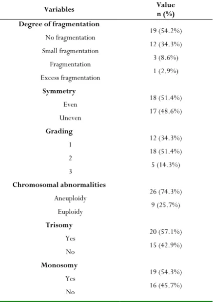
Thirty-five of 3PN embryos at cleavage stage from 12 patients were analyzed with the general characteristics presented in Table 2. A total of 74.3% (26 of 35) of the samples showed aneuploidy, 25.7% (9 of 35) embryos were euploidy, 54.3% (19 of 35) of embryos contained trisomy chromosome, meanwhile 57.1% (20 of 35) of embryos were monosomy (Table 2). The morphological criteria of 3PN embryos were
evaluated based on the degree of fragmentation and symmetry. In addition, embryos were graded according to the scoring system. This study showed that about 54.2% embryos were detected to have no fragmentation, 51.4% embryos had even symmetry, and 51.4% of embryos were grade 2 (Table 2).
Table 2. Distribution of 3PN embryos characteristics
Variables Value n (%)
Degree of fragmentation
No fragmentation Small fragmentation
Fragmentation Excess fragmentation
Symmetry
Even Uneven
Grading
1 2 3
Chromosomal abnormalities
Aneuploidy Euploidy
Trisomy
Yes No
Monosomy
Yes No
19 (54.2%) 12 (34.3%) 3 (8.6%) 1 (2.9%)
18 (51.4%) 17 (48.6%)
12 (34.3%) 18 (51.4%) 5 (14.3%)
26 (74.3%) 9 (25.7%)
20 (57.1%) 15 (42.9%)
19 (54.3%) 16 (45.7%)
This table was created by the authors.
Furthermore, in this study, the association between sperm factor and aneuploidy of 3PN embryos was also evaluated using independent samples T-test. However, there was no significant association of sperm parameters including sperm volume (p=0.424), total motile sperm (p=0.342), sperm concentration (p=0.239), and normal sperm morphology (p=0.342) with aneuploidy of 3PN embryos (Table 3).
Table 3. The association between sperm parameters with aneuploidy rate of 3PN embryo
Parameter Euploidy (n=9) Aneuploidy (n=26) value p
Sperm volume (ml) Total motile sperm (%) Sperm concentration (107/ml) Normal sperm morphology (%)
3.47 ± 1.14 63.27 ± 1.14 4.28 ± 3.68 13.65 ± 2.34
3.16 ± 0.8 52.66 ± 26.70
5.88 ± 4.25 19.67 ± 5.42
0.424 0.342 0.239 0.342
Independent Samples T-test This table was created by the authors.
Table4. The chromosomal status of 3PN embryos in association with the degree of fragmentation, symmetry,
and grading
Variables Euploidy (%) n=9 Aneuploidy (%) n=26 value p
Degree of fragmentation No fragmentation Small fragmentation
Fragmentation Excess fragmentation
Symmetry Even Uneven Grading
1 2 3
4 4 1 0
5 4
3 6 0
15 8 2 1
13 13
9 12
5
0.741
0.774
0.320
Chi-square test
This table was created by the authors.
Discussion
Fertilization of an oocyte by sperm, followed by fusion of each pronuclear, formed two pronuclear, in general, and then cleavages to blastomere stage. However, in IVF process, it has been observed that there might be an aberrant number of pronuclear such as 3PN [9]. Triploidy was found approximately
in 4%-7% of IVF and 6% of ICSI zygotes [10]. Aneuploidy was
found in the majority of 3PN embryos. 3PN embryos were commonly discarded in routine IVF practice because they have been well known to be genetically abnormal. Despite this general belief, some studies that examined the chromosomal composition of 3PN embryos indicated that various amounts of them were found to be diploid [11]. PGD has been one of the
several techniques that has been applied for embryo selection developed in IVF, especially to select genetically normal embryo to transfer to the one with the highest implantation potential [12]. This study demonstrated the high frequency of
aneuploidy among 3PN embryos (74.3%) compared to euploidy (25.7%).
Since aneuploidy is responsible for pregnancy failure, current studies have been developed to identify the effect of severe sperm parameters’ impairment with aneuploidy [13]. However,
this study found that no significant association was observed between aneuploidy in 3PN embryos and sperm parameters including sperm volume, total motile sperm, sperm concentration, and normal sperm morphology. In accordance with these findings, previously conducted studies from infertile men have uncovered an increased frequency of aneuploidy sperm, particularly in patients with abnormal semen parameters [14]. It has been stated that pregnancy outcome after
ICSI procedure is also affected by sperm abnormalities including the injection of sperm carrying a chromosomal anomaly, male gamete structural defect, anomalies of sperm activating factors, and potential for incorporating sperm mitochondrial DNA [15].
It has not yet been confirmed the exact reasons of 3PN fertilization. Some previous studies have demonstrated that the incidence of 3PN embryos in IVF program is a result of severe sperm abnormalities [7]. On the other hand, some studies
believed that the extra pronuclear in 3PN embryos is most likely of maternal origin, since only one spermatozoon is injected into the oocyte at ICSI fertilization [16]. In addition, the
formation of 3PN embryo is associated with ovarian stimulation prior to IVF program, as well as the lengthy stimulation. The findings of this study showed that there was a significant difference in females’ age, recombinant FSH total dose, mean length of stimulation, females’ weight and body mass index (BMI) among the patients. A previous study showed that the oocytes from reproductively older patients displayed a highly significant increase in the incidence of aneuploidy [1]. In contrast to this, a study conducted by
Brancati et al. demonstrated that the increased maternal age is not a risk factor of aneuploidy [16].
3PN embryo formation has been clarified for two main features, no extrusion of second polar body and dispermic fertilization [17]. Since, in this study, the ICSI technique was
used in which only one spermatozoa was injected to oocyte, dispermic fertilization could be rule out. Therefore, the only possible explanation for this 3PN embryo formation was no extrusion of the second polar body. It has been known that 3PN embryo is associated with spontaneous miscarriage after the implantation. For this reason, recognizing 3PN embryo formation in the early period is crucial for preventing the transfer of the embryo that develops from 3PN oocytes during IVF program [14].
Conclusion
In conclusion, the findings of this study suggested that sperm parameters did not play a role in increasing the high rate of aneuploidy in 3PN embryo. Therefore, the incidence of aneuploidy in 3PN embryos was assumed most likely of maternal origin.
Acknowledgements
This work was supported by the research grant from PITTA Universitas Indonesia (Idr. 90.000.000).
Conflicts of interest
The authors declared that they had no conflict of interest.
References
Journal of Advanced Pharmacy Education & Research | Jan-Mar 2019 | Vol 9 | Issue1 37
2. O. Coban, M. Serdarogullari, Z. O. Sekerci, and E. Murat, “Evaluation of the impact of sperm morphology on embryo aneuploidy rates in a donor oocyte program,” Syst. Biol. Reprod. Med., vol. 00, no. 00, pp. 1–5, 2018.
3. A. Borini, N. Tarozzi, D. Bizzaro, M. A. Bonu, L. Fava, C. Flamigni, and G. Coticchio, “Sperm DNA fragmentation: Paternal effect on early post-implantation embryo development in ART,” Hum. Reprod., vol. 21, no. 11, pp. 2876–2881, 2006. 4. J. García-Ferreyra, D. Luna, L. Villegas, R. Romero,
P. Zavala, R. Hilario, and J. Dueñas-Chacón, “High aneuploidy rates observed in embryos derived from donated oocytes are related to male aging and high percentages of sperm DNA fragmentation,” Clin. Med. Insights Reprod. Heal., vol. 9, pp. 21–27, 2015. 5. R. Mazzilli, D. Cimadomo, A. Vaiarelli, A. Capalbo,
L. Dovere, E. Alviggi, L. Dusi, C. Foresta, F. Lombardo, A. Lenzi, H. Tournaye, C. Alviggi, L. Rienzi, and F. M. Ubaldi, “Effect of the male factor on the clinical outcome of intracytoplasmic sperm injection combined with preimplantation aneuploidy testing: observational longitudinal cohort study of 1,219 consecutive cycles,” Fertil. Steril., vol. 108, no. 6, p. 961–972.e3, 2017.
6. H. Q. Liao, Q. OuYang, S. P. Zhang, D. H. Cheng, G. X. Lu, and G. Lin, “Pronuclear removal of tripronuclear zygotes can establish heteroparental normal karyotypic human embryonic stem cells,” J. Assist. Reprod. Genet., vol. 33, no. 2, pp. 255–263, 2016.
7. M. Li, W. Zhao, X. Xue, S. Zhang, W. Shi, and J. Shi, “Three pro-nuclei ( 3PN ) incidence factors and clinical outcomes : a retrospective study from the fresh embryo transfer of in vitro fertilization with donor sperm ( IVF-D ),” Int J Clin Exp Med, vol. 8, no. 8, pp. 13997–14003, 2015.
8. M. W. Joergensen, I. Agerholm, J. Hindkjaer, L. Bolund, L. Sunde, H. J. Ingerslev, and K. Kirkegaard, “Altered cleavage patterns in human tripronuclear embryos and their association to fertilization method: A time-lapse study,” J. Assist. Reprod. Genet., vol. 31, no. 4, pp. 435–442, 2014.
9. R. Rungsiwiwut, P. Numchaisrika, V. Ahnonkitpanit, P. Virutamasen, and K. Pruksananonda, “Triploid
human embryonic stem cells derived from tripronuclear zygotes displayed pluripotency and trophoblast differentiation ability similar to the diploid human embryonic stem cells,” J. Reprod. Dev., vol. 62, no. 2, pp. 167–176, 2016.
10. M. W. Joergensen, R. Labouriau, J. Hindkjaer, M. Stougaard, S. Kolevraa, L. Bolund, I. E. Agerholm, and L. Sunde, “The parental origin correlates with the karyotype of human embryos developing from tripronuclear zygotes,” Clin. Exp. Reprod. Med., vol. 42, no. 1, pp. 14–21, 2015.
11. Z. Chen, J. Yan, and H. L. Feng, “Aneuploid analysis of tripronuclear zygotes derived from in vitro fertilization and intracytoplasmic sperm injection in humans,” Fertil. Steril., vol. 83, no. 6, pp. 1845– 1848, 2005.
12. C. Rubio, J. Bellver, L. Rodrigo, G. Castillón, A. Guillén, C. Vidal, J. Giles, M. Ferrando, S. Cabanillas, J. Remohí, A. Pellicer, and C. Simón, “In vitro fertilization with preimplantation genetic diagnosis for aneuploidies in advanced maternal age: a randomized, controlled study,” Fertil. Steril., vol. 107, no. 5, pp. 1122–1129, 2017.
13. A. E. Calogero, N. Burrello, A. De Palma, N. Barone, R. D’Agata, and E. Vicari, “Sperm aneuploidy in infertile men,” Reprod Biomed Online, vol. 6, no. 3, pp. 310–317, 2003.
14. V. A. Kushnir and J. L. Frattarelli, “Aneuploidy in abortuses following IVF and ICSI,” J. Assist. Reprod. Genet., vol. 26, no. 2–3, pp. 93–97, 2009.
15. M. Bonduelle, “Prenatal testing in ICSI pregnancies: incidence of chromosomal anomalies in 1586 karyotypes and relation to sperm parameters,” Hum. Reprod., vol. 17, no. 10, pp. 2600–2614, 2002. 16. F. Brancati, R. Mingarelli, and B. Dallapiccola,
“Recurrent triploidy of maternal origin,” Eur. J. Hum. Genet., vol. 11, no. 12, pp. 972–974, 2003.
17. E. Yalçınkaya, A. Özay, EG Ergin, Z. Öztel, and H.
Özörnek. Live birth obtained by transfer of a