Małgorzata Trocha, Anna Merwid-Ląd, Ewa Chlebda, Tomasz Sozański,
Jan Magdalan, Małgorzata Pieśniewska, Lidia Fereniec-Gołębiewska,
Halina Gliniak, Adam Szeląg
Influence of Simvastatin on Oxido-Redox Status
and Nitric Oxide Synthases Protein Concentration
in Rat Liver Subjected to “Cold” Ischemia
Wpływ simwastatyny na stan oksydoredukcyjny
i stężenie syntetaz tlenku azotu
w wątrobach szczurzych poddanych niedokrwieniu „zimnemu”
Department of Pharmacology, Wroclaw Medical University, Poland
Abstract
Background. Hydroxymethylglutaryl-CoA reductase inhibitors (statins) play a great role in oxido-redox status in the liver. “Cold” ischemia could be responsible for liver cell damage.
Objectives. The influence of simvastatin (SV) on oxido-redox status and nitric oxide synthases protein concentra-tion in rat liver in a “cold” ischemia model was evaluated.
Material and Methods. The study was conducted on rat livers. Group C and C24 rats did not receive SV and groups S and S24 received SV intragastrically (25 mg/kg) for 21 days. Alanine and asparagine aminotransferases (ALT, AST) activities were determined to exclude SV-induced liver injury. The livers of groups C and S were homogenized just after isolation and those of groups C24 and S24 were preserved for 24 hours in HTK solution (4°C) before homogenization. Superoxide dismutase (SOD) and catalase (CAT) activities as well as lipid peroxide (LPO) and endothelial and inducible nitric oxide synthase (eNOS and iNOS) concentrations were measured in the liver homogenates.
Results. SV did not alter ALT and AST activity. Significant increases in SOD and CAT activities and eNOS pro-tein concentration were observed in group S compared with group C. After preservation, a significant difference between S24 and C24 was observed only in the case of CAT. No significant differences in LPO and iNOS levels between the groups were found.
Conclusions. SV may exert a protective action on rat liver, which was seen as increased antioxidant parameter activities and eNOS level in the unpreserved group. After 24 hours of “cold” preservation, these properties of SV were maintained only in the case of CAT activity (Adv Clin Exp Med 2010, 19, 1, 43–50).
Key words: simvastatin, liver, rat, ischemia-reperfusion, oxido-redox status.
Streszczenie
Wprowadzenie. Inhibitory reduktazy hydroksymetyloglutarylo-CoA (statyny) odgrywają znaczącą rolę w wątro-bie, wpływając m.in. na jej stan oksydoredukcyjny. „Zimne” niedokrwienie, jakiemu są poddane narządy podczas przeszczepienia, może być odpowiedzialne za ich uszkodzenie.
Cel pracy. Zbadano wpływ simwastatyny (SV) na układ oksydoredukcyjny oraz syntetaz tlenku azotu w modelu wątroby szczurzej poddanej „zimnemu” niedokrwieniu.
Materiał i metody. Doświadczenie zostało przeprowadzone na wątrobach szczurzych. W grupach C i C24 zwierzę-ta nie otrzymywały SV, podczas gdy w grupach S i S24 – SV była podawana dożołądkowo (25 mg/kg) przez 21 dni. Aby wykluczyć działanie hepatotoksyczne leku, po tym okresie oznaczano aktywność aminotransferazy alaninowej i asparaginianowej (ALT, AST). Wątroby w grupach C i S były homogenizowane bezpośrednio po izolacji z orga-nizmu zwierzęcia. Wątroby w grupach C24 i S24 po izolacji były przechowywane przez 24 godz. w roztworze HTK w temperaturze 4°C, a następnie homogenizowane. W homogenatach wątroby oznaczano aktywności dysmutazy ponadtlenkowej (SOD) i katalazy (CAT) oraz stężenia nadtlenków lipidów (LPO) i syntetaz tlenku azotu – śród-błonkowej (eNOS) i indukcyjnej (iNOS).
Adv Clin Exp Med 2010, 19, 1, 43–50 ISSN 1230-025X
OrIGINAL PAPErS
The pathogenesis of liver damage during transplantation is very complex and occurs in two stages: initial injury is caused by ischemia, while later the damage is aggravated by reperfu-sion of the organ [1]. In this procedure, apart from “warm” ischemia, the liver also undergoes “cold” ischemia. This phase of ischemia leads to swell-ing and other changes in various cell structures, resulting in decreased cell function. ATP deple-tion, increased glycolysis, and higher Kupffer cell stimulation are also found [2]. It is generally believed that the main sites of injury in “cold” ischemia are endothelial cells, whereas “warm” ischemia damages predominantly hepatocytes.
The involvement of proinflammatory media-tors and reactive oxygen species (rOS) in cold ischemia is doubtful[3] because rOS are produced mainly during the reperfusion period. However, in this study some parameters of oxidative status in cold ischemia were analyzed. NADPH oxidase, xantine oxidase, myeloperoxidase and malondi-aldehyde, and lipid peroxides (LPOs) are widely used as indicators of oxidative damage. To mini-mize the damaging effects of rOS to the liver, nonenzymatic and enzymatic antioxidants are involved [4]. The enzyme system includes, for example, superoxide dismutase (SOD), catalase (CAT), and glutathione peroxidase (GPx). SOD converts superoxide radicals to hydrogen per-oxide (H2O2) and oxygen [2]. H2O2 and LPO are
subsequently reduced by the selenium-containing enzyme GPx to water and lipid alcohols, respec-tively. Additionally, CAT is a very effective intrac-ellular antioxidant enzyme, mainly located in per-oxisomes, which catalyzes the reaction of H2O2 to
water and molecular oxygen [5].
Experimental data suggest that nitric oxide (NO) is an important component of ischemia-reperfusion (I/r) injury and can modulate rOS metabolism by reaction with free radicals such as superoxide in the formation of peroxinitrite [2, 6, 7]. Endothelial nitric oxide synthase (eNOS) is responsible for the basal production of NO that maintains the normal vascular tone within the sinusoids [8]. Under I/r conditions, eNOS activ-ity was shown to decrease, resulting in microvas-cular failure [9, 10]. Conversely, a higher level of
NO produced by inducible nitric oxide synthase (iNOS) is involved in the inflammatory process and promotes I/r injury [2, 7, 11].
Inhibitors of the 3-hydroxy-3-methylglutaryl coenzyme A (HMG-CoA) reductase are very pop-ular lipid-lowering drugs. The mechanism of their hypolipemic action is quite well understood, but their pleiotropic effects are still being intensively examined [12–15]. It was also shown that statins exert a protective action on transplanted liver [16, 17].However, it is also important to consider the hepatotoxicity of these drugs as well as the fact that statins are administered to many patients who could be potential liver donors. Therefore, because of the many different controversial prop-erties of these drugs, additional studies should allow us to broaden the knowledge about statins and determine the function of liver preserved for transplantation.
The aim of this study was to determine the effect of an inhibitor of HMG-CoA reductase, simvastatin (SV), on oxido-redox status and NOS protein concentrations in rat liver submitted to “cold” ischemia.
Material and Methods
Animals
The study was carried out on adult male Wistar rats (261.33 ± 28.5 g) obtained from the Animal Laboratory of the Department of Pathological Anatomy, Wroclaw Medical University. The ani-mals were housed individually in chambers with a 12:12 h light-dark cycle and temperature main-tained at 21–23°C. Before the experiment the animals had free access to standard food and water. The experiment was performed with the consent of the First Local Ethics Commission for Experiments on Animals in Wroclaw.
Substances
Histidine-tryptophan-ketoglutarate (HTK) solution (Dr. Franz Köhler, Chemie GmbH, Germany), SV (GZF Polfa, Poland), heparin (amp.
Wyniki. Nie wykazano istotnego wpływu SV na aktywność ALT i AST. Znaczący wzrost aktywności SOD, CAT oraz stężenie eNOS obserwowano w grupie S w porównaniu z grupą C. Po okresie przechowywania narządu znaczą-cą różnicę obserwowano między grupami S24 i C24 jedynie w odniesieniu do aktywności CAT. Nie uwidoczniono żadnych znaczących różnic w stężeniach LPO i iNOS między badanymi grupami.
Wnioski. SV wykazuje ochronne działanie na wątroby niepoddane niedokrwieniu „zimnemu”, odpowiadając za znaczące zwiększenie stężenia eNOS oraz aktywność wskaźników antyoksydacyjnych. Po okresie przechowywania jedynie aktywność CAT utrzymywała się na istotnie wyższym poziomie w grupie otrzymującej badany lek (Adv Clin Exp Med 2010, 19, 1, 43–50).
25000 U; Polfa, Poland), thiopental (amp. 0.5 g; Biochemie, Austria), 0.9% sodium chloride tion (Polpharma S.A., Poland), and ringer solu-tion (Polfa Lublin S.A., Poland) were used in the study.
Experimental Procedure
The rats were divided into four groups. Group C (n = 11) was untreated and their liv-ers were not preserved, group C24 (n = 11) was untreated and preserved, group S (n = 11) was SV-treated and not preserved, and group S24 (n = 12) was SV-treated and preserved livers of rats. SV was administered intragastrically in one daily dose of 25 mg/kg diluted in 4 ml/kg saline solution for 21 days. In the untreated groups the animals were given only saline solution in the same volume. On the day before liver isolation, alanine and aspar-agine aminotransferase (ALT, AST) activities were measured to exclude SV-induced liver injury.Then the animals were anesthetized by intraperitone-al injection of thiopentintraperitone-al at a dose of 70 mg/kg. A middle incision was made to open the abdomi-nal cavity, the inferior caval vein was ligated above the right renal vein opening, and a cannula was introduced into the portal vein through which ringer solution supplemented with heparin was perfused. Then the chest was opened and a sec-ond cannula was inserted into the inferior caval vein, pointing towards the liver. In groups C and S the livers were homogenized just after isolation. In groups C24 and S24 the livers were prepared for cooling, i.e. they were isolated from the rat body, flushed with HTK solution at 4°C for 3 min (5ml/ min), and transferred to HTK solution (100 ml) maintained at 4°C. After 24-hour storage, the liv-ers were homogenized on ice using lysis buffer (140 mM NaCl, 10 mM EDTA, 10% glycerol, 1% NP40, 20 mM Tris base, pH = 7.5). The homogenized tis-sues were then centrifuged at 4°C at 14,000 rpm for 25 min and the supernatants were taken [18].
Blood Enzyme and Tissue
Antioxidant Analysis
The LPO concentration was assessed in the supernatants using the colorimetric method based on the reaction of malondialdehyde with thiobar-bituric acid [19]. Briefly, trichloroacetic acid was added to the samples. After 10 min of incubation the samples were centrifuged (3500 rpm, 10 min), the supernatants were poured out, and the sedi-ment was washed with 0.05 M sulfuric acid and centrifuged once again. In the next step, thiobar-bituric acid and sulfuric acid were added to the sediment and the samples were incubated for
30 min in a boiling water bath. Then the samples were cooled and extracted with butyl alcohol and centrifuged. The absorbance at 530 nm was mea-sured.
CAT activity was determined following the decrease in the initial H2O2 concentration
(30 mM used as a substrate) at 240 nm at 25°C for 60 s according to Johanson and Borg [20]. Briefly, 100 μl of supernatants isolated from rat liver homogenate were placed in a cuvette in phos-phate-buffered saline (50 mM) to a final volume of 2 ml. After adding 1 ml of H2O2, the decrease in
absorption at 240 nm for 60 s was followed. One unit of CAT was defined as the amount of enzyme that degraded 1 μl of H2O2 per minute [21].
SOD activity was determined spectropho-tometrically using a ransod kit (randox Labo-ratories). eNOS and iNOS protein concentrations were analyzed by ELISA using Quantikine kits (r&D Systems).Total protein concentrations and ALT and AST activity were assayed with a com-mercial method in a certified laboratory.
Statistical Analysis
Data are expressed as mean values ± SD. Statistical analysis of the effect of ischemia on AST, ALT, SOD, and CAT activities and LPO, eNOS, and iNOS concentrations were performed using multifactor analysis of variance (ANOVA). Specific comparisons were made with contrast analysis. p < 0.05 was considered to be statistically significant.
Results
After three weeks of treatment, SV did not alter ALT and AST activity. No significant differences between the untreated (C and C24) and treated (S and S24) groups were found (ALT: 7.59 ± 5.22
U/l/g vs. 7.57 ± 2.01 U/l/g and AST: 20.76 ± 11.92 U/l/g vs. 20.74 ± 4.37 U/l/g, p = ns. in all cases). No significant differences in LPO level among the examined groups were revealed (C: 4.82 ± 1.11,
S: 5.18 ± 1.55, C24: 5.16 ± 1.23, S24: 6.12 ± 1.30 mmol/ml, p = NS for all comparisons).
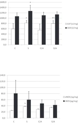
Treatment with SV evoked a significant increase in SOD activity in the unpreserved group. In the SV-treated group, SOD activity was higher than in untreated group (S vs. C, p < 0.05). “Cold” ischemia produced an insignificant decrease in SOD activity in the SV-treated group, and after preservation no difference between the SV-treated and untreated groups was found (S24 vs. C24, p = NS) (Fig. 1).
sig-nificant increase in CAT activity was observed in both the unpreserved and preserved groups. CAT activity was higher in the SV-treated group than in the untreated (S vs. C, p < 0.05). “Cold” isch-emia had no influence on this parameter in the untreated and treated groups (C vs. C24 and S vs. S24, p = NS in both cases). A significant difference between the preserved groups was observed (S24 vs. C24, p < 0.01) (Fig. 1).
The influence of SV on eNOS protein concen-tration was found in the unpreserved groups. Its concentration was higher in the SV-treated group than in the untreated group (S vs. C, p < 0.005). After preservation, no difference between the SV-treated and untreated groups in this param-eter was found (S24 vs. C24, p = NS). No influence
of either SV-treatment or “cold” ischemia on iNOS protein concentration was observed (p = NS for all comparisons) (Fig. 2).
Discussion
Based on many studies it is known that I/r may cause many pathological changes leading to organ damage. rOS are generated especially dur-ing reperfusion [4]. However, higher Kupffer cell activity observed during the “cold” ischemia peri-od [2] may suggest that in this phase there is also an imbalance between the levels of antioxidants and rOS. Many studies focused on searching such substances with protective properties in this kind
Fig. 1. Influence of SV and “cold” ischemia on SOD and CAT activities. Significant differences between groups are indicated with asterisks. Group C: untreated and unpreserved, group S: SV-treated and unpreserved, group C24: untreated and preserved, group S24: SV-treated and pre-served. Values are presented as means ± SD
Ryc. 1. Wpływ SV i „zimnego” nie-dokrwienia na aktywność SOD i CAT. Istotne statystycznie różnice między badanymi grupami zostały zaznaczone gwiazdką. Grupa C – zwierzęta niele-czone, wątroby nieprzechowywane, grupa S – zwierzęta leczone SV, wątroby nieprzechowywane, grupa C24 – zwierzęta nieleczone, wątroby przechowywane w 4°C, grupa S24 – zwierzęta leczone SV, wątroby przechowywane w 4°C. Wartości były przedstawione jako średnie ± SD
* *
**
Fig. 2. Influence of SV and “cold” isch-emia on iNOS and eNOS protein concen-trations. Significant differences between groups are indicated with asterisks. Group C: untreated and unpreserved, group S: SV-treated and unpreserved, group C24: untreated and preserved, group S24: SV-treated and preserved. Values are pre-sented as means ± SD
Ryc. 2. Wpływ SV i „zimnego” nie-dokrwienia na stężenie białka dla iNOS i eNOS. Istotne statystycznie różnice między badanymi grupami zostały zazna-czone gwiazdką. Grupa C – zwierzęta nieleczone, wątroby nieprzechowywane, grupa S – zwierzęta leczone SV, wątroby nieprzechowywane, grupa C24 – zwierzęta nieleczone, wątroby przechowywane w 4°C, grupa S24 – zwierzęta leczone SV, wątroby przechowywane w 4°C. Wartości były przedstawione jako średnie ± SD
of injury. Statins are suggested to be one of them. However, the antioxidant properties of this group differ among the particular drugs depending on their chemical structure, active metabolites, and some physicochemical properties; however, the potency of their antioxidant action does not seem to be related to the strength of their HMG-CoA reductase inhibitory action [13, 15, 22, 23]. There are many studies investigating the antioxi-dant properties of fluvastatin and its metabolites [22–24], but only a few studies with ambiguous results suggested antioxidant properties of SV [14]. One of them showed that SV exerted anti-oxidant action comparable to that of fluvastatin [25], but others demonstrated that SV, contrary to fluvastatin, lacked protective activity against DNA damage [22] or, as a lipophilic HMG-CoA reductase inhibitor, even evoked an increase in reactive nitrogen and oxygen species promoting oxidative stress [15]. It was also suggested that SV therapy might lead to reduced antioxidant capac-ity of tissues because of inhibition of important non-sterol compounds such as ubiquinone, which are derived from the same biosynthetic pathway as cholesterol [26].
Most experimental projects carried out in this field so far have assessed oxido-redox status after the completion of the entire I/r process, includ-ing “cold” and “warm” ischemia and reperfu-sion. There are also papers which demonstrate an impact of only “cold” ischemia on oxido-redox status, but in most of them, the organs are also submitted to reperfusion. Under these conditions, LPO concentrations have been demonstrated [27] to increase and a significant positive relation-ship between “cold” ischemia time and plasma LPO level during reperfusion was also noted [28]. Since current knowledge about oxido-redox status in livers after only cold ischemia is missing, the present study attempted to assess the influence of preservation, without reperfusion, on oxido-redox parameters in rat liver, and it failed to reveal an effect of “cold” ischemia on LPO concentration. Also, no influence of SV on this parameter was observed. In many studies, SV, decreasing LPO, exerted a protective impact on organ function and structure under I/r conditions, for example adriamycine-induced rat kidney injury [29], strep-tozotocin-induced diabetic nephropathy [30], or in hypertensive rats [31]. However, most of these experiments were conducted on organs other than liver and oxidative stress was induced by factors different from “cold” ischemia or consisted of two periods of ischemia and reperfusion. Lack of an SV effect on LPO concentration may result from the fact that “cold” ischemia did not evoke charac-teristic changes in total oxidative stress and
there-fore the postulated antioxidant properties of SV could not be fully revealed.
Decreases in SOD and CAT activities in rat livers were shown after the reperfusion period [32, 33]. The present study demonstrated that after three weeks of treatment, SV can produce significant increases in SOD and CAT activity, expressing antioxidant properties. However, after 24 hours of preservation, SOD activity decreased significantly and presented similar values to the untreated group. The activity of CAT remained on the same level in both groups, i.e. those submit-ted and not submitsubmit-ted to “cold” ischemia, which corresponds with the results of other experiments [34, 35]. Such result may mean that SV also has protective action in noxious conditions, but it is not as marked as in the group not subjected to “cold” ischemia and was found only in the case of CAT. On the other hand, no prooxidant proper-ties of this drug allow suggesting that livers from patients who were treated with SV are not injured and changes in outcome after transplantation should be equal to those cases in which the livers are from patients who did not take SV.
was observed. Since the influence of SV on eNOS protein expression or activity in “cold” ischemia, without reperfusion time, was not evaluated until now, it is difficult to compare the present results with other experiments. The present authors can only suspect that the decrease in eNOS protein concentration after preservation could indicate liver injury and the action of SV is not as obvious under such noxious conditions.
NO produced by iNOS in extensive amounts can react with superoxide and form peroxinitrite, augmenting liver injury [2]. It was show that upreg-ulation of this isoform plays a great role in the inflammatory process in the liver and inhibition of iNOS reduced liver injury [41]. Some authors described increased protein levels of the induc-ible isoform of NOS and iNOS mrNA expres-sion under I/r conditions [6, 42, 43]. However, in the present study, similarly to other studies [44], “cold” ischemia had no effect on iNOS level in liver homogenates. Such differences may result from the different experimental conditions, for example the different times of ischemia and rep-erfusion or different methods used for enzyme activity assessment. It is also possible that changes in synthase protein concentrations do not directly reflect increases or decreases in enzyme activity. Hines et al. [9] suggested that reduction in NO
synthesis during reperfusion may occur through the inhibition of enzyme function, not concentra-tion.
In summary, it can be concluded that the 24-hour cold preservation phase of transplanta-tion did not evoke the marked oxidative stress found just after reperfusion. Because of the scant knowledge about the influence of different drugs and xenobiotics commonly used by patients who could be potential organ donors on cold isch-emia, this study focused on the action of a very popular hypolipemic drug, SV, on rat liver under those conditions. The data obtained in the present study demonstrated the influence of SV on SOD and CAT activities after three weeks of treatment, which may reflect hepatoprotective properties of SV under normal conditions. Similarly, protective properties of SV on NO synthesis were revealed by elevation of eNOS protein concentration. After preservation, such beneficial action of SV was not expressed. Only CAT activity was significantly higher in the treated group. It is therefore likely that the liver of a donor treated chronically with SV is not harmed and may even be endowed with better tolerance to oxidative stress during trans-plantation. However, to make such conclusions, further studies determining liver function and animal survival after surgery are necessary.
References
[1] Cobreros A, Sainz L, Lasheras B, Cenarruzabeitia E: Hepatotoxicity of ethanol: protective effect of calcium chan-nel blockers in isolated hepatocytes. Liver 1997, 17, 76–82.
[2] Fan C, Zwacka RM, Engelhardt JF: Therapeutic approaches for ischemia/reperfusion injury in the liver. J Mol Med 1999, 77, 577–596.
[3] Kukan M, Haddad PS: role of hepatocytes and bile duct cells in preservation-reperfusion injury of liver grafts. Liver Transpl 2001, 7, 381–400.
[4] Scandalios JG: Oxidative stress: molecular perception and transduction of signals triggering antioxidant gene defenses. Braz J Med Biol res 2005, 38, 995–1014.
[5] Wassmann S, Wassmann K, Nickenig G: Modulation of oxidant and antioxidant enzyme expression and function in vascular cells. Hypertension 2004, 44, 381–386.
[6] Liu P, Yin K, Nagele R, Wong PY: Inhibition of nitric oxide synthase attenuates peroxynitrite generation, but augments neutrophil accumulation in hepatic ischemia-reperfusion in rats.J Pharmacol Exp Ther 1998, 284, 1139– 1146.
[7] Peralta C, Rull R, Rimola A, Deulofeu R, Roselló-Catafau J, Gelpí E, Rodés J: Endogenous nitric oxide and exogenous nitric oxide supplementation in hepatic ischemia-reperfusion injury in the rat.Transplantation 2001, 71, 529–536.
[8] Shah V, Haddad FG, Garcia-Cardena G, Frangos JA, Mennone A, Groszmann RJ, Sessa WC: Liver sinusoidal endothelial cells are responsible for nitric oxide modulation of resistance in the hepatic sinusoids. J Clin Invest 1997, 100, 2923–2930.
[9] Hines IN, Harada H, Flores S, Gao B, McCord JM, Grisham MB: Endothelial nitric oxide synthase protects the post-ischemic liver: potential interactions with superoxide. Biomed Pharmacother 2005, 59, 183–189.
[10] Serracino-Inglott F, Virlos IT, Habib NA, Williamson RCN, Mathie RT: Differential nitric oxide synthase expression during hepatic ischemia-reperfusion. Am J Surg 2003, 185, 589–595.
[11] Simonsen U, Rodriguez-Rodriguez R, Dalsgaard T, Buus NH, Stankevicius E: Novel approaches to improving endothelium-dependent nitric oxide-mediated vasodilatation. Pharmacol rep 2009, 61, 105–115.
[12] Wassmann S, Nickenig G: Interrelationship of free oxygen radicals and endothelial dysfunction – modulation by statins. Endothelium 2003, 10, 23–33.
[14] Hernández-Perera O, Pérez-Sala D, Navarro-Antolín J Sánchez-Pascuala R, Hernández G, Díaz C, Lamas S:
Effects of the 3-hydroxy-3-methylglutaryl-CoA reductase inhibitors, atorvastatin and simvastatin, on the expres-sion of endothelin-1 and endothelial nitric oxide synthase in vascular endothelial cells. J Clin Invest 1998, 101, 2711–2719.
[15] Parker RA, Huang Q, Tesfamariam B: Influence of 3-hydroxy-3-methylglutaryl-CoA (HMG-CoA) reductase inhibitors on endothelial nitric oxide synthase and the formation of oxidants in thevasculature. Atherosclerosis 2003, 169, 19–29.
[16] Kakkis JL, Ke B, Dawson S, Maggard M, Si M, Kaldas F, Cai W, Shau H, Seu P, Sauri H, Busuttil RW, Imagawa DK: Pravastatin increases survival and inhibits natural killer cell enhancement factor in liver transplanted rats. J Surg res 1997, 69, 393–398.
[17] Zivna H, Zivny P, Palicka V, Simakova E: Influence of high cholesterol diet and pravastatin sodium on the initia-tion of liver regenerainitia-tion in rats after partial hepatectomy. Nutriinitia-tion 2002, 18, 51–55.
[18] Morales AI, Vincente-Sanchez C, Jerkic M, Santiago JM, Sanchez-Gonzales PD, Perez-Barriocanal F, Lopez-Novoa JM: Effect of quercetin on metallothionein, nitric oxide synthase and cyclooxygenase-2 expression on experimental chronic cadmium nephrotoxicity in rats. Toxicol Appl Pharmacol 2006, 210, 128–135.
[19] Satoh K: Serum lipid peroxide in cerebrovascular disorders determined by a new colorimetric method. Clin Chim Acta 1978, 90, 37–43.
[20] Johanson LH, Borg HLA: A spectrophotometric method for determination of catalase activity in small tissue sample. Anal Biochem 1988, 174, 331–336.
[21] Zheleva A, Tolekova A, Zhelev M, Dobreva Z, Halacheva K, Popova S:In vivo antioxidant and prooxidant prop-erties of Amanita phalloides mushroom toxins. Trakia J Sci 2005, 3, 34–38.
[22] Imaeda A, Kaneko T, Aoki T, Kondo Y, Nakamura N, Nagase H, Yoshikawa T: Antioxidative effects of flu-vastatin and its metabolites against DNA damage in streptozotocin-treated mice. Food Chem Toxicol 2002, 40, 1415–1422.
[23] Watanabe T, Yasunari K, Nakamura M: Antioxidative Actions of Statins: Potential Mechanisms for Antiathersclerotic Effects. Mini rev Med Chem 2006, 6, 505–508.
[24] Demirbilek S, Tas E, Gurunluoglu K, Kondo Y, Nakamura N, Nagase H, Yoshikawa T: Fluvastatin reduced liver injury in rat model of extrahepatic cholestasis. Pediatr Surg Int 2007, 23, 155–162.
[25] Suzumura K, Yasuhara M, Narita H: Superoxide anion scavenging properties of fluvastatin and its metabolites. Chem Pharm Bull 1999, 47, 1477–1480.
[26] Passi S, Stancato A, Aleo E, Dmitrieva A, Littarru GP: Statins lower plasma and lymphocyte ubiquinol/ubiqui-none without affecting other antioxidants and PUFA. Biofactors 2003, 18, 113–124.
[27] Sumimoto K, Oku J, Dohi K, Kawasaki T: Lipid peroxidation in transplanted rat liver. Transplant Proc 1990, 22, 2023–2024.
[28] Serrano E, Diaz J, Acosta F, Palenciano CG, Parrilla P, Carbonell LF: relationship between cold ischemia time and lipid peroxidation in liver transplantation. Transplant Proc 2000, 32, 2648.
[29] Sonmez A, Yilmaz MI, Korkmaz A, Topal T, Caglar K, Kaya A, Eyileten T, Yenicesu M, Oguz Y, Basal S, Ipcioglu OM, Vural A: Hyperbaric oxygen treatment augments the efficacy of cilazapril and simvastatin regimens in an experimental nephrotic syndrome model. Clin Exp Nephrol 2008, 12, 110–118.
[30] Zhu B, Shen H, Zhou J, Lin F, Hu Y: Effects of simvastatin on oxidative stress in streptozotocin-induced diabetic rats: a role for glomeruli protection. Nephron Exp Nephrol 2005, 101, e1–e8.
[31] Carneado J, Alvarez de Sotomayor M, Perez-Guerrero C, Jimenez L, Herrera MD, Pamies E, Martin-Sanz MD, Stiefel P, Miranda M, Bravo L, Marhuenda E: Simvastatin improves endothelial function in spontane-ously hypertensive rats through a superoxide dismutase mediated antioxidant effect. J Hypertension 2002, 20, 429–437.
[32] Li L, Li CM, Zhang BY, Hu MD, Li XY, Ran JH, Huang M: Apoptosis of rat liver in cold preservation with custom-designed KYL solution. Hepatobiliary Pancreat Dis Int 2007, 6, 497–503.
[33] Somuncu S, Cakmak M, Dikmen G, Akman H, Kaya M: Ischemia-reperfusion injury of rabbit ovary and protec-tive effect of trapidil: an experimental study. Pediatr Surg Int 2008, 24, 315–318.
[34] Wassmann S, Laufs U, Müller K, Konkol C, Ahlbory K, Bäumer AT, Linz W, Böhm M, Nickenig G: Cellular antioxidant effects of atorvastatin in vitro and in vivo. Arterioscler Thromb Vasc Biol 2002, 22, 300–305.
[35] Luo JD, Zhang WW, Zhang GP, Zhong BH, Ou HJ: Effects of simvastatin on activities of endogenous antioxi-dant enzymes and angiotensin-converting enzyme in rat myocardium with pressure-overload cardiac hypertro-phy. Acta Pharmacol Sin 2002, 23, 124–128.
[36] Kurabayashi M, Takeyoshi I, Yoshinari D, Koibuchi Y, Ohki T, Matsumoto K, Morishita Y: NO donor ame-liorates ischemia–reperfusion injury of the rat liver with iNOS attenuation. J Invest Surg 2005, 18, 193–200.
[37] Brown GC, Foxwell N, Moncada S: Transcellular regulation of cell respiration by nitric oxide generated by acti-vated macrophages. FEBS Lett 1998, 439, 321–324.
[38] Lefer AM, Scalia R, Lefer DJ: Vascular effects of HMG Co-A-reductase inhibitors (statins) unrelated to choles-terol lowering: new concepts for cardiovascular disease. Cardiovasc res 2001, 49, 281–287.
[39] Sumi D, Hayashi T, Thakur NK, Jayachandran M, Asai Y, Kano H, Matsui H, Iguchi A: A HMG-CoA reductase inhibitor possesses a potent anti-atherosclerotic effect other than serum lipid lowering effects – the relevance of endothelial nitric oxide synthase and superoxide anion scavenging action. Atherosclerosis 2001, 155, 347–357.
[41] Hierholzer C, Harbrecht B, Menezes JM, Kane J, MacMicking J, Nathan CF, Peitzman AB, Biliar TR, Tweardy DJ: Essential role of induced nitric oxide in the initiation of the inflammatory response after hemorrhagic shock. J Exp Med 1998, 187, 917–928.
[42] Hsu CM, Wang JS, Liu CH, Chen LW: Kupffer cells protect liver from ischemia-reperfusion injury by an induc-ible nitric oxide synthase-dependent mechanism. Shock 2002, 17, 280–285.
[43] Wang LM, Tian XF, Song QY, Gao ZM, Luo FW, Yang CM: Expression and role of inducible nitric oxide syn-thase in ischemia-reperfusion liver in rats. Hepatobiliary Pancreat Dis Int 2003, 2, 252–258.
[44] Lu P, Chen DD, Tian Y, Zhang JH, Wu YH: The protection of the hepatic ischemic preconditioning is concerned with the NO/ET-1 system. Zhongguo Bingli Shengli Zazhi 2000, 16, 901–905.
Address for correspondence:
Małgorzata Trocha
Department of Pharmacology Wroclaw Medical University Mikulicza-radeckiego 2 50-345 Wrocław Poland
Tel.: +48 71 784 14 42
E-mail: [email protected]
Conflict of interest: None declared