R E S E A R C H
Open Access
Normalizing to GADPH jeopardises correct
quantification of gene expression in ovarian
tumours
–
IPO8 and RPL4 are reliable reference
genes
Zuzana Kolkova
1*, Arsen Arakelyan
2, Bertil Casslén
1, Stefan Hansson
1and Eva Kriegova
3Abstract
Background:To ensure a correct interpretation of results obtained with quantitative real-time reverse transcription-polymerase chain reaction (RT-qPCR), it is critical to normalize to a reference gene with stable mRNA expression in the tissue of interest. GADPH is widely used as a reference gene in ovarian tumour studies, although lacking tissue-specific stability. The aim of this study was to identify alternative suitable reference genes for RT-qPCR studies on benign, borderline, and malignant ovarian tumours.
Methods:We assayed mRNA levels for 13 potential reference genes–ABL1, ACTB, CDKN1A, GADPH, GUSB, HPRT1, HSP90AB, IPO8, PPIA, RPL30, RPL4, RPLPO, and TBP–with RT-qPCR in 42 primary ovarian tumours, using
commercially pre-designed RT-qPCR probes. Expression stability was subsequently analysed with four different statistical programs (GeNorm, NormFinder, BestKeeper, and the Equivalence test).
Results:Expression of IPO8, RPL4, TBP, RPLPO, and ACTB had the least variation in expression across the tumour samples according to GeNorm, NormFinder, and BestKeeper. The Equivalence test found variation in expression within a 3-fold expression change between tumour groups for: IPO8, RPL40, RPL30, GUSB, TBP, RPLPO, ACTB, ABL1, and CDKN1A. However, only IPO8 satisfied at a 2-fold change as a cut-off. Overall, IPO8 and RPL4 had the highest, whereas GADPH and HPRT1 the lowest expression stability. Employment of suitable reference genes (IPO8, RPL4) in comparison with unsuitable ones (GADPH, HPRT1), demonstrated divergent influence on the mRNA expression pattern of our target genes−GPER and uPAR.
Conclusions:We found IPO8 and RPL4 to be suitable reference genes for normalization of target gene expression in benign, borderline, and malignant ovarian tumours. Moreover, IPO8 can be recommended as a single reference gene. Neither GADPH nor HPRT1 should be used as reference genes in studies on ovarian tumour tissue.
Background
Most cases of ovarian cancer are diagnosed at an ad-vanced stage, with poor prognosis for the patients. Early stages of ovarian cancer are, on the other hand, more accessible to treatment and have much better prognosis. There is an ongoing search for biomarkers with capacity to detect in particular early stages of the disease in screening programs, since this would be the single most important step towards improving the prognosis. A
selective biomarker might, furthermore, be helpful in the preoperative assessment of ovarian lesions in order to employ optimal surgery.
Analysis of gene expression by quantitative real-time reverse transcription-polymerase chain reaction (RT-qPCR), a sensitive technique with broad dynamic range, is a frequent approach for the biomarker discovery in tumour tissue. However, in order to obtain reliable re-sults by RT-qPCR in heterogeneous clinical samples, the expression of a target gene needs to be normalized to a stably expressed reference gene (RG) to minimize the influence of variations in, e.g. extraction yield, reverse-transcription yield, and amplification efficiency [1]. * Correspondence:[email protected]
1
Department of Obstetrics & Gynaecology, Lund University, Skåne University Hospital Lund, Lund, SE 221 85, Sweden
Full list of author information is available at the end of the article
Stability of such reference genes has to be validated in benign and malignant tissues from the specific organ studied. Use of an unstable reference gene will inevi-tably produce erroneous results. Needless to say, this requirement applies also for ovarian tumours with dif-ferent difdif-ferentiation grades and histological types.
The traditionally used house-keeping gene, glyce-raldehyde-3-phosphate dehydrogenase (GADPH), was reported to display many diverse activities unrelated to its glycolytic function (e.g. apoptosis and DNA replica-tion) [2], and to be up-regulated in prostate cancer already in the 1990s [3]. The most commonly used RT-qPCR reference genes used for ovarian studies has been
GADPH (~40%), β-actin (ACTB) (~20%), ribosomal
RNA (18S and 28S rRNA) (~10%) and hypoxanthine phosphoribosyl transferase 1 (HPRT1) (<3%) [4]. More recent study has advised against the use of GADPH and ACTB as RG’s, due to their numerous pseudogenes present in the human genome [5].
Up to now, only two studies have focused on finding a reliable RG in normal ovarian tissue, and benign and malignant serous ovarian tumours. The obtained results, however, differ; Li et al. recommended combination of GUSB, PPIA, and TBP [4], whereas Fu etal. concluded that combination of RPL4, RPLPO, and HSP90AB1 (HSPCB) are more suitable [6]. Both studies were per-formed on Chinese populations, did not include bor-derline tumours, and used SYBR Green RT-qPCR technique.
The present study was performed on a Scandinavian population, included borderline tumours, used prede-signed commercial RT-qPCR probes, and applied four different statistical software programs. In addition to the above mentioned traditionally used and earlier re-commended RGs for ovarian tissue, we also selected four genes from a commercially printed array (ABL1, CDKN1A, IPO8, and RPL30). Thus, altogether 13 genes we included in the study. Finally, two target genes were chosen to demonstrate the divergent results, which may be obtained by normalizing their mRNAs to suitable vs. unsuitable RGs: G protein-coupled estrogen receptor (GPER), which has no differences in expression between benign and malignant ovarian tumours and urokinase plasminogen activator receptor (uPAR), which is up-regulated in malignant tumours.
Methods
Ovarian tumour tissue
Tissue samples (n = 42) were obtained from primary ovarian tumours during surgery at the Department of Obstetrics and Gynaecology, Lund University Hospital, during 2001–2007. None of the patients had received chemotherapy prior to the operation. The samples were cut in 5 × 5 × 5 mm cubes, quick frozen on dry ice, and
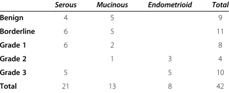
stored at −80°C until used. In addition to the routine histo-pathological examination, each specimen was re-evaluated by a second pathologist. Histological differen-tiation was classified as benign (n = 9), borderline (n = 11), and malignant (n = 22); the histological types were serous (n = 21), mucinous (n = 13), and endometrioid (n = 8) (Table 1). The mean age of included patients was 59 years (range 22–80) in the benign group, 55 years (35–86) in the borderline group, and 62 years (43–85) in the malignant group. The Ethical Review Board at Lund University Hospital approved the study design and in-formed consent was obtained from each patient.
Extraction of total RNA
Total RNA was extracted from about 125 mg frozen ova-rian tumour tissue. The tissue was homogenized in Trizol 50 mg/mL (Invitrogen, Carlsbad, CA) using rotating-knives (Polytron). All RNA samples were checked for con-centration and purity by NanoDrop Spectrophotometer ND-1000 (Saveen Werner, Limhamn, Sweden) having A260/280 and A260/230~ 2. RNA quality and integrity was verified by Agilent 2100 BioAnalyzer (Agilent Technolo-gies, Palo Alto, CA), i.e. all samples had RNA Integrity Number > 7.7.
cDNA synthesis
GeneAmp® RNA PCR kit (Applied Biosystems, Foster City, CA) was used for reverse transcription of total
RNA (0.2 μg) to cDNA. The final concentration of
cDNA was 1 μg/μL (+/− 7%) and A260/280ratio ~1.8 as assessed by NanoDrop. The cDNA samples were stored at−20°C until further use.
Quantitative RT-qPCR amplification
RT-qPCR was performed using a StepOnePlus™ cycler
(Applied Biosystems) under standard thermal cycling conditions (activation of contamination preventing en-zyme at 50°C for 2 min, enen-zyme activation at 95°C for 10 min, 40 cycles of denaturation at 95°C for 15 s, and annealing at 60°C for 1 min). PCR reactions were run in duplicates and negative controls were included in each amplification set. For each gene analysed, pre-manufactured real-time qPCR assays were used
(Ap-Table 1 Distribution of the primary ovarian tumours according to histopathology
Serous Mucinous Endometrioid Total
Benign 4 5 9
Borderline 6 5 11
Grade 1 6 2 8
Grade 2 1 3 4
Grade 3 5 5 10
plied Biosystems or Integrated DNA technologies, Inc., Coralville, IA, USA) (Table 2), with probes spanning exon junctions and not detecting genomic DNA. Using one malignant tumour sample and a universal human reference RNA (Stratagene, La Jolla, CA, USA), quanti-fication experiments were performed using two stan-dard curves from 10-fold serial dilutions of the cDNA (80–0.08 ng).
Identification of new potential reference genes
In order to identify new candidate reference genes in ova-rian tumour tissue, we employed a commercial array (TaqMan® Express Endogenous Control Plate, cat no 4396840, Applied Biosystems) consisting of 32 potential RGs (18S, GADPH, HPRT1, GUSB, ACTB, B2M, HMBS, IPO8, PGK1, RPLPO, TBP, TFRC, UBC, YWHAZ, PP IA, POLR1A, CASC3, CDKN1A, CDKN1B, GADD45A, PUM1, PSMC4, EIF2B1, PES1, ABL1, ELF1, MT-AT6, MRPL19, POP4, RPL37A, RPL30, RPS17).
We analysed one benign and one malignant sample of ovarian tumour, which were selected based on the greatest difference in expression of traditionally used RGs (ACTB, GADPH, and HPRT1), as measured by RT-qPCR. The difference between the threshold cycles (ΔCt) of the two samples was then calculated for each of the
32 genes in the array. Four genes with the lowest ΔCt were selected for inclusion in our main study.
Statistical analysis
Descriptive statistics, F-test for Ctvariance equality and Kolmogorov-Smirnov test for normality of log-trans-formed relative expression values were calculated by software SPSS 19.0 (SPSS Inc, Chicago, IL). The Equiva-lence test [7-9] and statistical applets BestKeeper [10], geNorm [11], and NormFinder [12] were used for ana-lysis of genes expression stability. GeNorm calculates a gene-stability measure, M-value, as the average pair-wise variation of a particular gene to all other candidate refe-rence genes [11]. On the other hand, the stability value calculated with NormFinder combines estimated both intra-group and inter-group variations [12]. Genes with the lowest M-values have the most stable expression (least variability). Relative expression values for target genes were analysed by Kruskal-Wallis and Mann– Whitney tests, and the log-transformed values by one-way ANOVA. P < 0.05 was considered significant.
Results
Selection of best RGs from the commercial gene array
In order to select optimal candidate RGs for this study on ovarian tumours, ΔCtbetween one benign and one
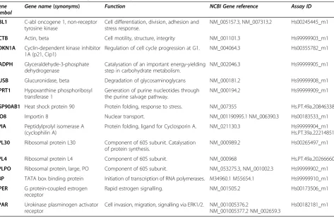
Table 2 Reference genes, target genes and assays used
Gene
symbol Gene name
(synonyms) Function NCBI Gene reference Assay ID
ABL1 C-abl oncogene 1, non-receptor tyrosine kinase
Cell differentiation, division, adhesion and stress response.
NM_005157.3, NM_007313.2 Hs00245445_m1
ACTB Actin, beta Cell motility, structure, integrity NM_001101.3 Hs99999903_m1
CDKN1A Cyclin-dependent kinase inhibitor 1A (p21, Cip1)
Regulation of cell cycle progression at G1. NM_004064.3 Hs00355782_m1
GADPH Glyceraldehyde-3-phosphate dehydrogenase
Catalysation of an important energy-yielding step in carbohydrate metabolism.
NM_002046.3 Hs99999905_m1
GUSB Glucuronidase, beta Degradation of glycosaminoglycans NM_000181.2 Hs99999908_m1
HPRT1 Hypoxanthine phosphoribosyl transferase 1
Generation of purine nucleotides through the purine salvage pathway.
NM_000194.2 Hs99999909_m1
HSP90AB1 Heat shock protein 90 Protein folding, response to stress. NM_007355 Hs.PT.49a.20846338
IPO8 Importin 8 Nuclear transport. NM_001190995.1 NM_006390.3 Hs00183533_m1
PPIA Peptidylprolyl isomerase A (cyclophilin A)
Protein folding, ligand for Cyclosporin A. NM_021130.3 Hs99999904_m1
Hs.PT.39a.22214851
RPL30 Ribosomal protein L30 Component of 60S subunit. Catalysation of protein synthesis.
NM_000989.2 Hs00265497_m1
RPL4 Ribosomal protein L4 Component of 60S subunit. NM_000968 Hs.PT.49a.20266660
RPLPO Ribosomal protein, large, PO Component of 60S subunit. NM_053275.3, NM_001002.3 Hs99999902_m1
TBP TATA box binding protein Initiation of transcription of RNA polymerases. M34960.1 M55654.1 Hs99999910_m1
GPER G protein-coupled estrogen
receptor
Rapid estrogen signalling. NM_001505.2 Hs00173506_m1
uPAR Urokinase plasminogen activator receptor
Cell invasion, migration, signalling via ERK1/2. NM_001005376.2
NM_001005377.2 NM_002659.3
malignant ovarian tumour sample with the greatest dif-ference in expression of the traditionally used RGs (ACTB, GADPH, and HPRT1), was measured by RT-qPCR and calculated for all 32 genes included in the ar-rays. The lowest ΔCt, i.e. the least variation, was found for CDKN1A (ΔCt: 0.47), ABL1 (0.76), RPL30 (0.83), RPS17 (1.09), MT-ATP6 (1.42), and IPO8 (1.71), whereas POP4 (6.11), GADPH (5.04), HPRT1 (4.91), POLR2A (4.41), CASC3 (3.48) had the highest ΔCt. The most abundant genes were 18S (mean Ct± SD: 12.11 ± 1.85) and MT-ATP6 (21.64 ± 1.00), the genes with lowest ex-pression were YWHAZ (31.42 ± 2.14) and TBP (31.37 ± 2.06). CDKN1A, ABL1, RPL30 and IPO8 were chosen to be included in our panel of potential reference genes.
Expression of selected candidate reference and target genes in primary ovarian tumours
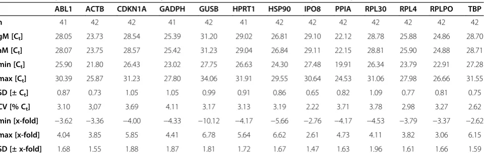
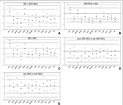
We analysed altogether 13 candidate reference genes (ABL1, ACTB, CDKN1A, GADPH, GUSB, HPRT1, HSP90AB, IPO8, PPIA, RPL30, RPL4, RPLPO, and TBP) and two target genes (GPER and uPAR) by RT-qPCR. Expression levels and variability of Ctvalues are shown for the RGs (Table 3). Of all genes, PPIA had the highest
(mean Ct± SD: 22.12 ± 0.82) and GUSB the lowest
(31.20 ± 0.99) level of mRNA (Figure 1). The amplifica-tion efficiencies of the TaqMan-based RT-qPCR assays were in the range 85–99% for all RGs, except ABL1 and HPRT1, which had 82% efficiency. The linear regression coefficient (r2) of the standard curves for all genes ranged between 0.998 and 1.
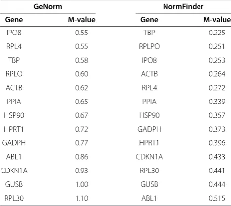
Gene expression stability calculated by GeNorm
Expression stability of the 13 candidate RGs was first assessed by GeNorm in the whole set of tumour sam-ples. The expression stability value (M-value) was calcu-lated based on the average pair-wise variation between all genes tested (Table 4). The genes with the lowest
M-value have the most stable expression and were ranked as follows: the most stable-IPO8 > RPL4 > TBP > RPLPO > ACTB > PPIA > HSP90 > HPRT1 > GADPH > ABL1 > CDKN1A > GUSB > RPL30.
Gene expression stability calculated by NormFinder
M-values were calculated for individual RGs using NormFinder that assessed the expression stability by combining estimated inter- and intra-group variation (Table 4). The genes were ranked according to expres-sion stability as follows: the most stable-TBP > RPLPO > IPO8 > ACTB > RPL4 > PPIA > HSP90 > GADPH > HPRT1 > CDKN1A > RPL30 > GUSB > ABL1. The five
best-ranked genes — TBP, RPLPO, IPO8, ACTB, and
RPL4 — turned out to be the same five most stable
genes found by GeNorm.
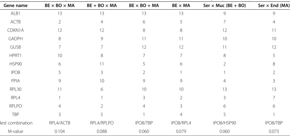
Moreover, NormFinder allowed stability analysis be-tween subgroups: 1) benign, 2) borderline, 3) malignant, 4) serous benign and borderline tumours 5) mucinous, benign and borderline tumours, 6) serous malignant tumours, and 7) endometrioid malignant tumours (Table 5). Combining the two most stable genes fur-ther improved the M-value in group-wise comparison. In all obtained combinations, IPO8 followed by RPL4 came out as the most stable genes.
Analysis of expression stability by BestKeeper and equivalence test
In the next step, candidate RGs were evaluated by BestKeeper and the Equivalence test for variations in ex-pression in the whole data set and between tumours groups as described above. IPO8 had the lowest stan-dard deviation (SD) of the Ct value across the groups (mean Ct± SD: 29.10 ± 0.65). The best-ranked genes by
GeNorm and NormFinder — IPO8, ACTB, TBP, RPL4,
and RPLPO — fulfilled the BestKeeper criteria for sta-bility variation of the Ct value with SD < 1 (Table 3).
Table 3 Descriptive and correlation analysis of the candidate RGs obtained by BestKeeper
ABL1 ACTB CDKN1A GADPH GUSB HPRT1 HSP90 IPO8 PPIA RPL30 RPL4 RPLPO TBP
n 41 42 42 41 42 41 42 42 42 42 42 42 42
gM [Ct] 28.05 23.73 28.54 25.39 31.20 29.02 26.81 29.10 22.12 28.78 25.88 24.86 28.70
aM [Ct] 28.07 23.75 28.57 25.42 31.23 29.04 26.84 29.11 22.15 28.81 25.90 24.88 28.71
min [Ct] 25.90 21.80 26.43 23.02 27.75 26.63 24.30 27.48 19.91 26.34 23.79 22.91 27.28
max [Ct] 30.39 25.87 31.23 27.80 34.06 31.91 29.55 30.64 24.53 31.06 27.98 26.66 31.55
SD [± Ct] 0.87 0.73 1.05 1.05 0.99 0.91 0.86 0.65 0.82 1.09 0.77 0.81 0.75
CV [% Ct] 3.10 3,07 3.69 4.11 3.17 3.13 3.19 2.22 3.71 3.78 2.98 3.27 2.62
min [x-fold] −3.62 −3.36 −4.00 −4.33 −10.12 −4.17 −5.66 −2.76 −4.17 −4.53 −3.79 −3.37 −2.62
max [x-fold] 4.04 3.85 5.85 4.41 6.78 5.64 6.62 2.61 4.73 4.11 3.82 3.06 6.15
SD [± x-fold] 1.68 1.55 1.88 1.87 1.81 1.72 1.67 1.47 1.63 1.96 1.61 1.66 1.59
GADPH had SD > 1 and hence did not meet the stability criteria.
Further, we applied the Equivalence test including both cut-offs of 2-fold and 3-fold expression change to iden-tify the best candidates according equivalent expression in group-wise comparison (Figure 2) [8]. The Equiva-lence test criteria at 3-fold expression change were ful-filled for IPO8, RPL4, RPL30, GUSB, TBP, RPLPO,
ACTB, ABL1, and CDKN1A in all subgroups (Table 6). GAPDH was stably expressed only in two out of the five subgroups, followed by HPRT1, HSB90AB1, and PPIA that were equivalently expressed in three subgroups using cut-off of 3. However, IPO8 was the only gene with equivalent expression within 2-fold change in all subgroups.
Interpretation of target genes expression
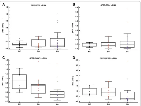
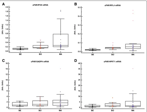
In order to show the effect of the unstable RGs on the final expression of target genes, GPER and uPAR mRNAs were related to either IPO8 and RPL4, or GADPH and HPRT1 mRNA. The choice of target genes was based on our previous observations that GPER mRNA expression did not show any variation between benign, borderline, and malignant ovarian tumour sam-ples [13], whereas uPAR mRNA was higher in borderline and malignant than benign ovarian tumour samples [14]. In accordance with our previously published results, the tissue content of GPER mRNA normalized to IPO8 or RPL4 mRNA showed no significant differences be-tween benign, borderline, and malignant tumour sam-ples. In contrast, GPER mRNA normalized to GADPH or HPRT1 mRNA was higher in benign and borderline tumours than in malignant tumours (Figure 3). uPAR mRNA normalized to IPO8 or RPL4 was significantly up-regulated in borderline and malignant tumours as compared to benign tumours, whereas when it was
Figure 1Expression levels of 13 candidate reference genes in benign (BE), borderline (BO), and malignant (MA) primary ovarian tumours.Values are given as the cycle threshold (Ct) and are inversely proportional to the amount of template. Expression levels of the genes
studied are shown as whiskers box plots.
Table 4 Ranking of 13 candidate RGs according to their expression stability by GeNorm and NormFinder
GeNorm NormFinder
Gene M-value Gene M-value
IPO8 0.55 TBP 0.225
RPL4 0.55 RPLPO 0.251
TBP 0.58 IPO8 0.253
RPLO 0.60 ACTB 0.264
ACTB 0.62 RPL4 0.272
PPIA 0.65 PPIA 0.339
HSP90 0.67 HSP90 0.357
HPRT1 0.72 GADPH 0.373
GADPH 0.77 HPRT1 0.396
ABL1 0.86 CDKN1A 0.433
CDKN1A 0.93 RPL30 0.441
GUSB 1.00 GUSB 0.444
normalized to GADPH or HPRT1 mRNA there were no differences between the tumour groups (Figure 4).
Discussion
Although RT-qPCR is the most commonly used method for assessing gene expression, in-depth studies of poten-tial reference genes and their expression pattern in ovar-ian tumour tissue are insufficient. The aim of this study was to identify the most stably expressed RGs, which can be recommended for normalization of RT-qPCR re-sults in benign, borderline and malignant ovarian tu-mour samples.
We analysed the traditionally used RGs, those reported as being suitable for ovarian tissue, and the four most promising genes from a commercial RG array. Alto-gether 13 potential reference genes were tested for sta-bility across groups of benign, borderline, and malignant primary ovarian tumours of different histological sub-types. Of the genes studied, IPO8, RPL4, TBP, RPLPO, and ACTB were found to be the most stable according to the statistical applets GeNorm, NormFinder and BestKeeper. Our findings on RPL4, RPLPO, and TBP in a Scandinavian population are in accordance with previ-ous reports in Asian populations [4,6]. In contrast, our results did not support PPIA as suitable RG, which has been observed previously [4]. With regard to the hetero-geneity of ovarian tumour materials and different rank-ing results produced by the commonly used statistical approaches, we decided to further employ the Equiva-lence test in our analysis. By applying strict criteria in the Equivalence test, i.e. only allowing a 2-fold change of expression, we could identify IPO8 expression as the most stable of all candidate genes tested.
We included IPO8 in our study because it showed low variation in expression between the benign and the ma-lignant sample in the commercial array. This gene was equivalently expressed across the tumour subgroups of different malignant potential and histology. IPO8 is a Ran-binding protein mediating nuclear import [15] and has been already reported stably expressed in lung tis-sues [16], gliomas [17], and colon cancer [18].
The second best RG for group-wise comparison, RPL4, encodes a protein that is a component of the 60S ribosome subunit [19]. Apart from ovarian tissue, it has previously been recommended as RG in combination with PGK1 for exfoliated cervical cells [20]. RPLPO, an-other gene from the ribosomal protein family, had stable expression in HPV-positive as in HPV-negative cervical samples [21] and in tamoxifen or estrogen treated breast cancer cells [22]. TBP, a key regulator of gene expres-sion, has previously been identified as a suitable RG for expression studies on human hepatitis B virus-related hepatocellular carcinoma [23], human renal cell carci-noma [24], and glioblastomas [17]. RPLPO and TBP also belonged to one of the most stably expressed genes in breast carcinomas [25].
Two other candidates that have not previously been tested as RGs in ovarian tumour tissue, ABL1 and CDKN1A, were selected from the commercial gene array. Both genes satisfied the Equivalence test at 3-fold expression change. ABL1, originally identified as a ho-mologue of the transforming gene of the Abelson mur-ine leukemia virus, is a proto-oncogene, which has been implicated in mitogenesis, regulation of gene transcrip-tion, and inhibition of apoptosis [26]. Nucleotide poly-morphism in the ABL1 gene has been associated with Table 5 NormFinder ranking of 13 candidate RGs and combinations of the two best in group-wise comparison
Gene name BE × BO × MA BE + BO × MA BE × BO + MA BE × MA Ser × Muc (BE + BO) Ser × End (MA)
ALB1 13 13 13 13 9 9
ACTB 2 4 6 5 7 4
CDKN1A 12 12 8 8 12 11
GADPH 8 9 11 11 10 10
GUSB 7 7 12 12 11 12
HPRT1 10 8 7 7 8 5
HSP90 6 11 5 6 2 8
IPO8 5 3 2 1 1 2
PPIA 9 10 9 9 4 3
RPL30 11 6 10 10 13 13
RPL4 1 1 3 2 3 7
RPLPO 4 2 4 3 6 6
TBP 3 5 1 4 5 1
Best combination RPL4/ACTB RPL4/RPLPO IPO8/TBP IPO8/RPL4 IPO8/HSP90 IPO8/TBP
risk of ovarian cancer [27]. CDKN1A (also known as p21) was initially described as an inhibitor of cancer cell proliferation [27]. However, recent studies suggest that it has dual functions since it also may promote tumour progression [28] and be associated with cisplatin resis-tance in ovarian cancer [29].
According to BestKeeper and Equivalence test criteria, we found that GADPH had the worst expression stability in our set of ovarian tumour samples. Similar unfavou-rable results were obtained for HPRT1. These observa-tions are in line with previous studies on other tissue
types that have discouraged use of GADPH and HPRT1 as RGs for clinical lung specimens [16] and renal cell cancer [24]. Most recently, a microarray study identified a group of genes highly correlated to GADPH up-regulation in various solid tumours, which were and proportionally associated with advanced stages [30]. Pre-vious reports on GADPH in ovarian tissue have either pointed out higher expression in malignant than in benign tumours and normal tissue [6], or not meeting the GeNorm stability criteria [4]. We further demon-strated that employment of GADPH or HPRT1 for
Table 6 Expression stability of the candidate RGs analysed by equivalence test
BE × BO + MA BE + BO × MA BE × MA Ser × Muc (BE + BO) Ser × End (MA) Total passes 2-fold/3-fold
ABL1 0 /1 0 /1 0 /1 1 /1 0 /1 1 /5
ACTB* 0 /1 0 /1 0 /1 1 /1 0 /1 1 /5
CDKN1A 0 /1 1 /1 0 /1 0 /1 0 /1 1 /5
GADPH 0 /0 0 /0 0 /0 0 /1 0 /1 0 /2
GUSB 0 /1 0 /1 1 /1 1 /1 0 /1 2 /5
HPRT1 0 /1 0 /0 0 /0 0 /1 0 /1 0 /3
HSP90 0 /1 0 /0 0 /0 0 /1 0 /1 0 /3
IPO8* 1 /1 1 /1 1 /1 1 /1 1 /1 5 /5
PPIA 0 /1 0 /0 0 /0 1 /1 0 /1 1 /3
RPL30 1 /1 0 /1 0 /1 0 /1 1 /1 2 /5
RPL4* 1 /1 0 /1 0 /1 0 /1 1 /1 2 /5
RPLPO* 0 /1 0 /1 0 /1 0 /1 1 /1 1 /5
TBP* 1 /1 0 /1 0 /1 0 /1 1 /1 2 /5
The expression within (1) or outside (0) 2-fold/3-fold expression change cut-off and the total number of meeting the cut-off criteria in the five subgroups. * Genes best-ranked by GeNorm, NormFinder and BestKeeper.
normalization resulted in erroneous conclusions on expression of target genes.
To our knowledge, this is the first report on RGs in ovarian tumours that include borderline tumours in addition to benign and malignant tumours. Since they are considered a non-invasive pre-stage of molecular type I ovarian cancer, it is important to include them in any study on biomarker discovery [31].
Ovarian cancer comprises tumours of different morph-ology and pathogenesis, which may have different gene expression profiles [32]. Therefore we wished to see whether the histology of ovarian tumours influences the stability of RGs. Thus, in contrast to the previous studies conducted exclusively on serous malignant tu-mours, our study also included mucinous and endo-metrioid tumours. However, small number of samples in some groups limited the comparisons that could be performed.
Conclusions
In conclusion, thorough statistical evaluation of our 13 candidate RGs identified IPO8 followed by RPL4 as the most suitable for the normalization of gene expression data in benign, borderline, and malignant ovarian tu-mours. For the first time, IPO8 is presented as the best normaliser for gene expression studies on ovarian tumour tissue with heterogeneous histology when used as a single RG. Neither GADPH nor HPRT1 should be used as RGs for ovarian tissue studies, because of poor expression stability. Normalizing to these genes may erroneously influence the quantification of the target gene(s) and hence reduce the reliability of the RT-qPCR results.
Abbreviations
RT-qPCR:Quantitative real-time reverse transcription-polymerase chain reaction; RG: Reference gene; IPO8: Importin 8; RPL4: Ribosomal protein 4; GADPH: Glyceraldehyde-3-phosphate dehydrogenase; HPRT1: Hypoxanthine phosphoribosyl transferase 1.
Competing interests
The authors declare that they have no competing interests.
Authors’contributions
ZK carried out the gene expression experiments and drafted the manuscript. AA performed the statistical analysis. BC drafted the manuscript. SH contributed methodological know-how. EK participated in the study design and drafted the manuscript. All authors read and approved the final manuscript.
Acknowledgements
This study was supported by the Swedish Cancer society, Skåne University Hospital and Region Skåne.
Author details
1
Department of Obstetrics & Gynaecology, Lund University, Skåne University Hospital Lund, Lund, SE 221 85, Sweden.2Institute of Molecular Biology, NAS RA 7 Hasratyan St, Yerevan 0014, Armenia.3Department of Immunology, Faculty of Medicine and Dentistry, Palacky University, Olomouc, Czech Republic.
Received: 10 May 2013 Accepted: 18 August 2013 Published: 30 August 2013
References
1. Bustin SA, Benes V, Garson JA, Hellemans J, Huggett J, Kubista M, Mueller R, Nolan T, Pfaffl MW, Shipley GL, Vandesompele J, Wittwer CT:The MIQE guidelines: minimum information for publication of quantitative real-time PCR experiments.Clin Chem2009,55(4):611–622.
2. Sirover MA:New insights into an old protein: the functional diversity of mammalian glyceraldehyde-3-phosphate dehydrogenase.Biochimica et biophysica acta1999,1432(2):159–184.
3. Chang TJ, Juan CC, Yin PH, Chi CW, Tsay HJ:Up-regulation of beta-actin, cyclophilin and GAPDH in N1S1 rat hepatoma.Oncol Rep1998,
5(2):469–471.
4. Li YL, Ye F, Hu Y, Lu WG, Xie X:Identification of suitable reference genes for gene expression studies of human serous ovarian cancer by real-time polymerase chain reaction.Anal Biochem2009,394(1):110–116. 5. Sun Y, Li Y, Luo D, Liao DJ:Pseudogenes as weaknesses of ACTB (Actb)
and GAPDH (Gapdh) used as reference genes in reverse transcription and polymerase chain reactions.PloS One2012,7(8):e41659.
6. Fu J, Bian L, Zhao L, Dong Z, Gao X, Luan H, Sun Y, Song H:Identification of genes for normalization of quantitative real-time PCR data in ovarian tissues.Acta biochimica et biophysica Sinica2010,42(8):568–574. 7. Stefan W:Testing Statistical Hypotheses of Equivalence and
Noninferiority; 2003.
8. Haller F, Kulle B, Schwager S, Gunawan B, von Heydebreck A, Sultmann H, Fuzesi L:Equivalence test in quantitative reverse transcription polymerase chain reaction: confirmation of reference genes suitable for normalization.Anal Biochem2004,335(1):1–9.
9. Kriegova E, Arakelyan A, Fillerova R, Zatloukal J, Mrazek F, Navratilova Z, Kolek V, du Bois RM, Petrek M:PSMB2 and RPL32 are suitable
denominators to normalize gene expression profiles in bronchoalveolar cells.BMC Mol Biol2008,9:69.
10. Pfaffl MW, Tichopad A, Prgomet C, Neuvians TP:Determination of stable housekeeping genes, differentially regulated target genes and sample integrity: BestKeeper–excel-based tool using pair-wise correlations. Biotechnol Lett2004,26(6):509–515.
11. Vandesompele J, De Preter K, Pattyn F, Poppe B, Van Roy N, De Paepe A, Speleman F:Accurate normalization of real-time quantitative RT-PCR data by geometric averaging of multiple internal control genes.Genome Biol2002,3(7). RESEARCH0034.
12. Andersen CL, Jensen JL, Orntoft TF:Normalization of real-time quantitative reverse transcription-PCR data: a model-based variance estimation approach to identify genes suited for normalization, applied to bladder and colon cancer data sets.Cancer Res2004,64(15):5245–5250. 13. Kolkova Z, Casslen V, Henic E, Ahmadi S, Ehinger A, Jirstrom K, Casslen B:
The G protein-coupled estrogen receptor 1 (GPER/GPR30) does not predict survival in patients with ovarian cancer.J Ovarian Res2012,5:9. 14. Borgfeldt C, Hansson SR, Gustavsson B, Masback A, Casslen B:
Dedifferentiation of serous ovarian cancer from cystic to solid tumors is
associated with increased expression of mRNA for urokinase
plasminogen activator (uPA), its receptor (uPAR) and its inhibitor (PAI-1). Int J Cancer2001,92(4):497–502.
15. Dean KA, von Ahsen O, Gorlich D, Fried HM:Signal recognition particle protein 19 is imported into the nucleus by importin 8 (RanBP8) and transportin.J Cell Sci2001,114(Pt 19):3479–3485.
16. Nguewa PA, Agorreta J, Blanco D, Lozano MD, Gomez-Roman J, Sanchez BA, Valles I, Pajares MJ, Pio R, Rodriguez MJ, Montuenga LM, Calvo A:Identification of importin 8 (IPO8) as the most accurate reference gene for the clinicopathological analysis of lung specimens.BMC Mol Biol2008,9:103. 17. Kreth S, Heyn J, Grau S, Kretzschmar HA, Egensperger R, Kreth FW:
Identification of valid endogenous control genes for determining gene expression in human glioma.Neuro Oncol2010,12(6):570–579. 18. Sorby LA, Andersen SN, Bukholm IR, Jacobsen MB:Evaluation of suitable
reference genes for normalization of real-time reverse transcription PCR analysis in colon cancer.J Exp Clin Cancer Res2010,29:144.
19. Klinge S, Voigts-Hoffmann F, Leibundgut M, Arpagaus S, Ban N:Crystal structure of the eukaryotic 60S ribosomal subunit in complex with initiation factor 6.Science2011,334(6058):941–948.
20. Steinau M, Rajeevan MS, Unger ER:DNA and RNA references for qRT-PCR assays in exfoliated cervical cells.JMD2006,8(1):113–118.
21. Daud II, Scott ME:Validation of reference genes in cervical cell samples from human papillomavirus-infected and -uninfected women for quantitative reverse transcription-PCR assays.CVI2008,15(9):1369–1373. 22. Shah KN, Faridi JS:Estrogen, tamoxifen, and Akt modulate expression of putative housekeeping genes in breast cancer cells.J Steroid Biochem Mol Biol2011,125(3–5):219–225.
23. Fu LY, Jia HL, Dong QZ, Wu JC, Zhao Y, Zhou HJ, Ren N, Ye QH, Qin LX:
Suitable reference genes for real-time PCR in human HBV-related hepatocellular carcinoma with different clinical prognoses.BMC Cancer
2009,9:49.
24. Jung M, Ramankulov A, Roigas J, Johannsen M, Ringsdorf M, Kristiansen G, Jung K:In search of suitable reference genes for gene expression studies of human renal cell carcinoma by real-time PCR.BMC Mol Biol2007,8:47. 25. Lyng MB, Laenkholm AV, Pallisgaard N, Ditzel HJ:Identification of genes for
normalization of real-time RT-PCR data in breast carcinomas.BMC Cancer
2008,8:20.
26. Colicelli J:ABL tyrosine kinases: evolution of function, regulation, and specificity.Sci Signal2010,3(139):re6.
27. Cunningham JM, Vierkant RA, Sellers TA, Phelan C, Rider DN, Liebow M, Schildkraut J, Berchuck A, Couch FJ, Wang X, Fridley BL, Gentry-Maharaj A, Menon U, Hogdall E, Kjaer S, Whittemore A, DiCioccio R, Song H, Gayther SA, Ramus SJ, Pharaoh PD, Goode EL:Cell cycle genes and ovarian cancer susceptibility: a tagSNP analysis.Br J Cancer2009,101(8):1461–1468. 28. Stivala LA, Cazzalini O, Prosperi E:The cyclin-dependent kinase inhibitor
p21CDKN1A as a target of anti-cancer drugs.Curr Cancer Drug Targets
2012,12(2):85–96.
29. Xia X, Ma Q, Li X, Ji T, Chen P, Xu H, Li K, Fang Y, Weng D, Weng Y, Liao S, Han Z, Liu R, Zhu T, Wang S, Xu G, Meng L, Zhou J, Ma D:Cytoplasmic p21 is a potential predictor for cisplatin sensitivity in ovarian cancer. BMC Cancer2011,11:399.
30. Wang D, Moothart DR, Lowy DR, Qian X:The expression of glyceraldehyde-3-phosphate dehydrogenase associated cell cycle (GACC) genes correlates with cancer stage and poor survival in patients with solid tumors.PloS One2013,8(4):e61262.
31. Romero I, Bast RC Jr:Minireview: human ovarian cancer: biology, current management, and paths to personalizing therapy.Endocrinology2012,
153(4):1593–1602.
32. Gomez-Raposo C, Mendiola M, Barriuso J, Hardisson D, Redondo A:
Molecular characterization of ovarian cancer by gene-expression profiling.Gynecol Oncol2010,118(1):88–92.
doi:10.1186/1757-2215-6-60
Cite this article as:Kolkovaet al.:Normalizing to GADPH jeopardises correct quantification of gene expression in ovarian tumours–IPO8 and RPL4 are reliable reference genes.Journal of Ovarian Research