R E S E A R C H A R T I C L E
Open Access
Current expectations of the arterial switch
operation in a small volume center: a
20-year, single-center experience
Man-shik Shim
1, Tae-Gook Jun
1*, Ji-Hyuk Yang
1, Pyo Won Park
1, I Seok Kang
2, June Huh
2and Jin Young Song
2Abstract
Background:We reviewed our 20-year experience with arterial switch operation (ASO) for transposition of the great arteries (TGA) or double outlet right ventricle with subpulmonary ventricular septal defect (Taussig-Bing anomaly) to assess the early and long-term outcomes.
Methods:Between January 1995 and December 2014, 139 consecutive patients who underwent ASO for TGA or
Taussig-Bing anomaly were included in this retrospective study. The median age at the operation was 9 (0–485) days, and 97 patients (70 %) underwent ASO less than 2 weeks. The median weight was 3.3 (2.1-10.3) kg. The patients were divided into three groups; simple TGA (n= 78) included patients with TGA with intact ventricular septum, complex TGA (n= 46) included those who had TGA with ventricular septal defect or other anomalies, and Taussig-Bing anomaly (n= 15). Median follow-up duration was 72.5 (0.4-230) months.
Results:There were 3(2.2 %) in-hospital deaths. One patient (0.7 %) underwent early reoperation due to coronary insufficiency. Late deaths occurred in 3 (2.2 %) of 136 survivors. The Kaplan-Meier’s survival rate was 97.6 ± 1.4 % at 15 years. Twenty-three patients (16.9 %) required 26 reintervention. The freedom from reintervention rates were 82.5 ± 3.7 % at 5 years and 75.8 ± 4.7 % at 10 years, respectively. Median interval between ASO and first reintervention was 22.8 (6.4-89.2) months. The multivariate analysis showed that diagnosis of Taussig-Bing anomaly (hazard ratio, 7.09;P< 0.001) and side by side great artery relationship (hazard ratio, 7.98;P= 0.001) were independent risk factors for reoperation. Five patients (3.9 %) had developed at least moderate neo-aortic regurgitation during the follow-up and one patient underwent reoperation mainly for neo-aortic regurgitation. By multivariate analysis, Taussig-Bing anomaly was the risk factor for at least moderate neo-aortic regurgitation (P= 0.035).
Conclusions:ASO can be performed with a low risk of early mortality and satisfactory long-term outcomes even in a small volume center. Close long-term surveillance is mandatory to detect structural or hemodynamic changes.
Keywords:Congenital heart defect, Arterial switch operation, Transposition of the great arteries, Double outlet right ventricle with subpulmonary ventricular septal defect, Taussig-Bing anomaly
* Correspondence:[email protected]
Dr. I Seok Kang and Dr. Jin Young Song confirmed that they are co-authors of the manuscript.
1
Department of Thoracic and Cardiovascular Surgery, Samsung Medical Center, Sungkyunkwan University School of Medicine, 50 Irwon-dong, Kangnam-gu, Seoul 135-710, Korea
Full list of author information is available at the end of the article
Background
The arterial switch operation (ASO) has become the procedure of choice for the transposition of great arter-ies (TGA) and double outlet right ventricle (DORV) with subpulmonary ventricular septal defect (VSD) (Taussig– Bing anomaly, TBA). Since the first successful ASO re-ported by Jatene et al. in 1975 [1], there have been a steadily improvements in diagnosis, surgical techniques, and perioperative management. The result of ASOs has been improved and the reported mortality has fallen to the range of 0%–6%, even when the complex group is included [2–5]. However, anatomic variations of the cor-onary artery, combined arch anomalies, low birth weight, and age presentation over 4 weeks are still considered risk factors [3, 4, 6].
The purpose of this study was to review the ASO at a single institution with a small volume. This report focuses on the short- and midterm results according to the anatomic subtype and surgical techniques. In ad-dition, it intends to determine the current risk factors for mortality and morbidity.
Methods
We conducted a retrospective review of patients who had an ASO with the diagnosis of d-TGA or DORV (TBA) from January 1995 to October 2014. A total of 139 consecutive patients were included in the study. Patients with various forms of TGA undergoing pallia-tive ASO, double-switch operation, and half-turned switch operation were excluded from this study. Patient characteristics are shown in Table 1. The patients were divided into 3 groups according to diagnosis. The simple TGA group (n= 78) included patients with TGA with an intact ventricular septum. The complex TGA group (n= 46) included those who had d-TGA with a VSD or other anomalies such as coarctation of the aorta, interrupted aortic arch, total anomalous pulmonary venous return, significant left ventricular outflow tract (LVOT) obstruction, and those who had l-TGA with situs inversus. The TBA group (n= 15) included pa-tients who were diagnosed with this anomaly. Per-mission to perform a retrospective review of medical records was obtained from the Institutional Review Board of Samsung Medical Center. The need for indi-vidual consent for the study was waived.
Definitions
Early death or reoperation was defined as death or reop-eration occurring within 30 days of ASO or before hos-pital discharge. Late death or reoperation was defined as death or reoperation occurring after discharge and more than 30 days after ASO. Reoperation was defined as an operation on the heart or great vessels performed after the ASO, excluding exploration for bleeding, wound
debridement, mechanical circulatory support, and pace-maker insertion. Reintervention included reoperation or catheter intervention performed after ASO.
Follow-up
Most patients underwent regular outpatient follow-up visits at a pediatric cardiology clinic. Complete clinical follow-up data, which included an echocardiogram, were available for 129 of the 136 survivors (94.9 %). Median follow-up duration was 72.5 (range 0.4–230) months. Although ischemic symptoms are the indication for the coronary angiography, all patients are too young to express their symptoms during early period after the ASO. So, we routinely performed the coronary angiog-raphy one year after the ASO.
Surgical techniques
After a standard median sternotomy, the thymus was re-moved. A large pericardial patch was harvested and fixed with a 0.625 % glutaraldehyde solution. Cardiopulmo-nary bypass (CPB) is instituted using bicaval cannulation as the usual pattern at temperatures between 25 °C and 32 °C. The ASO underwent at mild to moderate hypothermia (25 ~ 35 °C) while maintaining systemic perfusion. Routine selective cerebral perfusion or circu-latory arrest had not been used except in patients with combined aortic arch obstruction such as interrupted aor-tic arch and coarctation of aorta. When the arch recon-struction was anticipated, the arterial cannula is connected to the innominate artery via 3.5-mm Gore-Tex tube inter-position graft. During the arch repair, the selected ante-grade cerebral perfusion was conducted at >70 ml/kg/min at the lowest rectal temperature of 25 °C.
button transfer can be determined more easily and more accurately while the aorta is inflated. Additionally, the bleeding from anastomosis site can be confirmed more conveniently and it makes the procedure easier. After cardioplegic arrest, the ascending aorta was divided at the level of 10 mm above the sinotubular junction (slightly above the level of pulmonary artery bifurcation). The main pulmonary artery was divided at the level of bifurcation. After examination of the pulmonary valve, the marking sutures for commissures were inserted at the external surface of the proximal pulmonary root. The distal pulmonary artery was dissected more distally and a Lecompte maneuver was performed. The neoaorta reconstruction was completed with 8–0 or 7–0
polypropylene suture material. The aortic clamp was re-moved and bleeding controlled along the suture line. For coronary artery transfer, a generous coronary artery but-ton that included most of the corresponding sinus was harvested in the belief that it can allow a greater coron-ary artery length and prevent tension and torsion after coronary artery transfer [8]. During the coronary artery button preparation, we endeavored to minimize damage to the vasa vasorum of the coronary artery and to avoid transecting the proximal branches of coronary arteries. After inflation of the coronary sinus, we constructed tagging sutures on the corresponding sinus of each coronary artery button avoiding the previously inserted sutures marking the commissures. Small stab incisions
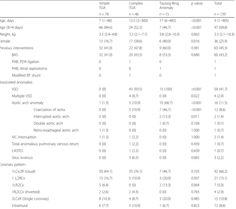
Table 1Demographic data and anatomic characteristics of 139 patients who underwent an arterial switch operation
Simple TGA
Complex TGA
Taussig-Bing
Anomaly p
value Total
n= 78 n= 46 n= 15 n= 139
Age, days 7 (1–48) 12.5 (2–300) 17 (6–485) <0.001 9 (1–485)
Age (≤14 days) 66 (84.6) 24 (52.2) 7 (46.7) <0.001 97 (69.8)
Weight, kg 3.3 (2.4–4.8) 3.3 (2.1–7.7) 3.8 (2.8–10.3) 0.063 3.3 (2.1–10.3)
Female 13 (16.7) 17 (38.6) 6 (40.0) 0.016 36 (25.9)
Previous interventions 32 (41.0) 22 (47.8) 9 (60.0) 0.381 63 (45.3)
BAS 32 (41.0) 20 (43.5) 8 (53.3) 0.680 60 (43.2)
PAB, PDA ligation 0 1 0 1
PAB, Atrial septostomy 0 0 1 1
Modified BT shunt 0 1 0 1
Associated anomalies
VSD 0 (0) 43 (93.5) 15 (100) <0.001 58 (41.7)
Multiple VSD 0 (0) 4 (8.7) 0 (0) 0.022 4 (2.9)
Aortic arch anomaly 1 (1.3) 5 (10.9) 10 (66.7) <0.001 16 (11.5)
Coarctation of aorta 0 (0) 5 (10.9) 7 (46.7) <0.001 12 (8.6)
Interrupted aortic arch 0 (0) 0 (0) 2 (13.3) 0.011 2 (1.4)
Double aortic arch 0 (0) 0 (0) 1 (6.7) 0.108 1 (0.7)
Retro-esophageal aortic arch 1 (1.3) 0 (0) 0 (0) 1.000 1 (0.7)
IVC interruption 1 (1.3) 1 (2.2) 0 (0) 1.000 2 (1.4)
Total anomalous pulmonary venous return 0 (0) 1 (2.2) 0 (0) 0.439 1 (0.7)
LVOTO 0 (0) 1 (2.2) 0 (0) 0.439 1 (0.7)
Situs inversus 0 (0) 3 (6.5) 0 (0) 0.065 3 (2.2)
Coronary pattern
1LCx;2R (Usual) 50 (64.1) 35 (76.1) 7 (46.7) 0.103 92 (66.2)
1 L;2RCx 13 (16.7) 5 (10.9) 3 (20.0) 0.507 21 (15.1)
1LR;2Cx 5 (6.4) 0 (0) 2 (13.3) 0.064 7 (5.0)
1R;2LCx (Inverted) 2 (2.6) 2 (4.3) 0 (0) 0.765 4 (2.9)
2LCxR (Single coronary) 8 (10.3) 4 (8.7) 3 (20.0) 0.485 15 (10.8)
Intramural 6 (7.7) 5 (10.9) 1 (6.7) 0.823 12 (8.6)
were made just outside the tagging sutures after clamping of the aorta, the orifices were widened into a C-shape to construct a trapdoor while being careful not to damage the aortic valve, and the coronary artery buttons were anastomosed with 8–0 polypropylene suture material. The neopulmonary artery root was reconstructed with a pantaloon-shaped autologous peri-cardial patch and then the root was anastomosed to the distal main pulmonary artery. When VSD closure was needed, the VSD was closed with a bovine pericardial patch or Dacron patch before the ASO.
Statistical analysis
Data were collected retrospectively. Continuous vari-ables are expressed as median (and range), and categor-ical variables are expressed as percentages. To compare the 3 groups, a one-way analysis of variance (ANOVA) and Kruskal–Wallis test were used for normal and skewed continuous variables, respectively, and chi-square and Fisher exact tests were used for categorical variables. Fac-tors associated with early mortality were analyzed by mul-tiple logistic regression. Kaplan–Meier survival analyses with a log-rank test were used to analyze late survival reintervention, reoperation, neoaortic insufficiency, and event-free survival. A Cox proportional hazards model with a forward stepwise procedure was used to evaluate risk factors for late survival, reoperation, neoaortic insuffi-ciency, and event-free survival. Variables withp< 0.20 in the univariate analysis constituted the starting set of co-variates and variables with p≥0.05 were excluded from each stepwise selection. Variables analyzed in the univari-ate analysis were age, sex, body weight, previous palliative surgery, previous balloon atrial septostomy, diagnosis with TBA, aortic arch obstruction (coarctation of aorta, inter-rupted aortic arch), unusual coronary artery, single coron-ary artery, intramural coroncoron-ary artery, CPB time, aortic cross-clamp time, postoperative open sternum, and side-by-side great artery relationship. All statistical analyses were conducted using IBM SPSS Statistics for Windows (version 22; IBM SPSS, Armonk, NY, USA). A p< 0.05 was considered significant.
Results and discussion
Perioperative characteristics
The median CPB time and mean aortic cross-clamp time were 196.5 (121–501) min and 120.5 (53–300) min, respectively. The sternum was left open in 42 of 139 patients (30.2 %) and delayed sternal closure was com-pleted at a median of 3 (2–6) days. The duration of post-operative mechanical ventilation was 4.8 (0.6–52.9) days. The median length of stay in an intensive care unit (ICU) and the median hospital stay were 8.8 (0.8–92.8) days and 15 (1–123) days, respectively.
Early outcomes
There were 3 (2.2 %) in-hospital deaths. The first patient was diagnosed preoperatively as having TGA with an in-tact ventricular septum and a coronary artery with an intramural course. He died within the first 24 hours after surgery which is presumed to be caused by myocardial ischemia. The second who had TGA with large VSD underwent the ASO at 61 days old due to delayed diag-nosis. She preoperatively had pulmonary hypertension and pulmonary congestion. The pulmonary hypertensive crisis was developed on the fourth postoperative day. Medical treatments including adjustment of mechanical ventilation setting, inhaled nitric oxide, additional seda-tives and pulmonary vasodilators was initiated immedi-ately and seem to be effective. However, intractable pulmonary hypertensive crisis was developed a few hours later on the fifth postoperative day and the patient died of right heart failure. These 2 cardiac deaths oc-curred in the earlier period before 2001. The third who had TGA with VSD died of unknown origin of septic shock. He suffered from capillary leak syndrome immedi-ately after the ASO and symptoms (such as hypotension, generalized edema, oliguria) worsen over time in contrast to his preserved heart function. With suspecting infection as a cause, general management of sepsis including broad-spectrum antibiotics began. However, his condition had become worse and disseminated intravascular coagulation (DIC) and vegetation on right atrium were developed. Finally, he died of multi-organ failure on the 48th postop-erative day. A multivariate logistic regression analysis demonstrated that low body weight (<3 kg) at surgery was the only risk factor for early hospital mortality (odds ratio, 16.7;p= 0.030; Nagelkerke R2, 0.372).
catheter-related infection in 1, and pericardial effusion in 1. There was no atrioventricular block.
Late outcomes
Late survival
Among the 136 patients surviving the initial operation, there were 3 (2.2 %) later deaths. Among them, the earl-ier two patients had TGA with an intact ventricular septum. Both patients were successfully discharged with-out complications on eleventh and ninth postoperative day, respectively. But they died suddenly and unexpect-edly 2 months after the ASO. Unfortunately, they died outside our hospital and we could not get a detailed his-tory. So, we could not identify their true cause of death. Nevertheless, considering the abruptness of the deaths in those patients with no problems during postoperative hospital course, we presume that the cause of death were coronary ischemia. The third patient who had TBA with coarctation of the aorta and a single coronary artery including the intramural course of the conal branch underwent ASO and coarctoplasty. This patient also underwent left subclavian artery interposition be-tween the aorta and the conal branch because of coron-ary injury. After 5 months, reoperation was performed for severe aortic insufficiency. At 14 months after the initial repair, we found that severe aortic insufficiency re-curred at a routine check-up and decided to reoperate electively. However, he died abruptly the day before hos-pital admission. Although the exact cause of death was unknown, he had symptoms of acute upper respiratory infection for a few days. Presumably, some infectious condition such as myocarditis might aggravate myocar-dial depression. The overall actuarial survival rates were 98.5 ± 1.1 % at 1 year and 97.6 ± 1.4 % at 15 years (Fig. 1). There were no significant risk factors for late mortality.
Late reintervention
During the follow-up period, 23 patients (16.9 %) subse-quently required 26 reintervention including 21 reopera-tions in 19 patients (15.4 %) and 5 balloon angioplasties in 5 patients (3.7 %). Primary reasons for reintervention according to the groups are presented in Table 2. Mul-tiple reinterventions were required in one patient who had TBA with an interrupted aortic arch. He underwent a first reoperation involving right ventricular outflow tract (RVOT) with a Gore-Tex monocuspid patch at 6 months after the initial surgery and underwent a sec-ond reoperation for RVOT stenosis using a monocuspid transannular homograft patch 2 years after reoperation. Seven years later, balloon angioplasty was performed for pulmonary artery stenosis and he finally underwent pul-monary valve replacement 9 years after the second sur-gery. Balloon angioplasty was performed for supravalvar pulmonary artery stenosis in 3 patients, for pulmonary valve stenosis in 1, and for residual coarctation of aorta in 1.
As shown in Fig. 2, The overall freedom from reinter-vention rates were 95.9 ± 1.8 % at 1 year, 82.5 ± 3.7 % at 5 years, and 75.8 ± 4.7 % at 10 years, respectively. One-and 5-year freedom from reintervention rates were signifi-cantly worse (p< 0.001) in the TBA group than those in the other groups: 79.4 ± 10.6 % at 1 year and 15.6 ± 13.2 % at 5 years in the TBA group, 98.5 ± 1.5 % at 1 year and 90.2 ± 3.8 % at 5 years in the simple TGA group, and 97.5 ± 2.5 % at 1 year and 88.5 ± 5.5 % at 5 years in the complex TGA group. There was no significant difference in the incidence of reintervention between the simple and complex TGA groups (p= 0.952). The median in-terval between ASO and the first reintervention was 22.8 (6.4–89.2) months and median interval between ASO and the first reoperation was 25.6 (6.4–89.2) months.
The most common primary indication for reoperation was pulmonary tract pathology (n= 12) including pulmon-ary valve stenosis and RVOT stenosis in 6 patients, supraval-var pulmonary artery stenosis in 5, and pulmonary artery aneurysmal dilatation in 1. The second most common pri-mary indication was systemic tract pathology (n= 5) includ-ing LVOT stenosis in 2 patients, aortic valve regurgitation in 1, aortic root dilatation in 1, and retroesophageal aortic arch in 1. Two patients underwent coronary artery ostial angio-plasty because of coronary ostial stenosis at 16.3 and 62.9 months, respectively, after the ASO. We performed a patch enlargement of coronary ostium using a native pul-monary artery patch. Both patients remained asymptomatic without a stenosis until recently. The first reoperation pro-cedures are detailed in Table 3. The follow-up periods after the reoperation were 9 years and 6 months, respectively. Thirteen of the 19 patients (68 %) who underwent reopera-tion required concomitant procedures for minor coexisting
lesions and the most frequently performed concomitant procedure was pulmonary artery angioplasty (n= 10). In-cluding the primary procedures, 16 patients (84.2 %) under-went pulmonary artery angioplasty during their first reoperation. The interval between the ASO and the first re-operation was not significantly different between groups ac-cording to the location of primary lesions (p= 0.584). Details of the first reoperation procedures are presented in Table 2. A multivariate analysis showed that diagnosis of TBA (haz-ard ratio, 7.09;p< 0.001) and side-by-side great artery rela-tionship (hazard ratio, 7.98; p= 0.001) were independent risk factors for reoperation.
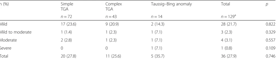
Neoaortic regurgitation
Among 136 early survivors, echocardiographic follow-up was completed in 129 patients (94.9 %). The median echocardiographic follow-up duration was 5.4 (0.02– 18.8) years. Two patients underwent a reoperation for neoaortic valve regurgitation. One patient underwent a repair at 8.5 months after the ASO, primarily for severe regurgitation, and the other underwent repair for
mild-to-moderate regurgitation concomitantly with another primary procedure. At the most recent echocardio-graphic follow-up, 36 of 129 (27.9 %) had at least mild neoaortic regurgitation; of these, the regurgitation was mild in 28 patients (77.8 %), mild to moderate in 3 (8.3 %), moderate in 4 (11.1%), and severe in 1 (2.8 %). The neoaortic regurgitation at the last follow-up is described in Table 4. The incidence of significant (at least moder-ate) neoaortic regurgitation was 3.9 %. The patient who underwent aortic valve repair for severe regurgi-tation remained with severe regurgiregurgi-tation at last follow-up and died before their second reoperation. Freedom from significant (at least moderate) neoaor-tic regurgitation was 97.0 ± 1.7 % at 5 years, 95.2 ± 2.5 % at 10 years, and 95.2 ± 2.5 % at 15 years. In multi-variate analysis, diagnosis of TBA was a risk factor for at least moderate neoaortic regurgitation (hazard ratio, 8.26; p= 0.035). We did not find a statistically significant difference in the development of significant (at least moderate) AR between open and closed cor-onary transfer techniques. However, despite of longer
Table 2Primary reasons for reintervention in 136 hospital survivors
Simple TGA
n= 77
Complex TGA
n= 44
Taussig–Bing Anomaly
n= 15
Total
n= 136 p
Reintervention 2 (2.8) 0 3 (20.0) 5 (3.7) 0.008
Supravalvar pulmonary stenosis 2 (2.6) 0 2 (13.3) 4 (2.9) 0.058
Residual CoA 0 0 1 (6.7) 1 (0.7) 0.110
Reoperation 7 (9.1) 5 (11.4) 7 (46.7) 19 (14.0) 0.003
Pulmonary tract lesion 4 (5.2) 3 (6.8) 5 (33.3) 12 (8.8) 0.008
Systemic tract lesion 1 (1.3) 2 (4.5) 2 (13.3) 5 (3.7) 0.043
Coronary artery lesion 2 (2.6) 0 0 2 (1.5) 0.631
Total 9 (11.7) 5 (11.4) 9 (60.0) 23 (16.9) <0.001
TGA, transposition of great arteries; CoA, coarctation of aorta
follow-up period, only 2 of 38 patients who under-went open coronary transfer developed significant AR. In terms of neo-AR, the older open technique is not inferior to closed technique. But the closed coronary transfer technique is easy to perform, convenient to confirm bleeding from anastomosis site, and reprodu-cible for unexperienced surgeons. We need more longer-term follow-up data to compare these tech-niques, because the number of events was small.
Event-free survival rate
The events in this study were defined as late death, reop-eration, and at least moderate neoaortic regurgitation. Event-free survival rate was 87.5 ± 3.2 % at 5 years, 71.8 ± 6.0 % at 10 years, and 68.0 ± 6.8 % at 15 years (Fig. 3). Event-free survival rate was similar (p = 0.763) for TGA groups, and the TBA group showed a significantly lower (p< 0.001) event-free survival than the TGA groups: 45.5 ± 15.9 % at 5 years in the TBA group, 93.9 ± 3.0 % at 5 years in the simple TGA group, and 89.2 ± 5.2 % at
5 years in the complex TGA group. Multivariate analysis showed significantly poorer event-free survival in patients with TBA (hazard ratio, 5.548;p< 0.001).
Discussion
The low early mortality (2.2 %) after ASOs found in this study is consistent with the findings of other recent stud-ies of large-volume centers [5, 6, 9–12]. Karamlou et al. [9] showed the small-volume center or surgeon performing the ASO can be associated with high early mortality and morbidity, and recently small-to-medium centers [13–15] still reported high early mortality. Karamlou et al. demon-strated that the surgeon volume more influence early out-come than the center volume [9]. We have three surgeons and each surgeons performed 2.32 cases/year. We are able to confirm that our center and surgeon volumes are low.
Popov et al. reported improvement in early out-comes, which were poorer during the initial period [16]. Among our 3 in-hospital deaths, cardiac deaths only occurred in 2 patients within the first 3 years.
Table 4Neoaortic regurgitation at the last follow-up
n (%) Simple
TGA
Complex TGA
Taussig–Bing anomaly Total p
n= 72 n= 43 n= 14 n= 129a
Mild 17 (23.6) 9 (20.9) 2 (14.3) 28 (21.7) 0.822
Mild to moderate 1 (1.4) 1 (2.3) 1 (7.1) 3 (2.3) 0.329
Moderate 2 (2.8) 1 (2.3) 1 (7.1) 4 (3.1) 0.557
Severe 0 0 1 (7.1) 1 (0.8) 0.109
Total 20 (27.8) 11 (25.6) 5 (35.7) 36 (27.9) 0.746
TGA, transposition of great arteries
a
Among the 136 hospital survivors, echocardiographic follow-up was completed in 129 patients Table 3Procedures performed during the first reoperation
Primary procedure Concomitant procedure Median interval between ASO and first reoperation, months (range)
p= 0.584 Pulmonary tract lesionin 12 patients RVOT widening with pulmonary
valvotomy, 4
PA angioplasty, 2 TV repair, 1
Residual VSD closure, 1
31.9 (6.4–89.2)
Transannular RVOT patch, 2 PA angioplasty, 1 TV repair, 1
PA angioplasty, 6 PV repair, 1
Systemic tract lesionin 5 patients LVOT widening, 2 PA angioplasty, 2 AV repair, 1
20.0 (8.5–39.0)
AV repair, 1 PA angioplasty, 1
Aortoplasty, 1
Aortoplasty, 1 PA angioplasty, 1
Distal aortic arch translocation, 1 PA angioplasty, 1
Coronary artery lesionin 2 patients Coronary ostial angioplasty, 2 PA angioplasty, 2 39.6 (16.3–62.9)
Besides the patient who died of sepsis, there was no cardiac death and extracorporeal membrane oxygen-ation support has not been necessary since 1998. The present study shows that ASO can be performed safely, even in a small center. As expected from other reports [12, 17], multivariate analysis revealed that low body weight (<3 kg) was the only significant risk factor for early death.
We could not determine the relationship between coron-ary anomalies and early mortality. Although coroncoron-ary anomalies have usually been considered a strong risk factor, recently some reports suggested that an unusual coronary anatomy is not a risk factor [12, 15, 16]. Because there have been many advances in surgical techniques in the past 2 de-cades, especially in those for coronary transfer, we believe that the technical advances and current experience may off-set the impact of coronary anomalies. In more than two-thirds of patients who underwent ASO more recently, cor-onary artery buttons were transferred after completion of neoaortic anastomosis, which made it easy to determine the optimal site for coronary transfer. We could not deter-mine the significance of these techniques because of time limitations and the small sample size. A larger sample size will be necessary for further studies to conclude a causal association. However, a coronary anomaly can make the procedure more difficult to perform. Postoperative myocardial ischemia is the most lethal complication. Myo-cardial ischemia is the most common cause of early death. Some recent studies reported that coronary anomaly remains a risk factor for early mortality [5, 12, 18, 19]. Although we should take note of coronary anatomy, our results and those of other recent studies imply that the continued development of surgical techniques for prevent-ing coronary stenosis may improve survival rates.
By contrast with earlier reports [19, 20] that complex TGA results in a higher mortality rate than simple TGA, we did not find any association between other anomalies and mortality. We performed aortic arch repair in 15 (10.8 %) patients simultaneously with the ASO and found concomitant aortic arch repair is not a risk factor. One-stage repair for the TGA associated with inter-rupted aortic arch or coarctation of aorta is reasonable.
Among 136 hospital survivors, there were 3 (2.2 %) late deaths, which showed a long-term survival rate of 97.6 % at 15 years, which was comparable to that found in other studies [11, 12]. Two patients died within 2 months of hospital discharge. Considering the abrupt-ness of the deaths in those patients with no problems during postoperative hospital course, the cause of their death might have been coronary ischemia. Reoperation for significant coronary artery stenosis was performed in 2 patients. Those patients had no symptoms or signs of coronary ischemia. Coronary stenosis was detected by routine coronary artery angiography. Previous studies showed that most patients have clinically silent coronary lesions, which were not evident on electrocardiography or echocardiography [21, 22]. Careful and prolonged regular follow-up using coronary angiography appears mandatory for optimal patient survival.
Although the ASO has been the treatment of choice for the TGA over the past 3 decades because of its excel-lent survival rate, a relatively high reintervention rate remains a problem. Our late reintervention rate and freedom from reintervention rate were similar to other centers [11–13, 15], and the reoperation rate was also comparable to others [20, 23]. The reoperation rates be-tween simple and complex TGA were similar, but higher for TBA.
We found that the most common indications for reoper-ation were pulmonary tract lesions, especially supravalvar pulmonary stenosis, as has also been reported by others [11–13, 16, 20, 23]. Because there are many anatomical and technical factors influencing pulmonary lesions, their incidence varies considerably [12, 19, 20, 23–25]. We usually reconstructed the neopulmonary root using a glutaraldehyde-fixed pantaloon autologous pericardial patch, and 12 (8.8 %) patients underwent pulmonary tract reoperation mainly because of pulmonary stenosis in 11 and pulmonary artery aneurysm in 1. Including 3 patients who underwent balloon angioplasty for pulmonary sten-osis, the reintervention rate was 11 %. Pulmonary reopera-tions were performed in the earlier postoperative period with a median interval of 31.9 months. One half of patients had RVOT obstruction with significant pulmonary valvar stenosis and underwent RVOT widening. Two patients underwent RVOT widening with transannular patch re-construction, which has the potential to require reopera-tion. One of these patients underwent a third reoperation for RVOT obstruction. It is important that the initial ASO be performed carefully to reduce the possibility of pulmon-ary stenosis by considering various factors. We completely mobilized the great arteries and extensively mobilized the pulmonary artery to the hilum to create a tension-free anastomosis. Great arteries are divided at the lower level of ascending aorta and at the higher level of the pulmonary artery to avoid forming a long neoaorta, and we used a pantaloon autologous pericardial patch as mentioned above. One recent study showed excellent midterm results with minimal supravalvar pulmonary stenosis [26].
We found that the most common indication for late re-operation for systemic tract lesions was LVOT obstruction, not neoaortic regurgitation. LVOT obstruction after the ASO is a rare complication with an incidence of 0.59 % [27]. LVOT obstruction occurred frequently in patients with TBA or preoperative LVOT anomalies or a significant pressure gradient [27]. We found that 2 patients under-went reoperation mainly because of LVOT obstruction. One had TBA, which had a malaligned conal septum, and the other posterior TGA with VSD had subaortic accessory chordae, which were resected during ASO. We note that patients with preoperative conditions (such as a significant pressure gradient, TBA, abnormal chordae insertion, or muscular hypertrophy), which can be a risk of LVOT ob-struction should have them properly managed simultan-eously during the first operation.
Like other studies that reported excellent neoaortic valve function [28, 29], the incidence of moderate or se-vere neoaortic regurgitation was 3.9 % and aortic valve repair was necessary in 1 (0.7 %) of these patients. How-ever, the development neoaortic regurgitation increases with time [28]. The median echocardiographic follow-up duration of 5.4 years in our study was shorter than in
other larger series [11, 28, 30, 31]. Therefore, further long-term follow-up data are necessary to clarify the de-velopment of neoaortic regurgitation.
This study has several limitations, including its retro-spective nature, limited sample size, and single center design. The small sample size and small number of events can influence the significance of our findings. Our follow-up period was relatively short for definitive evaluation of the definite long-term effect of the ASO for neoaortic valve function.
Conclusions
We showed that the ASO can be performed with good early results and favorable long-term outcomes even in a small-volume center. Adequate and precise coronary ar-tery transfer may improve survival rates regardless of coronary anomalies. The risk of late reintervention is low and TBA was identified as a risk factor for late rein-tervention. Although the incidence of development of significant neoaortic regurgitation is low in this study, a longer-term study will be necessary to evaluate the actual effect of ASO on neoaortic valve function. Close life-long surveillance is mandatory to detect structural or hemodynamic changes and to assess the true results of ASO.
Competing interests There is nothing to disclose.
Authors’contributions
MSS, TGJ, JYH participated in the design of the study, the statistical analysis and drafted the manuscript and gave final. PYP, ISK, JH, JYS participated in the data collection. All authors read and approved the final manuscript.
Author details
1Department of Thoracic and Cardiovascular Surgery, Samsung Medical Center, Sungkyunkwan University School of Medicine, 50 Irwon-dong, Kangnam-gu, Seoul 135-710, Korea.2Department of Pediatrics, Samsung Medical Center, Sungkyunkwan University School of Medicine, Seoul, Korea.
Received: 8 September 2015 Accepted: 9 February 2016
References
1. Jatene AD, Fontes VF, Paulista PP, de Souza LC, Neger F, Galantier M, et al. Successful anatomic correction of transposition of the great vessels. A preliminary report. Arq Bras Cardiol. 1975;28(4):461–64.
2. Prandstetter C, Hofer A, Lechner E, Mair R, Sames-Dolzer E, Tulzer G. Early and mid-term outcome of the arterial switch operation in 114 consecutive patients : A single centre experience. Clin Res Cardiol. 2007;96(10):723–9. doi:10.1007/s00392-007-0546-4.
3. Qamar ZA, Goldberg CS, Devaney EJ, Bove EL, Ohye RG. Current risk factors and outcomes for the arterial switch operation. Ann Thorac Surg. 2007;84(3):871–8. doi:10.1016/j.athoracsur.2007.04.102. discussion 8–9. doi:S0003-4975(07)00952-6.
4. Soszyn N, Fricke TA, Wheaton GR, Ramsay JM, d'Udekem Y, Brizard CP, et al. Outcomes of the arterial switch operation in patients with Taussig-Bing anomaly. Ann Thorac Surg. 2011;92(2):673–9. doi:10.1016/j.athoracsur.2011.04.032. 5. Stoica S, Carpenter E, Campbell D, Mitchell M, da Cruz E, Ivy D, et al.
6. Thrupp SF, Gentles TL, Kerr AR, Finucane K. Arterial switch operation: early and late outcome for intramural coronary arteries. Ann Thorac Surg. 2012;94(6):2084–90. doi:10.1016/j.athoracsur.2012.07.013.
7. Bove EL. Current technique of the arterial switch procedure for transposition of the great arteries. J Card Surg. 1989;4(3):193–9. 8. Kurosawa H, Van Mierop LH. Surgical anatomy of the infundibular septum
in transposition of the great arteries with ventricular septal defect. J Thorac Cardiovasc Surg. 1986;91(1):123–32.
9. Karamlou T, Jacobs ML, Pasquali S, He X, Hill K, O'Brien S, et al. Surgeon and center volume influence on outcomes after arterial switch operation: analysis of the STS Congenital Heart Surgery Database. Ann Thorac Surg. 2014;98(3):904–11. doi:10.1016/j.athoracsur.2014.04.093.
10. Khairy P, Clair M, Fernandes SM, Blume ED, Powell AJ, Newburger JW, et al. Cardiovascular outcomes after the arterial switch operation for D-transposition of the great arteries. Circulation. 2013;127(3):331–9. doi:10.1161/ circulationaha.112.135046.
11. Oda S, Nakano T, Sugiura J, Fusazaki N, Ishikawa S, Kado H. Twenty-eight years’experience of arterial switch operation for transposition of the great arteries in a single institution. Eur J Cardiothorac Surg. 2012;42(4):674–9. doi:10.1093/ejcts/ezs033.
12. Fricke TA, d'Udekem Y, Richardson M, Thuys C, Dronavalli M, Ramsay JM, et al. Outcomes of the arterial switch operation for transposition of the great arteries: 25 years of experience. Ann Thorac Surg. 2012;94(1):139–45. doi:10.1016/j.athoracsur.2012.03.019.
13. Manso PH, Amaral FT, Junior TJ, Jurca MC, Haddad J, Vicente WV, et al. Outcomes of patients after arterial switch operation: 18 years of experience in a single medium-volume center. Pediatric cardiology. 2015. doi:10.1007/ s00246-015-1213-6.
14. Parezanovic V, Mrdjen M, Illic S, Vulicevic I, Djukic M, Jovanovic I, et al. Mid-term results after complete surgical correction of transposition of the great arteries. Srp Arh Celok Lek. 2014;142(5–6):306–13.
15. Rodriguez Puras MJ, Cabeza-Letran L, Romero-Vazquianez M, Santos de Soto J, Hosseinpour R, Gil Fournier M, et al. Mid-term morbidity and mortality of patients after arterial switch operation in infancy for transposition of the great arteries. Revista espanola de cardiologia (English ed). 2014;67(3):181–8. doi:10.1016/j.rec.2013.06.021.
16. Popov AF, Tirilomis T, Giesler M, Oguz Coskun K, Hinz J, Hanekop GG, et al. Midterm results after arterial switch operation for transposition of the great arteries: a single centre experience. J Cardiothorac Surg. 2012;7:83. doi:10.1186/1749-8090-7-83.
17. Roussin R, Belli E, Bruniaux J, Demontoux S, Touchot A, Planche C, et al. Surgery for transposition of the great arteries in neonates weighing less than 2,000 grams: a consecutive series of 25 patients. Ann Thorac Surg. 2007;83(1):173–7. doi:10.1016/j.athoracsur.2006.07.042. discussion 7–8. 18. Pretre R, Tamisier D, Bonhoeffer P, Mauriat P, Pouard P, Sidi D, et al. Results
of the arterial switch operation in neonates with transposed great arteries. Lancet. 2001;357(9271):1826–30. doi:10.1016/s0140-6736(00)04957-6. 19. Prifti E, Crucean A, Bonacchi M, Bernabei M, Murzi B, Luisi SV, et al. Early and
long term outcome of the arterial switch operation for transposition of the great arteries: predictors and functional evaluation. Eur J Cardiothorac Surg. 2002;22(6):864–73.
20. Wetter J, Belli E, Sinzobahamvya N, Blaschzok HC, Brecher AM, Urban AE. Transposition of the great arteries associated with ventricular septal defect: surgical results and long-term outcome. Eur J Cardiothorac Surg. 2001;20(4):816–23.
21. Bonhoeffer P, Bonnet D, Piechaud JF, Stumper O, Aggoun Y, Villain E, et al. Coronary artery obstruction after the arterial switch operation for transposition of the great arteries in newborns. J Am Coll Cardiol. 1997;29(1):202–6.
22. Angeli E, Formigari R, Pace Napoleone C, Oppido G, Ragni L, Picchio FM, et al. Long-term coronary artery outcome after arterial switch operation for transposition of the great arteries. Eur J Cardiothorac Surg. 2010;38(6):714–20. doi:10.1016/j.ejcts.2010.03.055.
23. Raju V, Burkhart HM, Durham 3rd LA, Eidem BW, Phillips SD, Li Z, et al. Reoperation after arterial switch: a 27-year experience. Ann Thorac Surg. 2013;95(6):2105–12. doi:10.1016/j.athoracsur.2013.02.040. discussion 12–3. 24. Williams WG, McCrindle BW, Ashburn DA, Jonas RA, Mavroudis C, Blackstone
EH. Outcomes of 829 neonates with complete transposition of the great arteries 12–17 years after repair. Eur J Cardiothorac Surg. 2003;24(1):1–9. discussion−10.
25. Losay J, Touchot A, Serraf A, Litvinova A, Lambert V, Piot JD, et al. Late outcome after arterial switch operation for transposition of the great arteries. Circulation. 2001;104(12 Suppl 1):I121–6.
26. Swartz MF, Sena A, Atallah-Yunes N, Meagher C, Cholette JM, Gensini F, et al. Decreased incidence of supravalvar pulmonary stenosis after arterial switch operation. Circulation. 2012;126(11 Suppl 1):S118–22. doi:10.1161/ circulationaha.111.082867.
27. Leobon B, Belli E, Ly M, Kortas C, Le Bret E, Sigal-Cinqualbre A, et al. Left ventricular outflow tract obstruction after arterial switch operation. Eur J Cardiothorac Surg. 2008;34(5):1046–50. doi:10.1016/j.ejcts.2008.07.047. 28. Lange R, Cleuziou J, Horer J, Holper K, Vogt M, Tassani-Prell P, et al. Risk
factors for aortic insufficiency and aortic valve replacement after the arterial switch operation. Eur J Cardiothorac Surg. 2008;34(4):711–7. doi:10.1016/j. ejcts.2008.06.019.
29. Marino BS, Wernovsky G, McElhinney DB, Jawad A, Kreb DL, Mantel SF, et al. Neo-aortic valvar function after the arterial switch. Cardiology in the young. 2006;16(5):481–9. doi:10.1017/s1047951106000953.
30. Hazekamp MG. Long-term follow-up after the arterial switch operation: Not as perfect as we would have hoped? J Thorac Cardiovasc Surg. 2015;149(4):968. doi:10.1016/j.jtcvs.2014.12.026.
31. Kim JW, Gwak M, Shin WJ, Kim HJ, Yu JJ, Park PH. Preoperative Factors as a Predictor for Early Postoperative Outcomes After Repair of Congenital Transposition of the Great Arteries. Pediatric cardiology. 2014. doi:10.1007/ s00246-014-1046-8.
• We accept pre-submission inquiries
• Our selector tool helps you to find the most relevant journal • We provide round the clock customer support
• Convenient online submission • Thorough peer review
• Inclusion in PubMed and all major indexing services • Maximum visibility for your research
Submit your manuscript at www.biomedcentral.com/submit