Dr. A. Prameela Rani*
Univrsity College of Pharmaceutical Sciences,
ANU, Guntur. AP. India.
E-mail: [email protected] Mobile: 9440056759
Address for correspondence
A. Prameela Rani et al. / Journal of Global Trends in Pharmaceutical Sciences 5(2)-(2014) 1719 –1725
DESIGN AND OPTIMISATION OF ANASTRAZOLE LOADED CHITOSAN NANOPARTICLES BY EMULSIFICATION CROSSLINKING AND IONOTROPIC GLATION METHODS
INTRODUCTION:
Nanoparticles are defined as particulate dispersions or solid particles with a size in the range of 10-1000nm. The drug is dissolved, entrapped, encapsulated or attached to a nanoparticle matrix. Depending upon the method of preparation, nanoparticles, nanospheres or nanocapsules can be obtained [1]. Therapeutic nanoparticle (NP) technologies have the potential to revolutionize the drug development process and change the landscape of the pharmaceutical industry [2-6]. The emergence of nanotechnology has made a significant impact on clinical chemotherapy in the last two decades. Advances in biocompatible nanoscale drug carriers such as liposomes and polymeric nanoparticles have enabled more efficient and safer delivery of a myriad of drugs in site specific drug delivery. Advantages in nanoparticle drug delivery, particularly at the systemic level, include longer circulation half-lives, improved pharmacokinetics and reduced side effects [7].
In cancer treatments, nanoparticles can further rely on the enhanced permeability and retention effect caused by leaky tumor vasculatures for better drug accumulation at the tumor sites. Recent studies have
demonstrated that encapsulation of anti cancer drugs in a nanodroplet system would help in improving their cellular uptake and reducing their cytotoxic side effects [8-9].
Anastrozole 2,2_-[5-(1H-1,2,4-triazol-l-ylmethyl)-1,3-phenylene] bis(2-methylpropiononitrile) is a potent aromatase inhibitor and there is a need for an alternative to the oral method of administration to target cancer tissues, Anastrozole (Fig. 1), is a potent, thirdgeneration non-steroidal aromatase inhibitor for post-menopausal women in the treatment of breast cancer They have showed a great potential by serving as carriers for drugs, genes and imaging agents that would bind efficiently and selectively to specific targets on injured or neoplastic tissue. Nanocarriers may include liposomes, micelles, dendrimers and emulsification systems.[10].
Chitosan is the second most abundant natural polymer after cellulose obtained by deacetylation of chitin. Chitosan possess some ideal properties of polymeric carriers for nanoparticles such as biocompatible, biodegradable,nontoxic and inexpensive. These properties make chitosan a very attractive material as drug delivery carriers. Chitosan nanoparticles are prepared by the ionotropic gelation[11]. Based on the interaction between the negative groups of sodium tripolyphosphate (TPP) and Rendering positively charged amino (- NH2) and hydroxyl (-OH) groups, CS enables a high degree of chemical modification.[12].
The aim of presnt work was to formulate nanoparticles for anastrazole by ionotropic gelation technique and emulsification cross linking method .The nanoparticles are prepared with cationic group of amino group of chitosan with varying concentration based nanoparticles are loaded with anastrazole were prepared by with ngative group of sodium tripolyphosphate(TPP) by ionotropic gelation technique and emulsification cross-linking methods .All these formulated nanoparticles are charecterised for its particle size ,zeta potential ,drug Entrapment efficacy and in-vitro rlease kintics.By these two methods of formulation of nanoparticles ionotropic gelation method was found to be good,the particle size of all these formulations were found to be be 200,365,420,428 And 483.zeta potential of all formulations are -16.3±2.1 ,28.2±4.3,-10.38±3.6,-24.31±3.2 and 21.38±5.2.respectively. FT-IR studies indicated that there was no chemical interaction between drug and polymer and stability of drugThe in-vitro release behaviour from all the drug loaded batches was found to be zero order and provided sustained release over a period of 12 h and The values of n and r2for coated batch was 0.731 and 0.979.Since the values of slope (n) lies in between 0.5 and 1 it was concluded that the mechanism by which drug is being released is a Non-Fickian anomalous solute diffusion mechanism,
Keywords: Anastrazole, chitosan, sodium tripolyphosphate, emulsification crosslinking.
ABSTRACT
A. Prameela Rani*
1,
P. Sivannarayana
2,
K. Abbulu
3,
V. Saikishore
41
Univrsity College of
Pharmaceutical Sciences, ANU,
Guntur. AP.
2Vishwabharathi College of
Pharmaceutical Sciences,
Perecherla, Guntur-09, AP.
3
Nova College of
Pharmaceutical Education and
Research, Hyderabad
.
4
Bapatla College of pharmacy,
Bapatla, AP.
Journal of Global Trends in Pharmaceutical Sciences
Journal home page: www.jgtps.com
ISSN: 2230-7346
Figure 1: Anastrazole (2, 2_-[5-(1H-1, 2,
4-triazol-l-ylmethyl)-1, 3-phenylene] bis (2-methyl propiononitrile))
MATERIALS AND METHODS:
Anastrazole is an gift sample of celon laboratories PVT.LTD,Hydrabad,chitosan is an gift sample of uniloids biosciences pvt .LTD. ,sodium tri polyphosphate is purchased from LOBA CHEMIE PVT LTD,Arachis oil purchased from local market and All other chemicals solvents were in analytical grade.
PREPARATION OF NANOPARTICLES BY IONOTROPIC GELATION:
Anastrazole nanoparticles are prepared by ionic cross linking of chitosan solution with Tri poly phosphate (TPP) anions. Solutions were prepared by dissolving various concentrations of Chitosan ( 1-5mg/ml) and Trypolyphosphate (5 ml of 0.25 % W/V) in aqueous solution of acetic acid (2%v/v), and in double distillled water respectively. The nanoparticles were prepared in 20 different combinations of polymer and cross linking agent as per the composition range specified. Chitosan nanoparticles were spontaneously formed by the cross linking chitosan and tripolyphosphate under magnetic stirring at room temperature, while 8ml TPP aqueous solution was added drop wise into 20ml Chitosan solution containing 10 mg of Anastrazole using ethanol as a cosolvent ,The stirring was continued for about 1hr. The resultant nanoparticles suspensions were centrifuged at 20000x g for 1 hr using REMI C24 centrifug[13]. Out of which 5 formulations were found to be the better ones to investigate further and to reproduce a consistent formulation as shown in (table-1)
Preparation of nanoparticles by emulsification crosslinking:
Gels were prepared by dissolving various concentrations of Chitosan ( 1-5mg/ml) 2%(W/V) containing 10 mg of Anastrazole using ethanol as a cosolvent glacial acetic acid with magnetic stirrer until homogenous gel like solution was obtained. Half the quantity acetone equivalent to gel of was added into 15 ml of arachis oil and emulsified with magnetic stirrer. This emulsion was continuously stirred for half an hour for rapid formation of nanoparticles in the oil phase due to evaporation of acetone. To crosslink and separate the nanoparticles trypolyphosphate(5 ml of 0.25 % W/V) was added to the system. This was done by slowly adding with a micropipette [13]. The stirring was continued for about 1hr. The resultant nanoparticles suspensions are centrifuged at 20000x g for 30 min using REMI C24 centrifuge [14]. Excess of water was added to draw the nanoparticles into the aqueous phase. Out of which 5 formulations were found to be the better ones to
investigate further and to reproduce a consistent formulation as shown in (table-2)
FTIR STUDIES:
FTIR studies between anastrazole and the excipients were carried out to find interactions among the drug and polymers. Peaks of pure drug and combination with the polymer were checked for compatibility between them.
Particle size,Morphology and zetapotential:
The chitosan nanoparticles were observed with BECKMAN COULTER (Version 2.21 / 2.03) 6610LV. It was photographed using scanning electron gun operated with accelerating voltage of 0.3-30KV with a pre-centered tungsten hairpin filament shape and size were characterized simultaneously14.
Surface Charge Determination:
The zeta potential of the chitosan nanoparticles was measured by using a BECKMAN COULTER (Version 2.21 / 2.03) at 90° scattering angle recorded for 90 seconds. The sample was distributed in the proper suspending medium, specifically an aqueous solution of NaCl (0.9% w/v), filtered (0.2 µm) double-distilled water [15].
Percentage Yield :
The nanoparticles production yield was calculated by gravimetry. Fixed volumes of nanoparticles suspensions were centrifuged by cooling centrifuge (16,000×g, 1hr, 15 ºC) and sediments were dried. The process yield was calculated as follows.
Drug entrapment efficacy:
The entrapment efficiency of the nanoparticles was analyzed by gravimetric analysis (mass balance). The drug trapped in chitosan nanoparticles and the free drug, un entrapped drug was estimated to know total amout of the drug entrapped. The drug present in the formulated was extracted from the formulation and then analysed for the drug content. A known volume of the nanosuspension was filtered through 0.22µm whatmann filter paper. To the sediment obtained 2% of sodium citrate was added and centrifuged at 1000rpm for 15 min, to damage the chitosan crosslinks. Add 4% of acetic acid solution and till the sediment forms a clear solution. 5ml of methanol was taken into chitosan solution and vortexed for 10 min, colloidal solution was estimated under UV spectrophotometer at 221 nm16. The supernatant was also estimated as well to know the amount of the drug unentrapped.1ml of the methanol was added to 1ml of supernatant solution and filtered through 0.22µ m filter and absorbance at 221 nm was noted under UV spectrophotometer.
In vitro release study:
The receptor phase was 2:10 ratios of methanol and phosphate buffered saline (PBS, pH 7.4) respectively thermostatically maintained at 37°C, with each release experiment run in triplicate. Dialysis membrane with molecular weight cut off 12,000 to 14000 Daltons was used to separate receptor and donor phases. The latter consisted suspension of nanoparticles containing Anastrazole 1 mg mixed for 5 seconds to aid re-suspension in Phosphate Buffer Solution. Samples (1ml) from the receptor phase are taken at time intervals and an equivalent volume of phosphate buffer solution replaced into the receiver compartment. Diffusion of Anastrazole into the receptor phase was evaluated spectrophotometrically.
RESULTS AND DISCUSSION:
Preformulation Drug Excipient Compatability Studies The FT-IR graph peaks of anastrazole are compared to the peaks noted from the official compendium. The FT-IR studies of formulation shown to have same peaks as the pure drug which confirms the integrity of the compound which can be concluded that the drug not prone to have any interaction with the excipients. ( figure- 1&2.)
Particle size,Morphology and zetapotential:
Morphology study of the nanoparticles prepared by ionotropic gelation technique and emulsification crosslinking method were found be spherical with good structural composition having a definite boundary. The average particle sizes were found to for all the formulations shown in (Table-3). It was observed that the formulation with lower polymer and crosslinking agent no formation of particulate system was found. The formulations at particular ratio a proportional increase in the concentration increased size of the nanoparticles.
SEM Image for ionotropic gelation Technique
SEM image for emulsification crosslinking method
Determination of Surface Charge:
The nanoparticles prepared are maintained at ambient pH and temperature to prevent the degradation of the formulation. However the crosslinking agent being proton rich have influence on the zeta potential. (Table-3).
Entrapment Efficiency :
The entrapment efficacy of the drug in to the formulations is analysed by the gravemetric (mass balance analysis) which shows the results of each formulations. Formulation with higher chitosan concentrations was found to have more entrapment efficacy entrapping 43±2.90 % of the drug for ANIG -1.and for AN-EC 53±1.60
Chitosan unique molecular geometry plays an important role in the drug entrapment efficacy. Most of the drugs having positive charge are more suitable for the loading of drug into the chitosan network. The cationic drug Anastrazole is barely difficult task to have good entrapment. Interestingly this major hurdle is solved by the method of preparation. (Table-3)
7.5 InVitro Release Study:
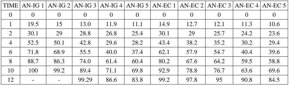
In vitro studies of optimized chitosan nano formulations were carried out for release pattern across cellophane membrane. The release patterns of AN-IG 1 , AN-IG 2, AN-IG 3,.AN-IG 4 and AN-IG 5 are 100.0 ,99.2 , 99.29 , 86.6 and 83.8 and The release patterns of AN-EC 1 , AN-EC 2, AN-EC 3,.AN-EC 4 and AN-EC 5 are 99.2 ,97.8 , 95 , 90.8 and 84.5 respectively respectively (Table -4)(graph-2&3). It was quite evident from the release profiles that show required drug release through nanoparticles for control relese of 10 hours in ionotropic gelation compared to emulsification crosslinking and AN-IG 1 was found to be good and best formulation and further research studies.
Study of Release Kinetics:
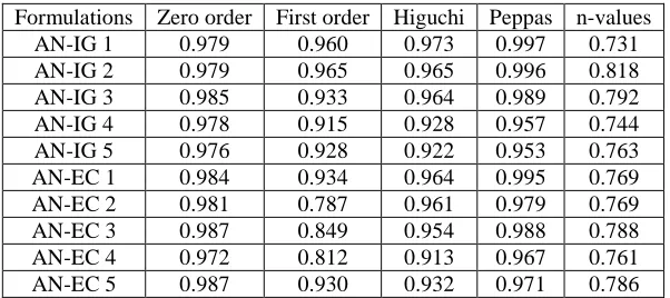
The results of In vitro release profile obtained from all the formulations were plotted to know the mechanism of drug release (graph-4, 5). The data were treated according to zero order release, first order release, higuchi’s and korsemeye peppa’s model equation. The release rate kinetics data for all the other equation can be seen in table - 5.
The equation, which is used to describe drug release mechanism, is:
CONCLUSION:
In this research work anastrazole nanoparticles are prepared by ionotropic gelation and emulsification crosslinking. The nanoparticles prepared by the Ionotropic gelation technique using chitosan as polymer and trypolyphosphate as cross linking agent were found to be nano range with acceptable physical and chemical natures. Based on percentage yield, drug entrapment
efficiency, particle size morphology, zeta potential and in
vitrorelease, formulation AN-IG 1(with polymer
crosslinking agent ratio 1:5) was found to be the optimal formulation to sustain the drug release effectively in 10hr it is quite evident for further reaserch (transdermal patch) to release drug in 24hrs.
Table 1: Formulation ratios of Drug-chitosan-Sodium tripolyphosphate
Batch code Amount of drug (mg/ml) Conc.of chitosan(mg/ml) Conc of cross linliking agent ( 0.25 % W/V)
AN-IG 1 10 1 5ml
AN-IG 2 10 2 5ml
AN-IG 3 10 3 5ml
AN-IG 4 10 4 5 ml
AN-IG 5 10 5 5ml
Table 2: Formulation ratios of Drug-chitosan-Sodium tripolyphosphate
Batch code Amount of drug (mg/ml) Conc.of chitosan (mg/ml) Volume of arachis oil (ml) Conc of cross linliking agent (0.25 % W/V)
AN-EC 1 10 1 15 ml 5ml
AN-EC 2 10 2 15ml 5ml
AN-EC 3 10 3 15ml 5ml
AN-EC 4 10 4 15ml 5 ml
AN-EC 5 10 5 15ml 5ml
Table 3: Particle Size, Zeta Potential, Entrapment Efficiency of Chitosan Nanoparticles prepared by ionotropic gelation
Formulations Average Particles size (nm) Zeta potential mV Entrapment efficiency %
AN-IG1 200±15.6 -16.3±2.1 43±2.90
AN-IG 2 365±17.9 -28.2±4.3 48±3.12
AN-IG 3 420±23.6 -10.38±3.6 69±1.63
AN-IG 4 428±20.4 -24.31±3.2 73±3.45
AN-IG 5 483±23.5 -21.38±5.2 79±3.53
AN-EC 1 350±23.6 10±2.1 53±1.60
AN-EC 2 365±17.9 25.2±4.3 58±2.32
AN-EC 3 423±15.6 3.38±3.6 69±3.63
AN-EC 4 465±23.5 14.31±3.2 73±5.45
AN-EC 5 468±20.4 21.38±5.2 83±2.43
Table 4: In vitro drug relase of anastrazole loaded chitosan nanoparticles
TIME AN-IG 1 AN-IG 2 AN-IG 3 AN-IG 4 AN-IG 5 AN-EC 1 AN-EC 2 AN-EC 3 AN-EC 4 AN-EC 5
0 0 0 0 0 0 0 0 0 0 0
1 19.5 15 13.0 11.9 11.1 14.9 12.7 12.1 11.3 10.6
2 30.1 29 28.8 26.8 25.4 30.1 29 25.7 24.2 23.6
4 52.5 50.1 42.8 29.6 28.2 43.4 38.2 35.2 30.2 29.4
6 71.8 68.9 55.5 40.0 37.4 62.1 57.9 54.7 40.4 39.6
8 88.7 86.3 74.0 61.4 60.4 80.2 67.6 64.2 59.5 58.8
10 100 99.2 89.4 71.1 69.8 92.9 78.8 76.7 63.6 69.6
Table 4: Drug Release kinetics of, zero, First order , Higuchi & Peppas plot, n values of selected formulations
Formulations Zero order First order Higuchi Peppas n-values AN-IG 1 0.979 0.960 0.973 0.997 0.731 AN-IG 2 0.979 0.965 0.965 0.996 0.818 AN-IG 3 0.985 0.933 0.964 0.989 0.792 AN-IG 4 0.978 0.915 0.928 0.957 0.744 AN-IG 5 0.976 0.928 0.922 0.953 0.763 AN-EC 1 0.984 0.934 0.964 0.995 0.769 AN-EC 2 0.981 0.787 0.961 0.979 0.769 AN-EC 3 0.987 0.849 0.954 0.988 0.788 AN-EC 4 0.972 0.812 0.913 0.967 0.761 AN-EC 5 0.987 0.930 0.932 0.971 0.786
Figure 1: FTIR spectra for anastrazole pure drug
Figure 2: FTIR spectra for combination of anastrazole with polymers
Graph 2: Drug Release Profiles of Nanoparticles prepared by ionotropic gelation
Graph 3: Drug Release Profiles of Nanoparticles prepared by Emulsion crosslinking
Graph 4: Higuchi plot of Nanopartricles prepared by ionotropic gelation
REFERENCES:
1. Nanoparticles – A Review,VJ Mohanraj and Y Chen, Trop J Pharm Res, June 2006; 5 (1),561-573.
2. Allen TM, Cullis PR (2004) Drug delivery systems: entering the mainstream. Science 303:1818–1822
3. Wagner V, Dullaart A, Bock A-K, Zweck A (2006) The emerging nanomedicine landscape. Nat Biotechnol 24(10):1211–1217
4. Farokhzad OC, Langer R (2009) Impact of nanotechnology on drug delivery. ACS Nano 3(1):16–20
5. Petros RA, DeSimone JM (2010) Strategies in the design of nanoparticles for therapeutic applications. Nat Rev Drug Discov 9(8):615–627 6. Shi J, Votruba AR, Farokhzad OC, Langer R
(2010) Nanotechnology in drug delivery and tissue engineering: from discovery to applications. Nano Lett 10(9):3223–3230
7. Davis ME, Chen ZG, Shin DM. Nanoparticle therapeutics: an emerging treatment modality for cancer. Nat. Rev. Drug Discov. 7(9), 771–782 (2008).
8. Zhang L, Gu FX, Chan JM, Wang AZ, Langer RS, Farokhzad OC. Nanoparticles in medicine: therapeutic applications and developments. Clin. Pharmacol. Ther. 83(5), 761–769 (2008).
9. Peer D, Karp JM, Hong S, Farokhzad OC, Margalit R, Langer R. Nanocarriers as an emerging platform for cancer therapy. Nat. Nanotechnol. 2(12), 751–760 (2007).
10. Geisler, J., King, N., Dowsett, M., Ottestad, L., Lundgren, S., Walton, P., Kormeset, P.O., Lonning, P.E., Influence of anastrozole
(Arimidex), a selective, non-steroidal aromatase inhibitor, on in vivo aromatisation and plasma oestrogen levels in postmenopausal women with breast cancer. Br. J. Cancer 74,1286–1291. 11. Charles Lovelyn, Anthony A. Attama Current
State of Nanoemulsions in Drug Delivery, Journal of Biomaterials and Nanobiotechnology, 2011, 2, 626-639.
12. Yateendra et al., design and development of riluzole loaded chitosan nanoparticles by emulsification crosslinking, Int J Pharm Pharm Sci, Vol 4, Issue 4, 244-248.
13. Dhanya KP, santhi K, Dhanaraj SA and Sajeeth CI: Formulation and evaluation of chitosan nanospheres as a carrier for the targeted delivery of lamivudine to the brain. International Journal of Comprehensive Pharmacy 2011; 5 (13).
14. Simar PK, Rekha Rao, Afzal Hussain, Sarita Khatkar: Preparation and Characterization of rivastigmine Loaded Chitosan Nanoparticles. J. Pharm Sci. & Res 2011; 3(5): 1227- 1232.
15. Calvo P, Remunan-Lopez C, Vila-Jato JL, Alonso MJ: Novel hydrophilic Chitosan–Polyethylene Oxide nanoparticles as protein carriers. J. Appl. Polym. Sci 1997; 63: 125–132.
16. Sangeetha S, Harish G, Malay Samanta K: Chitosan-Based nanospheres as Drug Delivery Systems for Cytarabine. International Journal of pharma and Bio Sciences 2010; 1(2).
17. Kumar et al.,Development and validation of spectrophotometric method for estimation of anastrozole bulk and pharmaceutical dosage formulation. International Journal of Advances in Pharmaceutical Sciences 1 (2010) 329-333.
How to cite this article:
A. Prameela Rani*1, P. Sivannarayana2, K. Abbulu3, V. Saikishore4: Design and Optimisation of Anastrazole loaded Chitosan Nanoparticles by Emulsification Crosslinking and Ionotropic Glation methods, 5(2): 1719 -1725. (2014)
![Figure 1: ylmethyl)-1, 3-phenylene] bis (2-methyl propiononitrile)) Anastrazole (2, 2_-[5-(1H-1, 2, 4-triazol-l-](https://thumb-us.123doks.com/thumbv2/123dok_us/8751410.1748673/2.595.110.228.50.120/figure-ylmethyl-phenylene-bis-methyl-propiononitrile-anastrazole-triazol.webp)