e- ISSN 0975 – 9328 Print ISSN 2229 – 7472
International Journal of Phytopharmacology
Journal homepage: www.onlineijp.com
ANTIFUNGAL ACTIVITY OF ETHANOLIC
OCIMUM SANCTUM
EXTRACTS AGAINST FUNGAL PATHOGEN ISOLATED FROM
SINUSITIS SUBJECTS
Harison Masih*, Surendra Pratap Yadav, Jyotsna kerin Peter, Swati Goswami
Department of Microbiology & Fermentation Technology,
Sam Higginbottom Institute of Agriculture Technology & Sciences, Allahabad, U.P, India.
ABSTRACT
Ocimum sanctum ethanol leaf extract was evaluated for its in vitro antifungal activity for isolates against Allergic fungal rhino sinusitis (AFS) positive subjects. Out of 25 clinical samples collected from different hospitals in Allahabad, seventeen subjects were diagnosed positive for noninvasive rhino sinusitis in the year 2003-2005. The fungal pathogens isolated were identified as Aspergillus niger (64%), Penicillium sp. (44%), Alternaria sp. (64%), Fusarium sp. (36%),
Exophiala sp. (40%). Ninety percent of A. niger infections were reported among 30-39 year age group old victims, male being infected the most was reported. Antimycotic activity of the extract was screened using agar well diffusion method with
O. sanctum ethanol leaf extract by taking concentration of 500mg/ml DMSO which had maximum zone of inhibition for
Fusarium sp. (19.33mm) and minimum for Alternaria sp. (8mm). These results support inhibition of AFS pathogens and thus
can be used for treatment of AFS, being economical, with no side effects and no pain.
Key words: Allergic/Eosinophilic fungal rhinosinusitis, Ocimum sanctum, Aspergillus, Penicillium, Alternaria, Fusarium, Exophiala.
INTRODUCTION
Fungi are ubiquitous, and our exposure to these organisms occurs on a daily basis with their common route of entry inside the body through sinonasal cavity. Four distinct histological categories of fungal sinusitis (FS) have been recognized: allergic, noninvasive fungal colonization (mycetoma or "fungus ball"); chronic invasive; and acute fulminant. Histological features, which are highly sensitive and specific for the diagnosis of allergic FS, includes: eosinophilic mucin with Charcot-Leyden crystals and collections of degenerating eosinophils. Allergic mucin is very characteristic but not pathognomonic for allergic fungal sinusitis(Mohammad, 2005). Allergic/Eosinophilic fungal rhinosinusitis (AFS) was first described in 1981 by Millar and for the past 25
Corresponding Author
Harison Masih
Email: [email protected]
years has been the subject of intense study and debate (Millar, 1981) AFS is a benign non invasive sinus disease, believed to be an allergic reaction to aerosolized environmental fungi. It is a form of chronic sinusitis characterized by nasal obstruction, sinus pain, rhinorrhoea, and frequent orbital symptoms (Mallick, 2008). AFS causing fungal pathogens includes
Aspergillus flavus, A. fumigatus, Fusarium sp. Rhizopus sp., Bipolaris sp. and Candida albicans (Tilak, 2012). Fungi commonly identified in electron microscopy were from the dematiaceous family and Aspergillus sp. Dematiaceous fungi include the genera Alternaria,
Bipolaris, Cladosporium, Culvularia, Drechslera and
Helminthosporium (Gupta, 2012). The Ayurvedic
Pharmacopoeia of India recommends the use of the leaves and seeds in rhinitis and influenza. Ursolic acid, isolated from leaves, exhibited significant protection of mast cell membrane by preventing granulation and decreased histamine release.
Ocimum sanctum as sacred armoury used to cure upper respiratory tract infections and bronchitis since long the mankind evolution. Ocimum sanctum leaf is carminative, stomachic, antispasmodic, antiasthmatic, antirheumatic, expectorant, stimulant, hepatoprotective, antiperiodic, antipyretic and diaphoretic (Khare, 2007). In vitro
antifungal activity of its leaves also reported against
Aspergillus niger, A. fumigates (Bansod and Rai, 2008).
Ocimum sanctum against some well known fungal
etiological agents as Candida albicans (Ahmad and Beg, 2001; Geeta et al., 2001; Kaya et al., 2008; Sharma, 2010), Fusarium solani sp. Melongenae (Joseph et al., 2008), Aspergillus flavus and aflatoxin B1 (AFB1) production (Reddy et al., 2009), Aspergillus niger
(Tewari and Robert, 2009; Mohan et al., 2011),
Aspergillus repens, Curvularia lunata and Fusarium
moniliforme were also reported(Amadi et al., 2010).
Being easily accessible, economical with diverse medicinal magic this plant was taken under to study its antifungal prospects to cure AFS. Our study is focused on identification of human fungal isolates from sinusitis patients and in vivo antifungal activity of O. sanctum
ethanol leaf extract.
METHODS AND MATERIALS Sample Collection and Processing
Nasal samples were collected from AFS positive subjects from Swaroop Rani Nehru Medical Hospital and Jeevan Jyoti Hospital, Allahabad, U.P. Awareness that fungi are colonizing the mucus prompted development of a simple noninvasive procedure to obtain maximum mucus for testing. Two puffs of phenylephrine hydrochloride (1%) were sprayed into each nostril to produce vasoconstriction which increased nasal lumen and consequently the yield from nasal lavage. After the injection of saline, the patients were asked to exhale forcefully through the nose and the return was collected in a sterile pan. The collected fluid was placed into centrifuge tubes and processed maintaining the sterile condition.
The specimen processed following the protocol of Ponikau et al., (1999). Briefly the collected specimen was suspended into an equal volume of diluted dithiothreitol and vortexed for 30 seconds. The mixture was allowed to stand at room temperature for 15 minutes followed by centrifugation at 3000g for 10 minutes. Retained sediment is vortexed for 30 seconds.
Isolation and Identification of Pathogenic Fungal Sinusitisfrom Clinical Samples
One-half ml of the prepared sediment was inoculated on the Sabouraud’s dextrose agar (SDA) media and incubated at 25oC for 15 days. Obtained fungal cultures were examined for their colony morphology considering color and texture. Microscopic characteristics
observed using lacto phenol cotton blue staining technique for their mycelia and spore arrangement. Sugar fermentation test was performed using glucose and sucrose followed by acid and gas production.
Extract Preparation
Ocimum sanctum ethanol leaf extract (OSELE)
was prepared using 10g of dried leaves soaked in 100 ml ethanol at 200 rpm for 24 hrs. at ambient temperature concentrated using vacuum rotary evaporator. Stock solutions of crude OSELE was prepared by diluting 0.2 g dried extracts with 1 ml of 10% dimethyl sulphoxide (DMSO) solution (Rathod et al., 2012).
Antimicrobial Susceptibility Test by Using Well Diffusion Method
Antifungal activity of OSELE was performed using agar well diffusion assay on pre-prepared SDA plates (Perez et al., 1990). Prepared wells (10 mm diameter) were filled with OSELE (0.1 ml, of 500mg/ml DMSO). Incubation period of 24-48 hours at 280C was maintained for observation of antifungal activity of plant extract. The antifungal activity was evaluated by measuring zones of inhibition (in mm) of fungal growth surrounding the plant extracts. The experiment was carried out in triplicates.
RESULT AND DISCUSSION
Twenty five clinical samples were examined for the incidence of the pathogenic fungal isolates collected from different hospitals of Allahabad city. Out of these, five positive fungal pathogens were isolated and identified as Aspergillus niger, Penicillium sp., Alternaria
sp., Fusarium sp., Exophiala sp. The maximum
incidences of AFS was foundin male patients with 20-29 years of age. Similarly seventeen patients were identified between 2003 and 2005 that were diagnosed with noninvasive fungal rhino sinusitis. Fourteen patients (82%) had allergic fungal mucin with polyps, while two patients had positive fungal cultures and chronic rhino sinusitis without polyps. One patient (5.8%) had a positive fungal stain, negative culture and evidence of chronic rhino sinusitis. Eleven patients (65%) had a positive fungal culture, and 10 patients (59%) had a positive fungal stain. Eight out of the ten (80%) patients with a positive fungal stain subsequently had positive fungal growth on culture. Three of 11 (27%) patients with a positive fungal culture had a negative fungal stain. Identified organisms included Aspergillus, Alternaria,
Bipolaris, Curvularia and Cladosporium. Fourteen
neutrophils. The most characteristic feature was the presence of allergic fungal mucin, which was grossly thick and dark with a consistency of peanut butter or wet clay. This fungal mucin consisted of inspissated basophilic, purple-colored mucous with large numbers of degenerated eosinophils. Fungal stains (GMS and PAS) demonstrated that the fungal hyphae were sparse and not always found. There were never fungal hyphae within the mucosal tissue (Mowry et al., 2008).
A total of 55 sputum samples from 41 patients were analyzed. Each sample was obtained on a separate visit and was independently analyzed. A total of 10 culture plates were inoculated with each sample. Yeasts were isolated from 1 plate from all but one of the samples (98%) that were inoculated on 1 plate, and 29% of samples (16 of 55) yielded A. fumigates . There were no differences between recovery rates of yeasts from homogenized sputum compared to sputum plug detection
of A. fumigatus was highly dependent on quantity of
sputum inoculated onto the culture plate. Of the 16 samples that were A. fumigatus positive on 1 plate, growth of the fungus was not detected with diluted-homogenized sputum samples. Not considering
differences due to the media, 19% and 44% of A.
fumigatus positive samples were detected using 10μl and
100μl of homogenized sputum, respectively, and 94% when a neat sputum plug was inoculated. One sample was culture negative with a sputum plug, but culture positive with 100μl homogenized sputum. In the majority of cases (11 of 16) filamentous fungal growth was observed after two days incubation at 37°C, with fungi detected in three samples after 4 – 6 days of incubation and growth was not detected with two until the 7th day (Pashley et al., 2012).
In vitro antifungal activity was performed using agar well diffusion method which gave excellent result in form of maximum zone of inhibition for Fusarium sp. followed by Penicillium sp., Aspergillus niger, Exophilala and Alternaria. Sharma et al. (2012), reported that the
Ocimum tenuiflorum was found to be effective against
pathogenic fungal isolates such as Aspergillus niger
(7mm) and Candida albicans (0.19mm) respectively. Kumar et al. 2013, reported that the Ocimum sanctum
extractwas effective against Fusarium sp. (8mm) showed maximum inhibition which conformed to the present results.
Figure 1. Incidence of fungal sinusitis subjects with respect to sex
Figure 2. Incidence of fungal sinusitis subjects with respect to age
Figure 4. A. Alternaria sp.,B. Aspergillus niger, C. Penicillium sp. ,D. Fusarium sp., E. Exophiala sp.
Figure 5. Antimicrobial activity of Ocimum sanctum against Human fungal isolates
Table 1. Human fungal isolates from samples collected from sinusitis subjects from different hospitals S.
No Subjects Sex Age
Aspergillus niger
Penicillium sp.
Exophiala Sp.
AlternariaSp
. Fusarium Sp.
1 1 M 12 Y N Y N Y
2 2 M 9 Y N N Y N
3 3 M 15 N Y N Y Y
4 4 M 20 N Y Y Y N
5 5 M 22 Y N N Y N
6 6 F 35 N Y Y N N
7 7 F 22 N N N Y Y
8 8 M 32 Y Y Y N N
9 9 M 20 Y N N Y N
10 10 F 11 Y N N Y Y
11 11 F 23 N Y N N N
12 12 F 21 Y N Y Y Y
13 13 F 15 Y Y N Y N
14 14 M 19 N Y N Y N
15 15 M 15 Y N N N Y
16 16 M 35 Y N Y N N
18 18 M 24 Y Y N Y N
19 19 F 21 Y N Y Y N
20 20 F 22 Y N N N Y
21 21 M 20 Y Y N Y N
22 22 M 21 N Y Y Y N
23 23 M 36 Y N N N Y
24 24 F 34 Y N Y N N
25 25 M 45 N Y N Y Y
Total no. of fungal isolates 16 11 10 16 9
Table 2. Incidence of fungal sinusitis subjects with respect to sex
Sex No. of sample Aspergillus niger Penicillium sp. Exophiala sp. Alternaria sp. Fusarium sp.
Male 15 10(66.66% ) 8 (53.33%) 5 (33.33%) 10(66.66% ) 5(33.33%)
Female 10 6(60%) 3(30%) 5(50%) 6(60%) 4(40%)
Total 25 16(64%) 11(44%) 10(40%) 16(64%) 9(36%) Due to organism Fcal= 7.31<Ftab=9.60,NS= Non Significant
Due to sex Fcal=29.004>Ftab=12.2; CD=6.1198; S=Significant
Table 3. Incidence of fungal sinusitis subjects with respect to age
Sex No. of sample Aspergillus niger Penicillium sp. Exophiala sp. Alternaria sp. Fusarium sp.
0-9 1 1(50%) N.A. N.A. 1(50%) N.A.
10-19 6 4(66.6%) 3(50%) 1(16.66%) 4(66.66%) 4(66.66%)
20-29 11 7(63.63%) 4(36.36%) 4(36.36%) 9(81.81%) 3(27.27%)
30-39 6 4(66.6%) 3(50%) 5(83.33%) 1(16.66%) 1(16.66%)
40-49 1 N.A. 1(33.33%) N.A. 1(33.33%) 1(33.33%)
Total 25 16(64%) 11(44%) 10(40%) 16(64%) 9(36%) Due to organism Fcal=0.947<Ftab=3.01; NS=Non Significant
Due to age Fcal= 8.9140>Ftab=3.01, C.D= 2.0699; S= Significant
N.A.= Not available
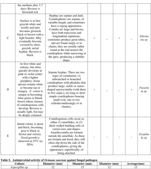
Table 4. Identification of fungal isolates from clinical samples
Fungal
Culture Morphological Identification
Biochemical identification w.r.t. Sugar fermentation
Culture Identifi
ed
Glucose Sucrose Acid
producti on
Gas produc
tion
Acid produc tion
Gas produc
tion Colony Morphology Microscopic
Characteristics
A
Surface is black with white border; thallus deep, Hypha erect
white to cream, cluster of black conidia. Reverse
white to cream.
Septate hyphae. Conidiophores long, smooth, may be brownish
near top. Phialides radiate around entire vesicle, biseriate,
with the metulae twice as long as the phialides. Conidia are
round and dark`
+ _ + _
Aspergi llus niger
B
Thermally dimorphic at 25-30oC on SDA, colony flat, powdery to velvety, tan, later becoming reddish yellow or white edge;
bluish gray-green in the centre. A
deep-reddish soluble pigment diffuses into
Smooth conidiophores with four or five terminal metulae, each
metula bearing four to six phialides. Conidia are smooth or
slightly rough and round to oval and form chains ; short, narrow extensions connect the conidia
_ _ _ _
the medium after 3-7 days; Reverse is
brownish red.
C
Surface is at first grayish white and woolly and later becomes greenish black or brown with a
light boarder. May eventually become covered by short,
grayish, aerial hyphae. Reverse is
black.
Hyphae are septate and dark. Conidiophores are septate, of variable length, and sometimes
have a zigzag appearance. Conidia are large and brown,
have both transverse and longitudinal septations, sometimes produce germ tubes,
and are found singly or in chains; they are usually rather
round at the end nearest the conidiophore while narrowing at
the apex, producing a clublike shape.
+ _ + _ Alterna
ria sp.
D
At first white and cottony, but often quickly develops at pink or violet center
with a lighter periphery. Some species remain white
or become tan or orangey . F. solani is
unique in becoming blue-green or bluish brown where clusters of conidiogenous cells
develop. Reverse is usually light, but may
be deeply coloured.
Septate hyphae. There are two types of conidiation: (i) unbranched or branched conidiophore with phialides that
produce large, sickle or canoe-shaped macroconidia (with three
to five septa.); (ii) long or short simple conidiophores bearing
small oval, one or two cellednconidiasingly or in
clusters.
+ _ _ + Fusariu
m sp.
E
Initial colony is moist and black, becoming
gray to black or brown and velvety.
Good growth is observed at 25oC on
SDA.
Conidiogenous cells occur as either (1) annellides, or (2) thick-walled budding cells of
varied sizes and shapes. Annelloconidia are formed outside the annellide. As these are formed and freed, they will often slip down the side of the
conidiophores, giving the appearance, superficially, of
being attached.
_ _ _ _ Exophia
la sp.
Table 5. Antimicrobial activity of Ocimum sanctum against fungal pathogen
Culture Diameter (mm) Diameter (mm) Diameter (mm) Average(mm)
Aspergillus sp. 12 15 12 13
Penicillium sp. 20 18 18 18.66
Alternaria Sp. 8 8 8 8
Exophiala Sp. 13 12 13 12.66
Fusarium Sp. 20 18 20 19.33
*included well size of 5mm diameter, due to oraganisium: F(cal) 29.351>F(tab)4.5; CD=6.605; S=Significant.
CONCLUSION
Present study revealed that the potent bioactive component from O. sancutum can be used to cure AFS thus providing relief to subjects without any surgical pain. It is economical and easily available and could be potent pharmaceutical drug.
ACKNOWLEDGEMENTS
The authors wish to thank Honorable Vice-Chancellor, SHIATS, and HOD, Department of Microbiology andFermentation Technology and thanks to Swaroop Rani Nehru Medical Hospital and Jeevan Jyoti Hospital, Allahabad for their immense help in sample collection from the subjects.
REFERENCES
Ahmad I and Beg AZ. Antimicrobial and phytochemical studies on 45 Indian medicinal plants against multi-drug resistant human pathogens. Journal of Ethno pharmacology, 74, 2001, 113–123.
Amadi JE, Salami SO, Eze CS. Antifungal properties and phytochemical screening of extracts of African Basil (Ocimum
gratissimum L.). Agricultureand BiologyJournal of North America, 1(2), 2010, 163-166.
Bansod S and Ray M. Antifungal Activity if Essential Oils from Indian Medicinal Plants against Human pathogenic Aspergillus fumigates and A. niger. World Journal of Medicinal Sciences, 3 (2), 2008, 81-88.
Geeta VDM, Kedlaya R, Deepa S, Ballal M. Activity of Ocimum sanctum (the traditional Indian medicinal plant) against the enteric pathogens. Indian Journal of Medical Sciences, 55(8), 2001, 434-438.
Gupta AK, Shah N, Kameswaran M, Rai D, Janakiram TM, Chopra H, Nayar R ,Soni A, Mohindroo NK, Rao CMS ,Bansal A , Meghnadh KR , Vaid N ,Patel HM, Sood S, Kanojia S, Charaya K, Pandhi SC, Mann SBS .Allergic Fungal Rhinosinusitis. Clinical RhinologyAn International Journal, 5(2) 2012, 72-86.
Joseph B, Dar MA, Kumar V. Bioefficacy of Plant Extracts to Control Fusarium solani F. Sp. Melongenae Incitant of Brinjal Wilt. Global Journal of Biotechnology & Biochemistry, 3(2), 2008, 56-59.
Kaya I, Yiğit N, Benli M. Antimicrobial Activity of Various Extracts of Ocimum basilicum L. and Observation of the Inhibition Effect on Bacterial Cells by Use of Scanning Electron Microscopy. African Journal of Traditional,
Complementary and Alternative medicines, 5(4), 2008, 363-369.
Khare CP. Indian Medicinal Plants An Illustrated Dictionary, Springer Science, BusinessMedia, LLC. 2007, 445-446. Kumar A, Rahal A , Chakraborty S, Tiwari R, Latheef SK and Dhama K. Ocimum sanctum (Tulsi) a miracle herb and
boon to medical Science. A Review International Journal of Agronomy and Plant Production, 4(7), 2013, 1580-1589.
Mallick S et al. Allergic fungal sinusitis. Kathmandu University Medical Journal, 6(2), 2008, 235-238. Millar JW, Johnston A and Lamb D. Allergic Aspergillosis of the maxillary sinuses. Thorax, 36, 1981, 710.
Mohammad JA, Azarpira N, Pourjafar M and Khademi B. Allergic Fungal Sinusitis Presenting as a Paranasal Sinus Tumor.
Iranian Journal of Allergy, Asthma and Immunology, 4(4), 2005, 193-195.
Mohan L, Amberkar MV and Kumari M. Ocimum sanctum (Tulsi). International Journal of Pharmaceutical Sciences
Review and Research, 7(1), 2011, 951-53.
Mowry SE, Bhuta S and Wang MB. Pathologic Features of Non-Invasive Fungal Rhinosinusitis. The Open
Otorhinolaryngology Journal, 2, 2008, 7-12.
Pashley CH, Fairs A, Morley JP, Tailor S, Agbetile J, Bafadhel M, Brightling CE and Wardlaw AJ. Routine processing procedures for isolating filamentous fungi from respiratory sputum samples may underestimate fungal prevalence.
Medical Mycology, 50, 2012, 433–438.
Perez C, Paul, M and Bezique P. An Antibiotic assay by the agar well diffusion method. Alta Biomedical Group Experiences, 1990, 15, 113.
Ponikau JU, Sherris DA, Kern, EB, Homburger HA, Frigas E, Gaffey TA and Roberts GD. The Diagnosis and Incidence of Allergic Fungal Sinusitis. Mayo Clinic Proceedings, 74, 1999, 877-884.
Rathod GP, Kotecha BM, Sharma R, Amin H and Praiapati PK. In vitro Antibacterial study of two commonly used medicinal plants in Ayurveda: Neem (Azadirachta indica L.) and Tulsi (Ocimum sanctum L.). International Journal of
Pharmaceutical & Biological Archieves, 3(3), 2012, 582-586
Reddy KRN, Reddy CS and Muralidharan K. Potential of botanicals and biocontrol agents on growth and aflatoxin production by Aspergillus flavus infecting rice grains. Food Control, 20, 2009, 173–178.
Sharma A, Meena A and Meena R. Antimicrobial activity of plant extracts of Ocimum tenuiflorum. International Journal of
Pharmacology Technology Research, 4(1), 2012, 176-180.
Tewari BB and Robert S.Studies on the Interaction of Natural Antifungals with Metal Ferrocyanides and Their Medicinal Applications. Natural Science, 7(3), 2009, 1-7.