Original Article
Development of a Multiplex RT-PCR Assay for Detection of the
Causal Agents of Citrus Tristeza and Cachexia Diseases with
Coamplification of Plant mRNA as an Internal Control
Naderpour M1*, Sadeghi L1, Nouri Z1,2, Kavand A1
1Seed and Plant Certification Research Institute, Karaj, Iran.
2. Department of Biotechnology, Faculty of Agriculture, Science and Research Unit, Islamic Azad University, Tehran, Iran.
Abstract
Background and Aims: Plant certification programs need reliable, fast, cheap and sensitive methods for detection of systemic pathogens with special interest in virus and viroid detection. Reverse transcriptase-polymerase chain reaction (RT-PCR) has been documented as an alternative assay for certification of plant propagating materials. The main object of the present study was the optimization of a multiplex RT-PCR assay for simultaneous detection of Citrus tristeza virus (CTV) and Hop stunt viroid (HSVd), the casual agents of citrus tristeza and cachexia, together with the plant mRNA as an internal control for citrus certification in the country.
Materials and Methods: Total RNA was extracted from healthy; CTV- and HSVd- infected citrus tissues and subjected to cDNA synthesis by M-MuLV H- reverse transcriptase, followed by optimization of simplex, diplex and multiplex RT-PCR (s-, d-, and mRT-PCR). Amplified fragments were further sequenced for evaluation of the accuracy of the assays. Results: CTV, HSVd and the plant internal control (Nad 5 gene) were successfully amplified in all assays and the sequence information revealed the accuracy of all assays in citrus certification programs.
Conclusion: Results from the developed s-, d-, and mRT-PCR assays revealed these detection methods as excellent candidates for citrus certification.
Keywords: Citrus Tristeza Disease; Multiplex RT-PCR Assay; Cachexia Disease
Introduction
itrus (Citrus spp.) is an economically important crop in Iran with an estimated cultivated area of 291 thousands hectare and production of 4.5 million tons annually. Citrus is mainly cultivated in northern and southern parts of Iran. The distribution of Plant pathogens, limiting citrus cultivation in different parts of
the country, mainly viroids and viruses belonging to different taxonomic groups have been the subjects of many researches.
Viroids are small infectious agents of single stranded, circular, uncapsidated RNA of about 250-400 nucleotides and have highly self-complementary sequences (5, 7). Viroids are classified into two families Avsunviroidae and Pospoviroidae based on their secondary structures and replication sites within the host cells. Citrus viroids belong to the family Pospoviroidae and classified into six groups Citrus bent leaf viroid (CVd-I), Citrus exocortis viroid (CEVd) and Hop stunt viroid
C
*Corresponding author: Masoud Naderpour, Ph.D., Seed and Plant Certification Research Institute, P. O. Box 31535-1516, Karaj, Iran.
Email: [email protected]
(HSVd), both belong to CVd-II, Citrus dwarfing viroid (CVd-III), Citrus bark cracking viroid (IV), V and CVd-OS, on the basis of their biological and physical properties. Many citrus diseases including wood pitting, gum pucket, gummy pitting, yellow corcky vein and gummy bark are associated with viroids (6, 16, 17, 19), but two of them, exocortis and cachexia (xyloprosis) are economically important diseases (9).
As a member of Closterovirus genus, Citrus tristeza virus (CTV) is one of the main devastating viruses limiting citrus production all over the world. The virus particles are monopartite and filamentous and the genome is single stranded RNA of about 19.3 kb.
Different certification methods have been developed and applied in citrus certification programs worldwide. Biological indexing, the gold standard in certification programs, is a well known way of classical indexing of health status of plant propagating materials. The program has many disadvantages, such as the expression of symptoms requires months or a year, even more, relating to specific plant-virus interactions and it can also be affected by environmental conditions. Moreover, biological indexing requires expert personnel for symptom diagnosis and all these make it an expensive process.
Nucleic acid hybridization systems and sequential polyacrylamide gel electrophoresis (sPAGE) are other methods for citrus certification programs (4, 12, 14, 15), but their efficiency relies mainly on virus and viroid multiplication in plant tissues, which rarely happens in screen house or green house conditions.
Reverse transcriptase-polymerase chain reaction (RT-PCR) technique is a quick, simple and reliable method for screening a large number of propagating material for the presence of viruses and viroids in the virus-free nuclear stocks and the virus-tested propagating materials, compared to the conventional bioassays, hybridization and sPAGE methods (1, 8, 9, 10, 18)
Due to the lack of official certification programs in citrus industry in the country, this
research was carried out in the Seed and Plant Certification Research Institute (SPCRI) with the objectives of development of simplex (sRT-PCR) and subsequently, diplex and multiplex RT-PCR (dRT-PCR and mRT-PCR) systems for virus-tested certification of citrus propagating materials and to include the coamplification of plant mRNA in all systems as a reliable candidate for analyzing the RT-PCR accuracy.
Methods
Source of plant material
HSVd-infected plant tissues were received from Dr. Rahimian, H., Sari University, Iran. CTV- infected plant tissues were collected from symptomatic trees of different citrus orchards in Sari. Infectivity of plant tissues for CTV were analyzed by Double Antibody Sandwich -Enzyme linked Immunosorbent Assay (DAS-ELISA) using commercial antibodies received from Bioreba, Switzerland, according to the manufacturer's instructions. The CTV-positive samples were used for RNA extraction and subsequent RT-PCR assays. Total RNA extraction
Total RNA was extracted from 100 or 250 mg viroid- and CTV-infected plant tissues using RNeasy Plant mini kit (Qiagen, Germany) according to the manufacturer's instruction or by phenol-chloroform precipitation methods. Briefly, for the phenol-chloroform precipita-tion, 250 mg of infected plant tissues were grinded in liquid nitrogen, lysed in 500 µL of the extraction buffer [100 mM Tris HCl (pH 8.5), 100 mM NaCl, 50mM EDTA (pH 8.0), 2% SDS], followed by addition of 500 µL phenol-chloroform and centrifugation for 10 minutes. Five hundered microlitres of chloroform was added to the supernatant and centrifuged for 10 minutes. Fourty microlitres of NaAc (pH 5.6) and 1 ml of EtOH (96%) were added to 400 µL supernatant, incubated 30 minutes at -20°C and centrifuged 15 minutes at 4°C. The resultant pellet was washed out with EtOH and air dried. Then resuspended in 600 µL distilled water, was added to 300 µL LiCl (8M), incubated overnight at -20°C and centrifuged for 15
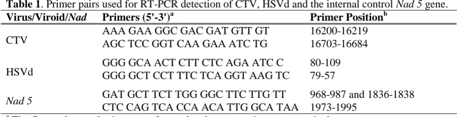
Table 1. Primer pairs used for RT-PCR detection of CTV, HSVd and the internal control Nad 5 gene.
Virus/Viroid/Nad Primers (5'-3')a Primer Positionb
CTV AAA GAA GGC GAC GAT GTT GT AGC TCC GGT CAA GAA ATC TG 16200-16219 16703-16684
HSVd GGG GCA ACT CTT CTC AGA ATC C GGG GCT CCT TTC TCA GGT AAG TC 80-109 79-57
Nad 5 GAT GCT TCT TGG GGC TTC TTG TT 968-987 and 1836-1838
CTC CAG TCA CCA ACA TTG GCA TAA 1973-1995
a
The first and second primers are forward and reverse primers, respectively.
b
Primer positions for HSVd, CTV and Nad 5 gene belong to the published complete genome sequence, coat protein (CP) and exones I and II on Nad 5 gene, respectively.
minutes at 4°C. The pellet was washed out with EtOH, dried and resuspended in 20 µL distilled water. All centrifugation was done at 13000 rpm.
Specific primers
Previously published primer pairs (Table 1) for PCR detection of CTV, HSVd and the internal control Nad 5 gene (Harper unpublished; 2, 11, 13) were used in this study. Concerning to the primer positions within the published genome sequences of Nad5 gene (D37958), CTV (U16304) and HSVd (GU825979), these primer pairs were expected to amplify 181, 504, and 296 nucleotides, respectively. First strand cDNA synthesis
cDNA was synthesized using the M-MuLV H -reverse transcriptase (Invitrogen, Carlsbad, California, USA) following the manufacturer's instructions. In brief, 1 µL of total RNA was mixed with 1 µL (conc. 10 pmol) of random hexamer primer (for CTV and Nad 5 gene reverse transcription) or strand-specific primer (for HSVd reverse transcription) and 6 µL of RNase-free water and kept 10 minutes at 70°C. The mixtures were then incubated on ice or room temperature. Twelve microlitres of reverse transcription mixture [4 µL of 5x RT buffer complete (250 mM TrisHCl, pH 8.3), 500 mM KCl, 15 mM MgCl2, 50 mM DTT), 1 µL of dNTP mix (10 mM of each dNTP), 20 units RNase inhibitor, 200 units of reverse transcriptase and 6.75 µL RNase-free water] were added to each reaction and incubated 90 minutes at 55°C followed by further incubation at 70°C for 10 minutes to inactivate the reverse transcriptase.
Simplex RT-PCR (sRT-PCR)
In conventional sRT-PCR, 1 µL of each specific forward and reverse primers (10 pmol) of CTV or HSVd (Table 1) were added separately to the PCR reaction mixture (25 µL) containing 2.5 µL PCR buffer (10X), 2 µL MgCl2 (25 mM), 2 µL dNTP (10mM), 2 µL cDNA (10% of the first strand reaction mix) and 0.2 µL (1 unit) Taq DNA polymerase (Fermentas, GmbH, Germany). In the case of Nad 5 gene, 0.5 µL of each primer was used. Polymerase chain reaction was done for 30 cycles using ThermoCycler Eppendorf (Germany) following an initial denaturation at 94°C for 3 minutes. The optimum temperatures for CTV, HSVd and mitochondrial Nad5 gene, as an internal control, were found at 58, 60 and 62 for 30s, respectively. Each cycle was consisted of a denaturation at 94°C for 35s, annealing temperatures as above, elongation at 72°C for 40s and a final extension at 72°C for 5 minutes for 35 cycles. The final product was analyzed by electrophoresis on 1.5% agarose gel in TAE buffer (0.04 M Tris-acetate, 1mM EDTA, pH 8.0) including GelRedTM and visualized by UV light.
Diplex RT-PCR (dRT-PCR)
Diplex RT-PCR assay was optimized separately for HSVD-Nad 5 and CTV-Nad 5 amplification. For the amplification of the first pair target, a PCR reaction mixture containing PCR master mixture (10 µL, Fermentas, GmbH, Germany), HSVd and Nad 5 primer pairs (1 µL and 0.5 µL of 10 pmol, respectively), cDNA (1.5 µL of each target) and H2O (4 µL) in a total volume of 20 µL was
prepared and subjected for target amplification in ThermoCycler Eppendorf (Germany). For CTV-Nad 5 amplification, a PCR reaction mixture (25 µL) containing 2.5 µL PCR buffer (10X), 2.2 µL MgCl2 (25 mM), 2 µL dNTP (10mM), 2 µL cDNA, primer pairs of CTV and Nad 5 (1 µL and 0.5 µL of 10 pmol, respectively) and 0.2 µL (1 unit) Taq DNA polymerase (Fermentas, GmbH, Germany) was used under the following PCR program: initial denaturation at 94°C for 3 min. Followed by 30 cycles of denaturation at 94°C for 35s, annealing at 60°C for 40s, elongation at 72°C for 40s and a final extension at 72°C for 5 minutes. The final products were analyzed by electrophoresis on 1.5% agarose gel buffered in TAE (0.04 M Tris-acetate, 1mM EDTA, pH 8.0) including GelRedTM and visualized by UV light.
Multiplex RT-PCR (mRT-PCR)
The mRT-PCR was optimized for the simultaneous detection of all three targets (CTV, HSVd and Nad 5) in one PCR reaction. The mRT-PCR reaction was performed in a total volume of 25 µL containing 2.5 µL of the
mixture of each of forward and reverse primers of HSVd, CTV and Nad 5, 2.5 µL PCR buffer (10X), 2 µL MgCl2 (25 mM), 2 µL dNTP (10mM), 1.5 µL of each of CTV and HSVd cDNA and 0.3 µL (1.5 unit) Taq DNA polymerase (Fermentas, GmbH, Germany). Amplification was performed in 35 cycles in the ThermoCycler Eppendorf (Germany). After an initial denaturation at 94°C for 3 min., each cycle was consisted of a denaturation at 94°C for 35s, annealing temperatures at 60°C for 40s, elongation at 72°C for 40s and a final extension at 72°C for 5 minutes. The PCR products were analyzed by electrophoresis on 1.5% agarose gel buffered in TAE (0.04 M Tris-acetate, 1mM EDTA, pH 8.0) including GelRedTM and visualized by UV light.
Sequencing
Amplicons of the expected size of HSVd and CTV were excised from the agarose gel 1.5%, purified using the Gel Extraction Kit (QIAquick, Qiagen, Germany) and sequenced directly in Sinaclon BioScience, Iran.
Fig. 1. Agarose gel electrophoresis of fragments of CTV CP, HSVd whole genome and Nad 5 exones I and II amplified in s-, d-, and mRT-PCR. Total RNA was extracted from the infected citrus tissues and subjected to reverse transcription using M-MuLV H- reverse transcriptase. Corresponding cDNAs were further amplified by sPCR (Lanes 1-3), dPCR (lanes 4-5) and mPCR (lane 6). Lanes 1 (181 bp), 2 (504 bp) and 3 (296 bp) indicate healthy, CTV- and HSVd- infected citrus plants, respectively.
Results
Optimization of sRT-PCR
Different concentrations of primer pairs and other PCR reagents were tested separately by sRT-PCR to establish their ability to optimum amplification of the expected genome sizes of HSVd, CTV and Nad 5 and their best amplification conditions. These experiments showed that the primers are able to detect these pathogens and the internal control (Fig. 1), whereas no amplifications happened in the negative controls (data are not shown). As Shown in figure 1, samples containing Nad 5, CTV and HSVd amplified segments of 181, 504 and 296 nucleotids, respectively. Direct sequencing of the amplified fragments of HSVd and CTV and their comparison with the reference sequences (GU825979 and U16304) in the data basses revealed 93% and 99% similarity confirming the accuracy of RT-PCRs.
Optimization of diplex and multiplex RT-PCR (dRT-RT-PCR and mRT-RT-PCR)
For d- and mRT-PCR amplification of targets (HSVd, CTV and Nad 5), different concentration of the PCR reagents and primer annealing temperatures were tested and the amplification specificity and reliability in both d- and mRT-PCR were compared with those amplified in sRT-PCR. In all s-, d-, and mRT-PCR, samples containing CTV and HSVd amplified segments of 504 and 296 nucleotids, respectively. Moreover an amplicon of 181 bp was produced in all samples carrying plant RNA (Nad 5) regardless of the presence of these pathogens (Fig. 1).
Discussion
Co-amplification of the main virus (CTV) and viroid (HSVd) pathogens of Citrus spp. in Iran together with the internal control (Nad 5 gene) by RT-PCR was performed in this study. The results indicated the amplification of all three targets in s-, d-, and mRT-PCR that were in agree with the results found in the other studies (Harper unpublished; 2, 11, 13). Moreover, direct sequencing of the amplified fragments from CTV and HSVd genomes revealed the
reliability and accuracy of the developed RT-PCR system.
Plant propagating certification schemes demand sensitive, reliable, cheap and fast detection methods. Due to the advantages of RT-PCR system (single/multiplex) concerning on time, cost, reliability and sensitivity, it has been documented as an alternative system for certification of plant propagating materials. Disadvantages of this system including the presence of inhibitors such as polyphenolics, polysaccharides and endogenous ribonucleases in the plant extracts (20) and inefficient synthesis of viroid genome (3) could be easily removed by proper RNA extraction methods and using proper sequence primers. Moreover, the accuracy and reliability of the extracted RNA and subsequently cDNA synthesis could be analyzed by the coamplification of the internal control (Nad 5 gene) in both d- and m-RT PCR.
Comparing to the classical biological indexing on indicator plants that needs completely controlled conditions for expression of disease symptoms and expertise in diagnosis of symptoms as well as long times (months to a year or even more) and large number of seedlings, the RT-PCR systems seems to be more applicable in certification schemes. Moreover, bioassays demand other detection methods such as sPAGE and/or hybridization techniques to reassess the results.
To our knowledge, developing of detection methods for citrus certification schemes and moreover, using a target-specific RT-PCR system and developing s-, d-, and mRT-PCR for individual and simultaneous detection of CTV, HSVd and an internal control are previously unreported events for citrus certification in Iran. Thus, specifically, the mRT-PCR system reported in this study should be considered as the first option for simultaneous detection of these pathogens in Iran.
Acknowledgements
We are grateful to Dr. Rahimian (Sari University, Iran) for HSVd infected tissues of Citrus spp.
References
1. Almeyda-Leon IH, Rocha-Pena MA, Iracheta-Cardenas MM, Orona-Castro F, Kahlke CJ. A simple method for the multiple detection of citrus viroids. Agrociencia 2007; 87-93.
2. Bagherian SAA, Izadpanah K. Two novel variants of hop stunt viroid associated with yellow corky vein disease of sweet orange and split bark disorder of sweet lime. Julius-Kuhn-Archives 2010; 427: 105-113.
3. Bernard L, Duran-Vila N. A novel approach for detection and characterization of citrus viroids. Mol. Cell. Probes 2006; 20: 105-113.
4. Cohen O, Batuman O, Stanbekova G, Sano T, Mawassi M, Bar-Joseph M. Construction of a multiprobe for the simultaneous detection of viroids infecting citrus trees. Virus Gen. 2006: 33: 287-292.
5. Ding B. The biology of viroid-host interactions. Ann. Rev. Phytopathol. 2009; 47: 105-131.
6. Fernandez-Valiela M, V.Fortugno C, Corizzi F. Incidence of bud-union crease in citrus trees grafted on trifoliate rootstock in the Delta del Parana and San Pedro areas of Argentina. P. 182-186. In: Pierce, W. C. (ed.). Proceedings of 3rd Conference Intl. Org. Citrus Virlo., IOCV, Riverside, Calif.
7. Flores R, Randles JW, Bar-Joseph M, Diener TO. A proposed scheme for viroid classification and nomenclature. Arch. Virol. 1998; 143: 623-629.
8. Garnsey SM, Zies DL, Irey M, Sieburth PJ, Semancik JS, Levy L, Hilf ME. Practical field detection of citrus viroids in florida by RT-PCR. The 15th IOCV conference 2002; 219-229.
9. Guardo M, Marletta T, Boninelli G, Leonardi A, Sorrentino G, Caruso A. Detection of viroids in forced citrus cuttings. J. Plant Pathol. 2005; 87: 141-144.
10.Ito T, Ikei H, Ozaki K. Simultaneous detection of six citrus viroids and Apple stem grooving virus
from citrus plants by multiplex reverse
transcription polymerase chain reaction. J. Virol. Methods 2002; 106: 235-239.
11.Madhurababu K, da Graca JV, Skaria M. Molecular detection and prevalence of citrus viroids in Texas. Hort. Sci. 2007; 42: 600-604. 12.Malfitano M, Barone M, Duran-Vila N, Alioto D. Indexing of viroids in citrus orchards of Campania, Sothern Italy. J. Plant Pathol. 2005; 87: 115-121.
13.Menzel W, Jelkmann W, Maiss E. Detection of four apple viruses by multiplex RT-PCR assays with coamplification of plant mRNA as internal control. J. Virol. Methods 2002; 99: 81-92.
14.Mohamed ME, Bani Hashemian SM, Dafalla G,
Bove JM, Duran-Vila N. Occurrence and identification of citrus viroids from sudan. J. Plant Pathol. 2009; 91: 185-190.
15.Murcia N, Serra P, Olmos A, Duran-Vila N. A novel hybridization approach for detection of citrus viroids. Mol. Cell. Probes 2009; 23: 95-102.
16.Reddy GS, Dakshinamurti V, Reddy VRK. Yellow corky vein: First report of a new graft-transmissible disorder of Sathgudi in Andhra Pradesh. Indian Phytopathol. 1974; 27: 82-84. 17.Roistacher CN. Graft-transmissible diseases of citrus: Handbook for detection and diagnosis, FAO, Rome, Italy, 1991.
18.Roy A, Fayad A, Barthe G, Brlansky RH. A multiplex polymerase chain reaction method for reliable, sensitive and simultaneous detection of multiple viruses in citrus trees. J. Virol. Methods 2005; 47-55.
19.Schwarz RE, McClean APD. 1969.
Gum-pocket, a new virus disease of Poncirus trifoliate. Plant Dis. Rep. 1969; 53: 336-339.
20.Singh RP, Nie X, Singh M, Coffin R, Duplessis P. Sodium sulphite inhibition of potato and cherry polyphenolics in nucleic acid extraction for virus detection by RT-PCR. J. Virol. Methods 2002; 99: 123-131.