Evaluation value of coronary CTA for coronary plaque features and its
correlation with platelet function and serum biochemical indexes
Jin-Xia Yang
Radiology Department, General Hospital of Liaoyang Petrochemical Company in Liaoyang City Liaoning Province, Shenyang City, Liaoning Province, 111003, China
Journal of Hainan Medical University
http://www.hnykdxxb.com
ARTICLE INFO ABSTRACT
Article history:
Received
Received in revised form Accepted
Available online
Keywords:
Coronary CT angiography Coronary plaque
Platelet function Inflammatory factors Chemokines
Corresponding author: Jin-Xia Yang, Radiology Department, General Hospital
of Liaoyang Petrochemical Company, Shenyang City, Liaoning Province, 111003, China.
Tel: 0419-53577777-62127; 13604196609
Fund Project: Natural Science Foundation of Science and Technology Committee of Liaoning Province No: 972254.
1. Introduction
Coronary CT angiography (CTA) is the important tool for diagnosis of coronary heart disease, and many current studies have shown that the CTA can not only judge the degree of coronary stenosis, but can also make quantitative evaluation on plaque features[1]. Coronary heart disease patients with different plaque features are with different treatment outcome, and studies have found that acute coronary syndrome (ACS) is mainly secondary to
unstable soft plaque rupture while acute cardiovascular events less occur in the calcified plaque. So early clarifying the plaque features is of important significance in the establishment of treatment and the judgment of treatment outcome[2,3]. In the study, CTA was applied in the judgment of plaque features in patients with coronary heart disease in our hospital, and the correlation between different plaque CT value and specific disease severity was further analyzed.
2. Materials and methods
2.1. General information
A total of 450 patients with coronary heart disease treated in
Objective: To analyze the evaluation value of coronary CT angiography for coronary plaque features and its correlation with platelet function and serum biochemical indexes. Methods:
A total of 450 patients with coronary heart disease were divided into calcified plaque group (CT value≥130HU) (n=117), soft plaque group (CT value≤60HU) (n=150) and mixed plaque group (CT value 60-130HU) (n=183) by coronary CT angiography (CTA), and 100 healthy subjects who received physical examination in our hospital during the same period were selected as control group. Differences in platelet function and serum biochemical indexes were compared among four groups of patients, and the judgment value of atheromatous plaque CT value from CTA for the severity of coronary heart disease was analyzed. Results: Platelet function parameters MPV, TEG-MA, P-selectin, PDGF-BB and vWF levels in peripheral blood of soft plaque group were higher than those of the other three groups; inflammatory factors CRP, IL-6, IL-12, IL-18 and IL-23 content in serum were higher than those of the other three groups; chemokines MCP-1, CXCL16, Fractalkine and RANTES content in serum were higher than those of the other three groups; adipocytokines Leptin and RBP4 content in serum were higher than those of the other three groups while SFRP5 content was lower than those of the other three groups. Atheromatous plaque CT value in patients with coronary heart disease was directly correlated with platelet function and the content of serum biochemical indexes.
our hospital between June 2013 and May 2016 were selected as observation group, they were all diagnosed by coronary angiography and echocardiography, and patients with congenital heart disease and malignant tumor were excluded. 100 healthy subjects who received physical examination in our hospital during the same period were selected as control group. Observation group included 250 male cases and 200 female cases that were 45-72 years old; control group included 54 male cases and 46 female cases that were 43-71 years old. The two groups of research subjects showed no statistically significant difference in the distribution of gender and age.
2.2. Coronary angiography and results interpretation
Patients' heart rate before examination was controlled to be < 65 times/min, and those with rapid heart rate could orally take metoprolol. 64-slice spiral CT was used for examination, contrast agent (80 mL) was intravenously injected at 4 mL/s for scanning, imaging scanning was started 45 s after intravenous injection, and the image data were sent to a computer workstation for the multiplanar reformation that was carried out by two radiologists with intermediate professional title above. Finally, the obtained plaque CT value was referred to secondarily divide the included patients into calcified plaque group (n=117): CT value ≥130 HU and
(189.63±24.78) HU in average; soft plaque group (n=150): CT value
acuities ≤60 HU and (31.83±4.05) HU in average; mixed plaque group (n=183): CT value 60-130 HU and (94.36±10.52) HU in average.
2.3. Platelet function
Soon after admission, 2 mL of fasting cubital venous blood was collected from all subjects, and the platelet adhesion tester and platelet aggregation tester were used to detect platelet function indexes, including platelet count (PLT), mean platelet volume (MPV), maximal thrombus solidness (TEG-MA), P-selectin, platelet-derived growth factor BB (PDGF-BB) and von willebrand factor (vWF).
2.4. Serum biochemical indexes
Soon after admission, 2 mL of fasting cubital venous blood was collected from all subjects and centrifuged at 1 500 r/min for 10min to get supernatant, and the following indexes were detected: (1) inflammatory factors: ELISA was used to detect C-reactive protein (CRP), interleukin-6 (IL-6), interleukin-12 (IL-12), interleukin-18 (IL-18) and interleukin-23 (IL-23) content in serum; (2) chemokines: monocyte chemoattractant factor-1 (MCP-1), chemokine CXCL16, chemokine Fractalkine and T cell chemokine (RANTES); (3) adipocytokines: Leptin, secreted frizzled-related protein 5 (SFRP5) and retinol-binding protein 4 (RBP4).
2.5. Statistical methods
Data in the study was input in software SPSS23.0, measurement data was by t test, correlation analysis was performed by Pearson test and P<0.05 indicated statistical significant differences.
3. Results
3.1. Platelet function parameters
MPV, TEG-MA, P-selectin, PDGF-BB and vWF levels in peripheral blood of soft plaque group, mixed plaque group and calcified plaque group were significantly higher than those of control group. MPV, TEG-MA, P-selectin, PDGF-BB and vWF levels in peripheral blood of mixed plaque group were significantly higher than those of calcified plaque group. MPV, TEG-MA, P-selectin, PDGF-BB and vWF levels in peripheral blood of soft plaque group were significantly higher than those of mixed plaque group and calcified plaque group. Differences in platelet function parameters MPV, TEG-MA, P-selectin, PDGF-BB and vWF levels were statistically significant (P<0.05) while differences in PLT levels were not statistically significant among four groups (P>0.05), shown in Table 1.
3.2. Inflammatory factors
CRP, IL-6, IL-12, IL-18 and IL-23 content in serum of soft plaque group, mixed plaque group and calcified plaque group were significantly higher than those of control group. CRP, 6, IL-12, IL-18 and IL-23 content in serum of mixed plaque group were significantly higher than those of calcified plaque group. CRP, IL-6, IL-12, IL-18 and IL-23 content in serum of soft plaque group were significantly higher than those of mixed plaque group and calcified plaque group (P<0.05), shown in Table 2.
3.3. Chemokines
MCP-1, CXCL16, Fractalkine and RANTES content in serum of soft plaque group, mixed plaque group and calcified plaque group were significantly higher than those of control group. MCP-1, CXCL16, Fractalkine and RANTES content in serum of mixed plaque group were significantly higher than those of calcified plaque group. MCP-1, CXCL16, Fractalkine and RANTES content in serum of soft plaque group were significantly higher than those of mixed plaque group and calcified plaque group (P<0.05), shown in Table 3.
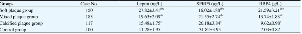
3.4. Adipocytokines
plaque group and calcified plaque group were significantly higher than those of control group while SFRP5 content were significantly lower than that of control group. Leptin and RBP4 content in serum of mixed plaque group were significantly higher than those of calcified plaque group while SFRP5 content was significantly lower than that of calcified plaque group. Leptin and RBP4 content in serum of soft plaque group were significantly higher than those of mixed plaque group and calcified plaque group while SFRP5 content was significantly lower than those of mixed plaque group and calcified plaque group (P<0.05), shown in Table 4.
3.5. Correlation between atheromatous plaque CT value and
coronary heart disease
Pearson test showed that atheromatous plaque CT value was significantly negatively correlated with platelet function parameters MPV, TEG-MA, P-selectin, PDGF-BB and vWF levels, inflammatory factors CRP, IL-6, IL-12, IL-18 and IL-23 content, chemokines MCP-1, CXCL16, Fractalkine and RANTES content as well as condition judgment indexes Leptin and RBP4 content, and significantly positively correlated with condition judgment index
SFRP5 (P<0.05).
4. Discussion
Coronary CTA can be used to determine the atheromatous plaque features in patients with coronary heart disease, different plaque components can show different CT values in the CT imaging, and the plaques that are rich in lipid core are generally with low CT value (<60 HU), commonly known as soft plaque[4]. In the study, the plaque CT values of the included patients with coronary heart disease were detected at first through CTA, and then used for qualitative plaque grouping, and it was found that 450 patients with coronary heart disease included 117 cases with calcified plaque (CT value ≥130 HU), 150 cases with soft plaque (CT value ≤60 HU) and 183 cases with mixed plaque group (CT value 60-130 HU). Studies both at home and abroad have confirmed that soft coronary plaque group of patients are with the highest incidence of rupture and acute myocardial infarction, whereas patients with calcified plaque are with more stable condition. In order to define the value of atheromatous plaque grouping by CTA for judging the overall condition of patients
Table 3
Comparison of chemokine content in serum among four groups.
Groups Case No. MCP-1 (pg/L) CXCL16 (μg/L) Fractalkine (ng/L) RANTES (ng/L)
Soft plaque group 150 22.57±3.18abc 3.42±0.48abc 78.95±8.11abc 1 215.82±150.95abc
Mixed plaque group 183 17.23±2.75ab 2.84±0.35ab 53.82±6.95ab 689.73±75.82ab
Calcified plaque group 117 15.43±1.84a 2.31±0.29a 39.74±4.11a 342.95±40.28a
Control group 100 13.27±1.94 1.87±0.23 30.27±3.89 214.83±25.96
a vs. control group, P<0.05; b vs. calcified plaque group, P<0.05; c vs. mixed plaque group, P<0.05.
Table 4
Comparison of condition judgment index content in serum among four groups.
Groups Case No. Leptin (ng/L) SFRP5 (μg/L) RBP4 (g/L)
Soft plaque group 150 27.82±3.41abc 16.02±1.88abc 21.59±3.21abc
Mixed plaque group 183 19.63±2.09ab 21.55±2.74ab 13.74±1.83ab
Calcified plaque group 117 15.48±1.75a 26.18±3.84a 9.62±0.98a
Control group 100 11.28±1.95 31.82±3.95 7.03±0.82
a vs. control group, P<0.05; b vs. calcified plaque group, P<0.05; c vs. mixed plaque group, P<0.05.
Table 1
Comparison of platelet function parameters among four groups.
Groups Case No. PLT (伊109/L) MPV (fL) TEG-MA (mm) P-selectin (μg/L) PDGF-BB (ng/L) vWF (伊103 mU/L)
Soft plaque group 150 191.52±23.48 8.45±0.91abc 73.29±7.85abc 254.95±30.17abc 261.29±30.28abc 489.55±56.78abc
Mixed plaque group 183 189.77±20.75 8.12±0.89ab 68.93±7.64ab 174.38±20.15ab 153.09±18.45ab 327.58±40.18ab
Calcified plaque group 117 185.46±21.38 7.79±0.85a 62.17±7.21a 69.73±7.21a 83.62±9.15a 243.19±25.76a
Control group 100 176.48±20.43 7.48±0.81 57.34±6.05 32.74±4.09 39.48±4.52 153.82±20.12
a vs. control group, P<0.05; b vs. calcified plaque group, P<0.05; c vs. mixed plaque group, P<0.05.
Table 2
Comparison of inflammatory factor content in serum among four groups.
Groups Case No. CRP (mg/L) IL-6 (ng/L) IL-12 (ng/L) IL-18 (ng/L) IL-23 (ng/L)
Soft plaque group 150 3.89±0.45abc 72.17±8.34abc 95.94±10.12abc 89.63±9.27abc 93.21±10.18abc
Mixed plaque group 183 2.18±0.25ab 45.83±6.12ab 75.48±8.11ab 56.92±6.15ab 59.74±7.12ab
Calcified plaque group 117 1.21±0.18a 31.29±3.85a 34.95±4.12a 20.45±2.78a 29.53±3.41a
Control group 100 0.78±0.09 20.38±2.79 11.18±1.79 7.39±0.82 13.84±1.76
with coronary heart disease, the platelet function, serum biochemical indexes and other clinical common disease evaluation projects were further compared among various groups of research subjects in the study.
The basis of atheromatous plaque formation in patients with coronary heart disease is vascular intima damage, and as intima damage increases, the blood coagulation system is activated and induces hypercoagulable state. Platelet hyperfunction is an independent risk factor for acute cardiovascular events in patients with coronary heart disease, and therefore, the platelet function state was compared among groups at first in the study first so as to make clear the relation between different plaque features and platelet function. PLT, MPV, TEG-MA, PDGF-BB and vWF are the clinical common platelet function indexes[5,6]. P-selectin is the molecule released by α particle after platelet activation, and is the most specific platelet activation marker. PDGF-BB exists in platelets in the form of α-particle, and can reflect the platelet release function. In the case of endothelial damage, vWF is released into the blood, and can reflect the degree of endothelial cell damage. MPV is an index reflecting megakaryocyte growth and thrombopoiesis, and TEG-MA mainly reflects the platelet aggregation function[7,8]. It was found in the study that MPV, TEG-MA, P-selectin, PDGF-BB and vWF levels were higher in coronary heart disease patients with different plaques features, and as plaque CT value decreased, MPV, TEG-MA, P-selectin, PDGF-BB and vWF levels further increased, indicating that soft plaque group of patients are with the highest platelet activity and also the highest probability of long-term acute coronary embolic events.
There are also many serum biochemical indexes directly related to coronary heart disease, and the levels of inflammatory cytokines and chemokines of all groups were further analyzed in the study. Chronic systemic inflammatory response has been confirmed to be throughout the whole course of coronary heart disease, and is also one of the main risk factors that add to the continuous increase of atheromatous plaque area and the occurrence of acute myocardial infarction[9,10]. Detection of the content of CRP, IL-6, IL-12, IL-18, IL-23 and other pro-inflammatory factors showed that CRP, IL-6, IL-12, IL-18 and IL-23 content in serum of patients with coronary heart disease were higher than those of healthy subjects, and as the plaque CT value decreased, the above factor levels further increased, indicating that soft plaque group of patients are with most serious systemic inflammatory state. High expression of chemokines is the basis for macrophages and lipid metabolites to accumulate in local coronary artery and form atheromatous plaque, and MCP-1 can mediate the activation of a variety of transcription factors and inflammatory mediators, leading to unstable plaque[11,12]. Chemokine CXCL16 can activate T lymphocytes and participate in the interaction between dendritic cells and T cells, and study has confirmed that it participates in the inflammatory reaction of atherosclerosis. Chemokine Fractalkine can be combined with receptor CX3CR1 and then prompt the directional migration of mononuclear cells and
NK cells, increase inflammatory response and prompt plaque to be unstable[13]. T cell chemokine (RANTES) can mediate mononuclear macrophages accross the endothelium as well as promote cellular inflammatory factor expression and atherosclerosis. It was found in the study that soft plaque group of patients were with higher MCP-1, CXCL16, Fractalkine and RANTES content in serum, indicating that high expression of chemokine is the core factor of the coronary artery disease aggravation and plaque instability.
Leptin, SFRP5 and RBP4 are new molecules discovered in recent years that are directly related to coronary heart disease[14]. It has been found in the mouse models that Leptin can promote the development of atherosclerosis, and Leptin levels in patients with coronary heart disease are significantly higher than those in patients without coronary heart disease. SFRP5 can inhibit Wnt5a so as to stop the inflammatory mediators of macrophages and fat cells from being functional, and its level is negatively correlated with the severity of coronary heart disease[15,16]. RBP4 can interfere with the glucolipid metabolism so as to promote inflammation, and many studies have also found that RBP4 is directly related to the insulin resistance, obesity, atherosclerosis and so on[17]. In the study, serum levels of the above three factors were tested, and it was found that Leptin and RBP4 content in patients with coronary heart disease were higher while SFRP5 content was lower, which is consistent with the functions of three factors mentioned above. Leptin and RBP4 content of soft plaque group were higher than those of mixed plaque group and calcified plaque group while SFRP5 content was lower than those of mixed plaque group and calcified plaque group, showing that coronary heart disease develops as the plaque CT value decreases.
Finally, the correlation between atheromatous plaque CT value under CTA and the condition in patients with coronary heart disease was analyzed, and it was found that atheromatous plaque CT value in patients with coronary heart disease was negatively correlated with platelet function parameters MPV, TEG-MA, P-selectin, PDGF-BB and vWF levels, inflammatory factors CRP, IL-6, IL-12, IL-18 and IL-23 content, chemokines MCP-1, CXCL16, Fractalkine and RANTES content as well as condition judgment indexes Leptin and RBP4 content, and positively correlated with condition judgment index SFRP5. It is thus clear that at the same time of quantitative judgment of atheromatous plaque, coronary CTA can intuitively and accurately judge the overall disease severity, and is expected to become the reliable means for long-term treatment guide and prognosis judgment in patients with coronary heart disease.
References
different characteristics detected by CT coronary angiography for patients with coronary heart diseases. J Shanghai Jiaotong Univ (Med Sci) 2014; 34(8): 1176-1179.
[3] Gan L, Feng C, Liu C, et al. Association between serum N-terminal pro-B-type natriuretic peptide levels and characteristics of coronary atherosclerotic plaque detected by coronary computed tomography angiography. Exp Ther Med 2016; 12(2): 667-675.
[4] Rodriguez-Granillo GA, Carrascosa P, Bruining N, et al. Defining the non-vulnerable and vulnerable patients with computed tomography coronaryangiography: evaluation of atherosclerotic plaque burden and composition. Eur Heart J Cardiovasc Imag 2016; 17(5): 481-491. [5] Liang H, Huang HJ, Zhou XL, et al. Changes of platelet function after
antiplatelet therapy and single nucleotide polymorphism of COX-1 and COX-2 in elderly patients. Chin General Pract 2014; 17(20): 2345-2348. [6] Jamasbi J, Megens RT, Bianchini M, et al. Differential inhibition of
human atherosclerotic plaque-induced platelet activation by dimeric GPVI-Fc and anti-GPVI antibodies: Functional and imaging studies. J
Am Coll Cardiol 2015; 65(22): 2404-2415.
[7] Wang JQ, Liu JG, Wang CL. Correlation analysis between visualization of hemorrheologic changes and platelet function in patients with acute
coronary syndrome after percutaneous coronary intervention. Chin J
Pathophysiol 2014; 30(2): 214-219.
[8] Jacobin-Valat MJ, Laroche-Traineau J, Larivière M, et al. Nanoparticles functionalised with an anti-platelet human antibody for in vivo detection ofatherosclerotic plaque by magnetic resonance imaging. Nanomedicine 2015; 11(4): 927-937.
[9] Sun LQ. Effect of Erigeron breviscapus on serum C-reactive protein as well as interleukin-6, -12, -18 and -23 in elderly patients with acute coronary syndrome. Chinese J Gerontol 2014; 34(1): 249-252.
[10] Matusiak A, Chałubiński M, Broncel M, et al. Putative consequences of
exposure to Helicobacter pylori infection in patients with coronary heart
disease in terms of humoral immune response and inflammation. Arch
Med Sci 2016; 12(1): 45-54.
[11] Zhu SS, Wang QC, Hu J. Clinical significance of serum chemokine CXCL16 detection before and after coronary heart disease and unstable angina treatment. Chin J Clin Rat Drug Use 2015; 8(3): 88-91.
[12] Zhang CY, Cao Z, Yang Y, et al. The serum level change of Fractalkine, monocyte chemotactic protein-1 and interleukin-8 in patients with coronary heart disease and its significance. J Bengbu Med Coll 2016;
41(4): 485-488.
[13] Tian J, Hu S, Wang F, et al. PPARG, AGTR1, CXCL16 and LGALS2 polymorphisms are correlated with the risk for coronary heart disease. Int
J Clin Exp Pathol 2015; 8(3): 3138-3143.
[14] Chen Y, Feng YM. Correlation between serum leptin level with coronary heart disease risk stratification and lesion degree of coronary artery. J
Hainan Med Univ 2015; 21(1): 32-35.
[15] Zeng R, Xu CH, Xu YN, et al. Association of leptin levels with pathogenetic risk of coronary heart disease and stroke: a meta-analysis.
Arq Bras Endocrinol Metabol 2014; 58(8): 817-823.
[16] Li YY, Dong WP, Lu H, et al. Correlation of serum Leptin, SFRP5 and RBP4 with coronary heart disease. Acta Univ Med Anhui 2016; 51(8): 1160-1163.