T E C H N I C A L N O T E
Open Access
Neural substrates associated with humor
processing
Jihye Noh
1, Ji-woo Seok
2, Suk-Hee Kim
3, Chaejoon Cheong
1and Jin-Hun Sohn
2*Abstract
Background:Humor is composed of a cognitive element related to the detection of humor and an affective
element related to the appreciation of humor. To investigate activated areas of the brain related to the two components of humor and to identify neural substrates associated with the degree of humor intensity, 13 participants were scanned while watching cartoons.
Findings:While watching humorous scenes, various areas of the brain were activated, including (1) the inferior gyrus, an area involved in reconciling ambiguous semantic content with stored knowledge, and (2) the temporal gyrus and fusiform gyrus, brain regions associated with the feeling of mirth. Further, humor intensity was positively correlated with BOLD signal magnitude in the nucleus accumbens, a region known to be involved in
psychologically and psychopharmacologically driven rewards.
Conclusions:Our findings demonstrate a two-component neural circuit model of humor processing and a key
region important in pleasurable feelings accompanied by humor.
Keywords:Emotion; Humor; Detection; Appreciation; Event-related fMRI
Findings
Humor plays a central and unique role in human life. Without humor, life would undeniably be less exhilarating. Humor provides an effective means of communicating ideas, attracting partners, boosting mood, and even coping in times of trauma and stress (Brownell and Gardner 1988; Dixon 1980; Garilovic et al. 2003; Martin 2001; Mobbs et al. 2003; Neuhoff and Schaefer 2002; Nezlek and Derks 2001).
Humor involves both cognitive and affective elements (Gardner et al. 1975). The cognitive element refers to ‘getting the joke’ which includes moments during which the perceiver attempts to comprehend disparities between a punch line and prior experience (Brownell et al. 1983). The affective element refers to‘enjoying a joke’; these are moments during which the perceiver experiences pure visceral, emotional responses depending on the hilarity of the experience (Shammi and Stuss 2003).
Studies have been conducted to identify biological neural systems related to humor. Gardner et al. (1975) examined patients with brain injury. They found that the left hemisphere of the brain is related to the integration of information that is required for understanding humor and that the right hemisphere is associated with the emo-tional processing of humor. In another study, patients with an injury in the right hemisphere showed a low physical reaction and emotional response to humor (Shammi and Stuss 1999).
Studies comparing activated areas of the brain respon-sible for the affective element and the cognitive element have also been conducted. Moran et al. (2004) examined a brain region that is activated during humor detection (cognitive element) and humor appreciation (affective element). The inferior frontal gyrus and posterior middle temporal gyrus were found to be activated during humor detection condition, while the insula and amygdala were activated during humor appreciation condition.
While numerous studies have focused on block-designed functional magnetic resonance imaging (fMRI) related to humor, few studies have explored event-related fMRI. Whether emotion-inducing stimuli should be presented in a block-designed paradigm or event-related paradigm * Correspondence:[email protected]
2Department of Psychology, Brain Research Institute, Chungnam National University, Daejeon, South Korea
Full list of author information is available at the end of the article
during fMRI remains controversial. Block-designed para-digms are often used because of their easy implementation and because randomization, jittering, and spacing of different stimulus categories is not necessary (Josephs et al. 1997). In blocked designs, stimulus presentation is lengthened and consecutive stimuli in a block are predictable (Zarahn et al. 1997). Prolonged exposure of stimuli may decrease emotional involvement and hence alter underlying brain activation. In emotion research, presentation duration is particularly important, not only from a methodological point of view but also in respect of differences in information processing (Buhler et al. 2008). Event-related designs are superior in terms of rapid estimation of the hemodynamic response func-tion to a short stimulus and are useful for emofunc-tion experiments, in part because of their ability to avoid the effects of confounding factors, such as habituation and anticipation (Rosen et al. 1998). In this study, we examined differences in brain activation associated with the two elements of humor, the cognitive element and the affective element, and identified brain areas positively correlated with the rating of funniness using an event-related fMRI paradigm.
Availability and requirements
Participants
Thirteen healthy, right-handed subjects participated in the experiment (mean age 24.8 ± 3.8 years, range 23−33 years, four men and nine women). No participant had a history of psychiatric or neurological disorders. The sub-jects were instructed to watch cartoons without laughing and to not move their heads.
Stimuli
Five cartoons were used in this study. All images were selected from a pilot study. The cartoons consisted of 7, 8, 9, 11, or 13 scenes. For fMRI study, cartoons were dis-played on a monitor and presented to the subject through a 45° angled mirror positioned above the head coil.
Experimental design
Stimuli were presented by showing five cartoons selected from a pilot experiment. Stimuli were presented according to an event-related fMRI paradigm with each cartoon being presented for 32 to 46 s. The subjects were instructed to press a button on a keypad immediately if they found the cartoon to be funny. The interval between the cartoons was 4 s. Following the scan, each subject was instructed to assess humor intensity. The subjects were asked to state whether they found the cartoon funny. If they did find it funny, they were asked to rate their perceived humor intensity on a scale of 1 to 7
(1 least funny, 7 most funny). The subjects were then asked to explain the meaning of the cartoons (Figure 1).
Image acquisition
fMRI experiments were conducted using an ISOL 3 T Forte scanner (ISOL Technology, Gyeonggi, Korea). During the presentation of visual stimuli, fMRI scanning was performed with the single shot Echo Planar Imaging sequence (repetition time (TR), 2,000 ms; echo time (TE), 28 ms; flip angle, 80°; field of view (FOV), 240 mm; matrix size, 64 × 64; slice thickness, 5 mm, no gap; and in-plane resolution, 3.75 mm, three dummy scans). Anatomical T1-weighted images were obtained with a 3-D FLAIR sequence (TR 280, TE 14, FA 60, FOV 240, matrix 256 × 256, 4-mm slice thickness).
Data analysis
Functional images were analyzed using SPM99 (http:// www.fil.ion.ucl.ac.uk/spm/software/spm99/). Data includ-ing head motion artifacts that could not be corrected were excluded from analysis. All functional images were rea-ligned with six movement parameters (translation; x,y, z and rotation; pitch, roll, yaw) to correct head motion. Echo-planar imaging (EPI) and T1-weighted images were coregistered and spatially normalized to the Montreal Neurological Institute template (MNI template) using an automated spatial transformation. Normalized images were smoothed using a 7-mm isotropic Gaussian kernel.
Following preprocessing, statistical analysis was per-formed. fMRI data were analyzed for each subject indi-vidually in the context of the general linear model (GLM) and theory of Gaussian random fields. Using subtraction and correlation procedures, activated areas in the brain while observing different pictures were color-coded by T-score.
Results and discussion
Behavioral results
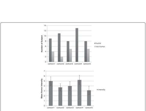
After fMRI experiments, all participants rated the intensity of humor. The number of subjects who experienced humor while watching each cartoon was 9 for cartoon #1, 11 for cartoon #2, 8 for cartoon #3, 13 for cartoon #4, and 8 for cartoon #5 (Figure 2, upper image). Participants who recognized the cartoons as funny reported an average of 3.5 points for cartoon #1, 3.2 points for cartoon #2, 2.5 points for cartoon #3, 5.2 points for cartoon #4, and 1.9 points for cartoon #5, respectively (Figure 2, lower image).
fMRI results
Figure 1Experimental design.Total experiment time was 45 min, including briefing and psychological assessment.
0 2 4 6 8 10 12 14
cartoon1 cartoon2 cartoon3 cartoon4 cartoon5
Number of Subject
humor
non-humor
0 1 2 3 4 5 6 7
cartoon1 cartoon2 cartoon3 cartoon4 cartoon5
Mean Humor Intensity
intensity
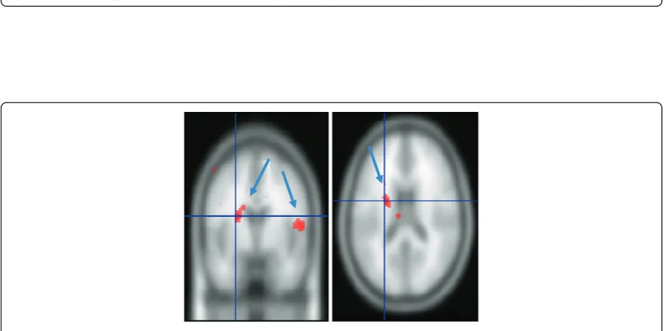
Figure 4Areas of the brain activated during cartoon scenes with different degrees of humor intensity.Activation in the left nucleus accumbens and right middle temporal gyrus is correlated with humor intensity.
and right middle frontal gyrus (p< 0.001, uncorrected; Figure 3).
Humor detection is critically dependent upon resolving incongruities between punch lines and expectations shaped by the storyline (Sul 1972). Consistent with this notion, frontal regions engaged during humor detection have been implicated in language tasks that encourage the retrieval and appraisal of relevant semantic knowledge (Binder et al. 1997, Price et al. 1999). Recent studies have indicated further specialization within the left inferior frontal cortex for reconciling ambiguous semantic content with stored knowledge (Gold and Buckner 2002, Thompson-Schill et al. 1997). Inferior frontal regions may resolve ambi-guities between these expectations and punch lines.
Regression analysis was used to examine the association between humor intensity (i.e., the degree of funniness as rated by each subject) and BOLD signal magnitude. This analysis revealed that humor intensity was associated with increased activation in the left nucleus accumbens and right middle temporal gyrus (p < 0.001, uncorrected; Figure 4).
Activation of the fusiform gyrus and anterior temporal region caused by electrical stimulation induced laughter accompanied by a feeling of mirth (i.e., positive emotion; Arroyo et al. 1993). The temporal area, including the temporoparietal junction, is involved in the integration of multisensory information and coherence building and inferring knowledge (Ferstl and von Cramon 2002; Goel et al. 1995). Additionally, the temporal lobe may contribute to generating, testing, and correcting internal prediction regarding external sensory events, which is crucial for resolving incongruity in humor processing (Samson et al. 2009). These regions may be involved in the incongruent or surprising (Brownell et al. 1983) elements of a joke and thus may play a pivotal role in the early stages of the humor network.
The nucleus accumbens has been implicated in psycho-logically and psychopharmacopsycho-logically driven rewards in various studies (Breiter et al. 2001; Knutson et al. 2001). Activation of the nucleus accumbens elicited by humor converges with findings from fMRI studies across a number of psychologically rewarding tasks, suggesting that this structure is involved in processing a diverse number of stimuli with rewarding characteristics (Aharon et al. 2001; Breiter et al. 2001; Erk et al. 2002; Rilling et al. 2002). Additionally, electrical stimulation of the nucleus accumbens results in laughter and giddiness (Okun et al. 2004). Although we cannot exclude other intervening factors (e.g., novelty), given the results of prior fMRI and physiological studies implicating the nucleus accumbens modulation in self-reported happiness (Knutson et al. 2001) and cocaine/amphetamine-induced euphoria in humans (Brieiter et al. 2001; Drevets et al. 2001), it is reasonable to conclude that nucleus accumbens activation
observed in this study reflects the hedonic feeling accom-panying humor.
Conclusions
In this study, using event-related fMRI, we identified areas of the brain that were activated during humor processing. We have presented evidence for differential systems underlying the cognitive and affective processes of humor and the brain region correlated with the degree of humor intensity.
Competing interests
The authors declare that they have no competing interests.
Authors' contributions
JN, JWS, JHS, and CC carried out fMRI experiments and analysis. JN and SHK drafted the manuscript. All authors read and approved the final manuscript.
Acknowledgements
This research has been supported by the Converging Research Center Program funded by the Ministry of Education, Science and Technology (2013K000332), the Korea Science and Engineering Foundation (No. 20120006577), and the Korea Basic Science Institute (T33408).
Author details
1Division of Magnetic Resonance Research, Korea Basic Science Institute, Ochang, Chungbuk, South Korea.2Department of Psychology, Brain Research Institute, Chungnam National University, Daejeon, South Korea.3Department of Professional Counseling and Psychotherapy, Graduate School of Health and Complementary Medicine, Wonkwang University, Iksan, Cheonbuk 570-749, South Korea.
Received: 2 December 2013 Accepted: 10 January 2014
References
Aharon I, Etcoff N, Ariely D, Chabris CF, O’Connor E, Breiter HC (2001) Beautiful faces have variable reward value: fMRI and behavioral evidence. Neuron 32(3):537–551
Arroyo S, Lesser RP, Gordon B, Uematsu S, Hart H, Schwerdt P, Andeasson K, Fisher RS (1993) Mirth, laughter and gelastic seizures. Brain 116(4):757–780 Binder JR, Frost JA, Hammeke TA, Cox RW, Rao SM, Preieto T (1997) Human brain
language areas identified by functional magnetic resonance imaging. J Neurosci 17(1):353–362
Breiter HC, Aharon I, Kahneman D, Dale A, Shizgal P (2001) Functional imaging of neural responses to expectancy and experience of monetary gains and losses. Neuron 30:619–639
Brownell HH, Gardner H (1988) Neuropsychological insights into humour. In: Durant J, Miller J (eds) Laughing matters: a serious look at humour. Longman Scientific and Technia, Harlow, UK
Brownell HH, Michel D, Powelson J, Gardner H (1983) Surprise but not coherence: sensitivity to verbal humor in right-hemisphere patients. Brain Language 18:20–27
Buhler M, Klein SV, Klemen J, Smolka MN (2008) Does erotic stimulus
presentation design affect brain activation patterns? Event-related vs. blocked fMRI designs. Behav Brain Funct 4(1):30
Dixon NF (1980) Humor: A cognitive alternative to stress. In Spielberger CD, Sarason IG (eds) Anxiety and stress, Hemisphere, Washington DC
Drevets WC, Gautier C, Price JC, Kupfer DJ, Kinahan PE, Grace AA, Price JL, Mathis CA (2001) Amphetamine-induced dopamine release in human ventral striatum correlates with euphoria. Society of Biological Psychiatry 49:81–96 Erk S, Spitzer J, Wunderlich AP, Galley L, Walter H (2002) Cultural objects
modulate reward circuitry. Neuroreport 13:2499–2503
Ferstl EC, von Cramon DY (2002) What does the frontomedian cortex contribute to language processing: coherence or theory of mind? Neuroimage 17:1599–1612 Gardner H, Ling PK, Flamm L, Silverman J (1975) Comprehension and
Gavrilobic J, Lecic-Tosevski D, Dimic S, Pejovic-Milovancevic M, Knezevic G, Priebe S (2003) Coping strategies in civilians during air attacks. Soc Psychiatry Psychiatr Epidemiol 38(3):128–133
Goel V, Grafman JNS, Sadato N, Hallett M (1995) Modeling other minds. Neuroreport 6:1741–1746
Gold BT, Buckner RL (2002) Common prefrontal regions coactivate with dissociable posterior regions during controlled semantic and phonological task. Neuron 35(4):803–812
Josephs O, Turner R, Friston K (1997) Event-related fMRI. Hum Brain Mapp 5:243–248 Knutson B, Adams CM, Fong GW, Hommer D (2001) Anticipation of increasing
monetary reward selectively recruits nucleus accumbens. J Neurosci 21:RC159 Martin RA (2001) Humor, laughter, and physical health: methodological issues
and research findings. Psychol Bull 127(4):504–519
Mobbs D, Grecius MD, Abdel-Azim E, Menon V, Reiss AL (2003) Humor modulates the mesolimbic reward centers. Neuron 40:1041–1048
Moran JM, Wig GS, Adams RB, Janata P, Kelley WM (2004) Neural correlates of humor detection and appreciation. Neuroimage 21(3):1055–1060 Netzlek JB, Derks P (2001) Use of humor as a copying mechanism, psychological
adjustment, and social interaction. Humor International Journal of Humor Research 14:395–413
Neuhoff CC, Scahaefer C (2002) Effects of laughing, smiling, and howling on mood. Psychol Rep 91:1079–1080
Okun MS, Bowers D, Springer U, Shapira NA, Malone D, Rezai AR, Nuttin B, Heilman KM, Morecraft RJ, Rasmussen SA, Greenberg BD, Foote KD, Goodman WK (2004) What’s in a smile? Intra-operative observation of contralateral smiles induced by deep brain stimulation. Neurocase 10(4):271–279
Price CJ, Green DW, von Studnitz RA (1999) Functional imaging study of translation and language switching. Brain 122(12):2221–2235 Rilling JK, Gutman DA, Zeh TR, Pagnoni G, Berns GS, Kilts CD (2002) A neural
basis for social cooperation. Neuron 35(2):395–405
Rosen BR, Buckner RL, Dale AM (1998) Event-related functional MRI: past, present and future. PNAS Proc Natl Acad Sci U S A 95(3):773–780
Samson AC, Hempelmann CF, Huber O, Zysset S (2009) Neural substrates of incongruity-resolution and nonsense humor. Neuropsychologia 47:1023–1033 Shammi P, Stuss DT (1999) Humour appreciation: a role of the right frontal lobe.
Brain 122:657–666
Shammi P, Stuss DT (2003) The effects of normal aging on humor appreciation. J Int Neuropsychol Soc 9:855–863
Suls J (1972) A two-stage model for the appreciation of jokes and cartoons. In: Goldstein PE, McGhee JH (eds) The psychology of humor: theoretical perspectives and empirical issue. Thieme Medical Publishers, New York, pp 81–100 Thompson-Schill SL, E’Esposito M, Aguirre GK, Farah M (1997) Role of left inferior
prefrontal cortex in retrieval of semantic knowledge: a reevaluation. PNAS 94(26):14792–14797
Zarahn E, Aguirre G, D’Esposito M (1997) A trial-based experimental design for fMRI. Neuroimage 6:122–138
doi:10.1186/s40543-014-0020-7
Cite this article as:Nohet al.:Neural substrates associated with humor processing.Journal of Analytical Science and Technology20145:20.
Submit your manuscript to a
journal and benefi t from:
7Convenient online submission
7Rigorous peer review
7Immediate publication on acceptance
7Open access: articles freely available online 7High visibility within the fi eld
7Retaining the copyright to your article