Harpenden, Herts, AL5 2JQ
Telephone: +44 (0)1582 763133 Web: http://www.rothamsted.ac.uk/
Rothamsted Research is a Company Limited by Guarantee Registered Office: as above. Registered in England No. 2393175.
Rothamsted Repository Download
A - Papers appearing in refereed journals
Chen, Z. C., Yokosho, K., Kashino, M., Zhao, F-J., Yamaji, N. and Ma, J.
F. 2013. Adaptation to acidic soil is achieved by increased numbers of
cis-acting elements regulating ALMT1 expression in Holcus lanatus. Plant
Journal. 76 (1), pp. 10-23.
The publisher's version can be accessed at:
•
https://dx.doi.org/10.1111/tpj.12266
The output can be accessed at:
https://repository.rothamsted.ac.uk/item/8qx8x
.
© Rothamsted Research. Licensed under the Creative Commons CC BY.
Adaptation to acidic soil is achieved by increased numbers of
cis
-acting elements regulating
ALMT1
expression in
Holcus lanatus
Zhi Chang Chen1, Kengo Yokosho1, Miho Kashino1, Fang-Jie Zhao2,3, Naoki Yamaji1and Jian Feng Ma1,* 1Institute of Plant Science and Resources, Okayama University, Chuo 2–20–1, Kurashiki, Japan,
2
State Key Laboratory of Crop Genetics and Germplasm Enhancement, College of Resources and Environmental Sciences, Nanjing Agricultural University, Nanjing 210095, China, and
3
Rothamsted Research, Harpenden, Hertfordshire AL5 2JQ, UK
Received 26 February 2013; revised 5 June 2013; accepted 12 June 2013; published online 16 June 2013.
*For correspondence (e-mail [email protected]–u.ac.jp).
SUMMARY
Yorkshire fog (Holcus lanatus), which belongs to the Poaceae family and is a close relative of the agro-nomic crop oat (Avena sativa), is a widely adaptable grass species that is able to grow on highly acidic soils with high levels of Al, but the mechanism underlying the high Al tolerance is unknown. Here, we characterized two accessions of H. lanatuscollected from an acid plot (soil pH 3.6, HL–A) and a neutral plot (pH 7.1, HL–N) in terms of Al tolerance, organic acid anion secretion and related gene expression. In response to Al (pH 4.5), the HL–A roots secreted approximately twice as much malate as the HL–N roots, but there was no difference in citrate secretion. Cloning of the gene HlALMT1 responsible for malate secretion showed that the encoded amino acid sequence did not differ between two accessions, but the expression level in the outer cell layers of the HL–A roots was twice as high as in the HL–N roots. This difference was not due to the genomic copy number, but was due to the number of cis-acting elements for an Al-responsive transcription factor (HlART1) in the promoter region of HlALMT1, as demonstrated by both a yeast one-hybrid assay and a transient assay in tobacco protoplasts. Furthermore, introduction of HlALMT1driven by the HL–A promoter into rice resulted in significantly more Al-induced malate secre-tion than introducsecre-tion of HlALMT1driven by the HL–N promoter. These findings indicate that the adapta-tion of H. lanatusto acidic soils may be achieved by increasing number ofcis-acting elements for ART1 in the promoter region of theHlALMT1gene, enhancing the expression ofHlALMT1and the secretion of malate.
Keywords: adaptation, aluminum tolerance, malate,ALMT1,Holcus lanatus.
INTRODUCTION
Soluble ionic aluminum in acid soils (mainly Al3+) rapidly
inhibits root elongation at the micromolar level, subse-quently affecting the uptake of water and nutrients and resulting in low crop productivity on such soils (Kochian, 1995; Ma, 2007; Ryanet al., 2011). However, some plants have evolved mechanisms to detoxify Al both internally and externally. There is a wide variation in Al tolerance between plant species and cultivars within a species. Several mechanisms for the variation in Al tolerance have been proposed (Kochian, 1995), among which variation in the secretion of organic acid anions, such as citrate, malate or oxalate, from roots appears to be the most common mechanism in both monocots and dicots (Ryan et al., 2001; Kochian et al., 2004; Ma, 2005, 2007; Magalhaes,
2006). These organic acid anions are able to chelate toxic Al ions, thereby detoxifying Al externally.
patterns differ among plant species. The proteins encoded byALMT1genes transport malate and are localized to the plasma membranes of root cells. In contrast, MATE1/ AACT1genes have been identified in barley (Hordeum vul-gare) (Furukawa et al., 2007), sorghum (Sorghum bicolor) (Magalhaes et al., 2007), Arabidopsis (Arabidopsis thali-ana) (Liuet al., 2009), rye (Secale cereale) (Yokoshoet al., 2010), maize (Zea mays) (Maron et al., 2010), rice bean (Vigna umbellata) (Yang et al., 2011), rice (Oryza sativa) (Yokosho et al., 2011) and wheat (Tovkach et al., 2013). The proteins encoded by theMATE1/AACT1genes are also localized to the plasma membranes, but transport citrate. High expression levels of these genes are associated with high Al tolerance in wheat and barley (Sasakiet al., 2006; Furukawa et al., 2007). Furthermore, recent studies have shown that alterations of the promoter regions are respon-sible for enhanced expression of ALMT1 (Sasaki et al., 2006; Ryan et al., 2010) and MATE1/AACT1 (Magalhaes et al., 2007; Fujiiet al., 2012; Tovkachet al., 2013).
In addition to Al-tolerance genes related to organic acid anion secretion, other Al-tolerance genes have been identi-fied, especially in rice. Identification of a transcription fac-tor for Al tolerance, ART1, revealed that multiple genes are involved in the high Al tolerance in rice (Yamaji et al., 2009). Functional analyses of several downstream genes regulated by ART1 showed that they are involved in both internal and external detoxification of Al at various cellular sites, such as the cell wall, plasma membrane and tono-plast (Huang et al., 2009, 2012; Xia et al., 2010; Yokosho et al., 2011; Chenet al., 2012).
Compared with the progress made in important crops and model plant species, little is known about the molecu-lar mechanisms of Al tolerance in plant species growing in their natural habitats. Yorkshire fog (Holcus lanatus), which belongs to the Poaceae family and whose closest relative is the agronomic crop oat (Avena sativa), is a highly adapt-able grass species found in wide environmental conditions (Grimeet al., 1988). For example,H. lanatuswas found in most plots in the long-term Park Grass Experiment at Rothamsted Research, which vary greatly in nutrient avail-ability and soil pH (from 3.6–7.5) (Silvertownet al., 2006). In the plots that are highly acidic due to long-term applica-tions of ammonium sulfate, H. lanatus is the dominant species (>90% of the total above-ground plant biomass in the plot), presumably due to its strong tolerance to Al toxicity. However, it is also found in plots receiving the same fertilizer treatment but limed to the neutral range of soil pH, where its biomass production decreases to<10% of the total due to competition from other plant species. In an effort to understand how H. lanatus adapts to highly acidic soils, we compared two accessions of plants from an acidic plot (pH 3.6, designated HL–A) and a limed plot (pH 7.1, designated HL–N) with regard to their Al tolerance, organic acid anion secretion and expression of related
genes. We found that the high Al tolerance in the acces-sion from the acidic plot results from an increased number of cis-acting elements for the transcription factor HlART1 in HlALMT1 gene, resulting in increased expression of HlALMT1and higher secretion of malate from the roots.
RESULTS
Comparison of tolerance to low pH and Al between two accessions ofHolcus lanatus
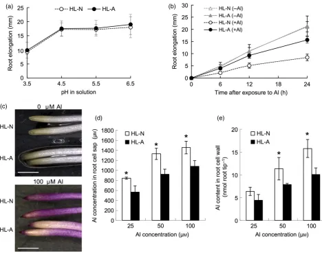
We first compared the root elongation at various pHs between HL–A from an acidic plot and HL–N from a neutral plot. However, there was no difference in the root elonga-tion between the two accessions at pHs ranging from 3.5 to 6.5 (Figure 1a). We then compared the Al tolerance between two accessions. In the presence of 50lMAl, root elongation of HL–N was inhibited to a great extent than that of HL-A at each time point (Figure 1b). The Al tolerance of HL–A was as high as for ajaponicarice cultivar, Nippon-bare, the most Al-tolerant species among small-grain cereal crops (Figure S1). Eriochrome cyanine R staining showed that HL–N accumulated more Al in the root tips than HL–A (Figure 1c). Consistent with this result, the Al content in the cell sap and cell wall of the root tips (0–1 cm) was also significantly higher in HL–N than in HL–A at each Al concentration (Figure 1d,e), indicating that an exclusion mechanism is involved in the higher Al tolerance of HL–A.
HL–A secreted more malate than HL–N in response to Al
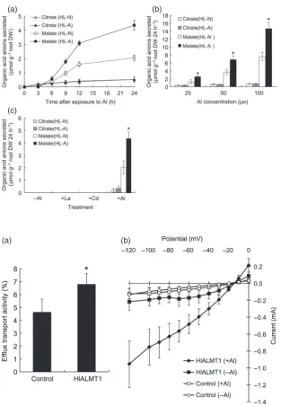
Next we investigated whether Al-induced secretion of organic acid anions is involved in higher Al tolerance in the HL–A accession. Both accessions secreted malate and citrate from the roots in response to Al (Figure 2a), but the amount of citrate secreted was small and did not differ sig-nificantly between the two accessions. In contrast, HL–A secreted approximately twice as much malate as HL–N (Figure 2a). The malate secretion induced by Al in HL–A (Figure 2) was comparable to that in rye and wheat, which utilize organic acid anion secretion for Al detoxifica-tion (Liet al., 2000). A dose–response experiment showed that the amount of malate secreted increased with increas-ing Al concentrations in both accessions, but HL–A secreted more malate than HL–N at all Al concentrations tested (Figure 2b). Furthermore, the secretion was only induced by Al, but not by Cd or La, indicating that the organic acid anion secretion is specific to Al (Figure 2c).
Cloning of a gene responsible for malate secretion,
HlALMT1
RACE to obtain the full-length cDNA of the putative malate transporter gene in both accessions ofH. lanatus. The iso-lated gene (designatedHlALMT1) consists of a 1532 bp long cDNA, including a 1389 bp ORF. There are five single nucle-otide polymorphisms between HL–A and HL–N (Figure S2a), but the encoded peptides (463 amino acids) were the same (Figure S2a). HlALMT1 shared 81, 43, 44 and 42% identity, respectively, with TaALMT1, GmALMT1, BnALMT1 and AtALMT1 (Figure S2b).
Transport activity of HlAMLT1
To investigate the transport activity of HlALMT1 for malate efflux, we expressed this gene in Xenopus oocytes and injected the oocytes with14C-labeled malate. Measurement of the 14C radioactivity showed that oocytes expressing HlALMT1 had a significantly higher efflux activity for
malate than the control in the absence of Al (Figure 3a), indicating that HlALMT1 is able to transport malate out of the cells.
To examine whether HlALMT1 is activated by Al, we per-formed two-electrode voltage clamp analysis. The results showed that the current in oocytes expressing HlALMT1 was much higher in the presence of Al than that in the absence of Al (Figure 3b). This result indicate that HlALMT1 is activated by Al.
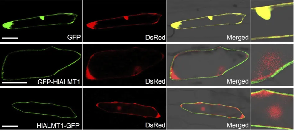
Subcellular localization of HlAMLT1
The subcellular localization of HlALMT1 was investigated by transiently expressingGFP–HlALMT1orHlALMT1–GFP together with the DsRedgene encoding red fluorescence protein in onion epidermal cells. DsRed was used as a marker which is localized at cytoplasm and nucleus. When (a)
(c)
(d) (e)
(b)
Figure 1. Physiological analysis of tolerance to low pH and Al in twoHolcus lanatusaccessions.
(a,b) Response to various pHs (a) and Al (b). Seedlings were exposed to a buffered solution at various pHs for 24 h or a solution containing 50lMAl for 0, 6, 12,
18 and 24 h. The root length was measured before and after the treatment. Values are meansSD (n=10).
(c) Eriochrome cyanine R staining. Roots exposed to 0 and 100lMAl for 24 h were stained in 0.1% Eriochrome cyanine R for 15 min. Scale bar=10 mm.
(d,e) Al concentration in the cell sap (d) and Al content in the cell wall (e) of root tips (0–1 cm). Seedlings of both accessions were exposed to a solution
contain-ing 25, 50 and 100lMAl for 24 h. The root tips were excised and fractionated into cell sap and cell wall. The Al concentration was determined by atomic
GFP alone was expressed, fluorescence was observed in the cytosol and nucleus, and completely overlapped with the DsRed signal (Figure 4, top panels). By contrast, the green fluorescence of both GFP–HlALMT1 (Figure 4, mid-dle panels) and HlALMT1–GFP (Figure 4, bottom panels) was only observed at the cell periphery distinct from the DsRed signal. These results indicate that HlALMT1 is local-ized to the plasma membrane.
Expression pattern ofHlALMT1inH. lanatus
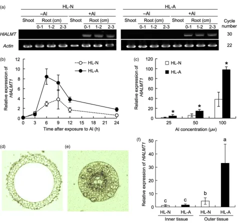
Expression ofHlALMT1was hardly detected in both roots and shoots of two accessions in the absence of Al (Figure 5a). However, expression in the roots but not in the shoots was greatly induced by Al in both accessions (Figure 5a). Spatial analysis showed thatHlALMT1
expres-sion in both the root tips (0–1 cm) and the mature regions (1–3 cm) was up-regulated by Al (Figure 5a). A time-course experiment showed that this induction occurred after 3 h exposure to Al (Figure 5b). The expression level of HlALMT1 was significantly higher in HL–A than in HL–N after between 6 and 24 h of Al exposure (Figure 5b). The expression of HlALMT1 increased with increasing Al concentrations in both accessions, but the expression level in HL–A was higher than that in HL–N at each Al concentra-tion (Figure 5c).
To investigate the tissue specificity ofHlALMT1 expres-sion, we used laser microdissection to separate the root tip tissues into outer and inner parts (Figure 5d,e). Expression ofHlALMT1in the outer tissues was higher than that in the inner tissues in both accessions (Figure 5f). There was no (a)
(c)
(b)
Figure 2. Time- and dose-dependent Al-induced
secretion of organic acid anions in twoHolcus
lanatusaccessions.
(a) Time-dependent secretion of organic acid anions. Seedlings were exposed to a solution
containing 50lMAl for 0, 3, 6, 9, 12 and 24 h.
(b) Dose–response. Seedlings were exposed to a
solution containing 25, 50 or 100lMAl for 24 h.
(c) Metal specificity. Seedlings were exposed to
a solution containing 50lMof Al, La or Cd for
24 h.
Root exudates were collected after each treat-ment, and the levels of organic acid anions were determined by an enzymatic method. Values are
meansSD (n=3). (b, c) The asterisks indicate
a significant difference between accessions (P<0.05 by Tukey’s test).
(a) (b)
Figure 3. Transport activity of HlALMT1 in
Xenopusoocytes.
(a) Basal transport activity for malate efflux in
Xenopus oocytes. HlALMT1 cRNA or water
(control) was injected into Xenopus oocytes.
After 1–day cultivation, the oocytes were
injected with14
C-labeled malate. The release of 14
C-labeled malate from the oocytes in the absence of Al was determined after 1 h. The percentage of total malate injected is shown.
Values are meansSD (n=3–4). The asterisk
indicates a significant difference compared with control (P<0.05 by Tukey’s test).
(b) Al-activated transport activity for malate efflux. Malate was injected into oocytes with or
withoutHlALMT1 expression, and the inward
current was recorded at membrane potentials between -120 and 0 mV in the presence and
absence of 100lMAl. Values are meansSD
difference in the expression level in the inner tissues between the two accessions, but expression in the outer tissues was much higher in HL–A than HL–N (Figure 5f). These results indicate that the difference in HlALMT1 expression between the two accessions arises from expression in the outer tissues.
Copy number of theHlALMT1gene
The differential expression ofHlALMT1in the two acces-sions may be due to a difference in the genomic copy number of HlALMT1. However, this possibility was ruled out because the two accessions have the same number of genomic copies of HlALMT1[normalized against that for a silicon transporter gene Lsi1 (low silicon rice 1; Os02 g0745100)] (Figure 6a) (Maet al., 2006).
We then compared the absolute mRNA level of HlALMT1. The transcript copy number calculated from the standard curves usingCTvalues was three times higher in
HL–A than in HL–N (Figure 6b). These results indicate that the expression levels of HlALMT1 in the two accessions may be attributed to the transcript copy number but not the genomic copy number.
Comparison of the promoter region ofHlALMT1in the two accessions
A possible mechanism for the differential expression of HlALMT1in the two accessions may be different promoter activities. To test this possibility, we isolated and com-pared the promoter region ( 2 kb) between the two acces-sions. Although there was no difference in the amino acid sequence ofHlALMT1between the two accessions (Figure
S2a), variations in the promoter region were found (Figure S3). However, these differences are not due to large dele-tions or inserdele-tions in the promoter region. In Arabidopsis, expression of AtMATE1 and AtALMT1 is regulated by a transcription factor, STOP1 (Liuet al., 2009; Sawakiet al., 2009). In rice, expression ofOsFRDL4(MATE) is regulated by ART1, a homolog of STOP1 (Yamajiet al., 2009), and its corecis-acting element sequence has recently been identi-fied as [GGN(T/g/a/C)V(C/A/g)S(C/G)] (Tsutsuiet al., 2011). Searches for the high-affinity ART1 cis-acting element
(GGTCCT, GGCCCT, GGTACT, GGTCGT, GGGCCT,
GGACCT or GGTGCT, the terminal T is unchanged) in the promoter region of HlALMT1 revealed that five elements are present in HL–A but only three in HL–N (Figure S3). The difference in the cis-acting element number between the two accessions is caused by single nucleotide substitu-tions.
Cloning and expression analysis ofHlART1
To examine whether the difference in the cis-acting element number explains the different expression levels of HlALMT1, we first cloned an ART1 homolog (HlART1) in H. lanatusbased on the conserved sequences ofART1and ART1-like genes in rice and Arabidopsis. A cDNA fragment of 360 bp was amplified, and a 1697 bp full-lengthHlART1 gene including a 1443 bp ORF was obtained from the cDNA of the two accessions of H. lanatus roots using 5′ and 3′ RACE. There was no difference in the nucleotide sequence of HlART1 between the two accessions (Figure S4a).HlART1encodes a peptide of 481 amino acids (Figure S4a), and the encoded protein shares 51.8 and 46%
iden-Figure 4. Subcellular localization of HlALMT1.
Constructs expressing GFP alone (top panels) or fusions betweenHlALMT1and green fluorescent protein gene (GFP) at the N–terminus (middle panels) or the
tity, respectively, with rice ART1 and Arabidopsis STOP1 (Figure S4b). HlART1 has four conserved C2H2 domains (Figure S4a), suggesting a role in the regulation of gene transcription.
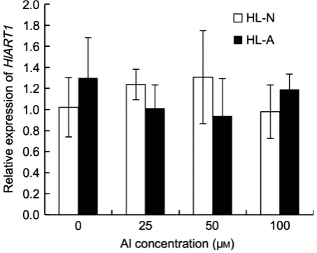
The expression level ofHlART1was similar in the roots of HL–A and HL–N, and was not affected by various con-centrations of Al in either accession (Figure 7).
Promoter binding and transcriptional activation assay
To investigate whether HlART1 interacts with the promoter of HlALMT1, we performed a yeast one-hybrid assay. In the absence of 3–amino-1,2,4–triazole (3AT, an inhibitor of HIS3 protein which catalyses histidine biosynthesis), there was no difference in growth among the transformed yeast (a)
(b) (c)
(d) (e)
(f)
Figure 5. Expression pattern ofHlALMT1inH. lanatus.
(a) Organ-dependent expression. Expression ofHlALMT1in the various root segments and shoots in the two accessions treated with or without Al. The PCR
cycle number is shown on the right.
(b) Time-dependent expression ofHlALMT1. Seedlings were exposed to a solution containing 50lMAl for 0, 3, 6, 9, 12 and 24 h. The expression level relative to
HL–N at 3 h is shown.
(c) Dose-dependent expression ofHlALMT1. Seedlings were exposed to a solution containing 25, 50 and 100lMAl for 24 h. The expression level relative to
HL–N at 25lMis shown.
(d,e) Outer tissues (d) and inner tissues (e) of the roots (7.5–12.5 mm) after laser microdissection.
(f) Tissue specificity ofHlALMT1expression. The expression level relative to the inner tissues of HL–N is shown.
Actinwas used as an internal standard. Values are meansSD (n=3). The asterisk in (c) and means with different letters in (f) indicate significant differences
cells (Figure 8a). However, in the presence of 20 or 50 mM 3AT, the HlART1-expressing cells showed better growth than pGADT7-expressing (control vector) cells (Figure 8a). Furthermore, growth of yeast cells containing the HL–A promoter was better than that of those containing the HL– N promoter. The results for rice ART1, which was included as a positive control, were similar to those for HlART1, suggesting that both rice ART1 and HlART1 probably bind to the samecis-acting element in the promoter region of HlALMT1. These results suggest that HlART1 interacts with the promoter region ofHlALMT1, and that the number of cis-acting elements is involved in regulation of the expres-sion level ofHlALMT1.
To quantify the promoter activity for the two accessions, we performed a transient assay in tobacco leaf protoplasts.
TheHlALMT1promoter from HL–A or HL–N was fused to the CaMV 35S minimal (–46) promoter (Fang et al., 1989) and GFP as a reporter gene, and then transformed into tobacco (Nicotiana tabacum) mesophyll protoplasts together with the constructs expressing HlART1 (effector gene) andDsRed(internal control) (Figure 8b). The expres-sion ofHlART1normalized against that ofDsRedwas simi-lar in all transformed protoplasts (Figure 8c). In contrast, expression of the reporter gene GFP normalized against that of DsRedwas higher in protoplasts expressing constructs carrying HlALMT1 promoters from either accession than those carrying the CaMV 35S minimal promoter (control) when introduced together with HlART1, indicating HlART1-dependent activity ofHlALMT1promoter. Furthermore, the GFP expression was twice as high in protoplasts carrying the HL–A promoter than in those carrying the HL–N pro-moter (Figure 8d). These results show that the HlALMT1 promoter from HL–A has a higher activity than that from HL–N.
Functional analysis of theHlALMT1promoter in transgenic rice
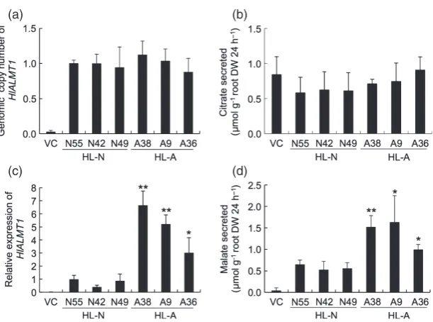
To further examine the role of the promoter region of HlALMT1 in the regulation of gene expression, we intro-ducedHlALMT1driven by promoters from the two acces-sions into rice. We obtained a number of transgenic lines and selected three independent lines with a single copy of HlALMT1 for further analysis (Figure 9a). The expression level ofHlALMT1was higher in the lines in which expres-sion was driven by the HL–A promoter than in those driven by the HL–N promoter (Figure 9b). Rice secretes citrate but not malate in response to Al (Maet al., 2002). There was no difference in citrate secretion between the trans-genic lines and the vector control when exposed to Al (Figure 9c). However, the amount of malate secreted from the roots was higher in the transgenic lines expressing HlALMT1 driven by the promoter from either accession
(a) (b)
Figure 6. Genomic and transcript copy number ofHlALMT1inH. lanatus.
(a) Genomic copy number in the two accessions. The values were normalized to theCTvalues ofLsi1.
(b) Transcript copy number. The seedlings were exposed to 50lMAl for 24 h prior to determination of the absolute expression level ofHlALMT1in the root tips
(0–1 cm) by quantitative real-time RT–PCR. The transcript copy number was calculated from standard curves usingCTvalues.
Values are meansSD (n=3). The asterisk indicates a significant difference compared with HL–N (P<0.05 by Tukey’s test).
Figure 7. Expression pattern ofHlART1.
Seedlings of both accessions were exposed to a solution containing 0, 25,
50 or 100lMAl for 24 h, and then the root tip (0–1 cm) were excised for
expression analysis. The expression level relative to HL–N (–Al) is shown.
compared with the vector control (Figure 9d), indicating thatHlALMT1 functions as a transporter of malate. More-over, the amount of malate secreted was significantly higher in the transgenic lines expressingHlALMT1driven by the HL–A promoter than that by the HL–N promoter (Figure 9d). The Al tolerance in the transgenic lines did not significantly change compared with vector control, prob-ably due to the basic high Al tolerance of rice (data not shown). These results further indicate that the different expression levels ofHlALMT1in the two accessions result from the difference in the promoter region.
DISCUSSION
Differential expression ofHlALMT1is responsible for natural variation in Al tolerance ofHolcus lanatus
Analysis of the HL–A accession ofHolcus lanatus, which has adapted to acidic soil, revealed a higher Al tolerance than the HL–N accession collected from a near-neutral soil from the neighboring experimental plot (Figure 1b). Physiological studies showed that this accession accumu-lated less Al in the roots and secreted more malate in response to Al exposure (Figures 1d,e and 2). Malate secretion from the roots has been established as a
mech-anism for Al tolerance in many plant species (Ma, 2000; Maet al., 2001).
The two accessions differ in the extent of malate secre-tion induced by Al (Figure 2), indicating that this difference is at least one of the reasons responsible for the natural variation in Al tolerance between the two accessions, although its contribution to Al tolerance is unknown.
There are two patterns of organic acid anion release, which differ in the timing of secretion (Ma, 2000; Maet al., 2001). In pattern I, no discernible delay is observed between addition of Al and the onset of secretion, while, in pattern II, organic acid anion secretion is delayed for several hours after exposure to Al. Our results show that malate secretion in H. lanatus follows pattern II (Figure 2a): requiring 6 h to release malate from the roots in both accessions. This pattern is different from that in wheat (Delhaizeet al., 1993), but similar to that observed in Arabidopsis, maize, rye and triticale (X Triticosecale Wittmack) (Pelletet al., 1995; Liet al., 2000; Maet al., 2000; Kobayashi et al., 2007), suggesting that different mecha-nisms for organic acid anion secretion are involved in dif-ferent plant species.
The Al-induced secretion of malate is mediated by ALMT1 in wheat, Arabidopsis, oilseed rape and soybean (a)
(b)
(c) (d)
Figure 8. Yeast one-hybrid assay and promoter activity assay.
(a) Yeast one-hybrid assay. The plasmids pGADT7, pGADT7-HlART1 or pGADT7-ART1
together with plasmids N (pHIS2.1+HlALMT1
promoter from HL–N) or A (pHIS2.1+HlALMT1
promoter from HL–A) were introduced into
yeast strain Y187 and cultured on synthetic complete (SC) medium without His in the
pres-ence of 0, 20 mMor 50 mM3–amino-1,2,4–
triaz-ole (3AT, a competitor of HIS3). Four serial 1:10 dilutions (from left to right) of yeast cell
sus-pensions starting from OD600=0.5 were
spot-ted on plates. The yeast was allowed to grow at
30°C for 3 days.
(b–d) Promoter activity assay in tobacco
protop-lasts. (b) Schematic diagram of the reporter, effector and internal control plasmids used in transient expression analysis. (c,d) Expression
levels of the effector gene (HlART1) (c) and the
reporter gene (GFP) (d) driven by the CaMV 35S
minimal promoter (Mini) or the HlALMT1
promoters from HL–N and HL–A. The reporter
vector, effector vector and internal control vec-tor were co-transformed into tobacco protop-lasts by the poly(ethylene glycol) method. The expression level was determined by
quantita-tive RT–PCR. The expression level relative to
that of Mini in the presence of HlART1 is
shown. Values are meansSD (n=3). Means
(Sasaki et al., 2004; Hoekenga et al., 2006; Ligaba et al., 2006; Lianget al., 2013). In the present study, we isolated a homolog of ALMT1 in H. lanatus. HlALMT1 showed the highest similarity to TaALMT1 (Figure S2b). Like other ALMT1s, HlALMT1 is localized to the plasma membrane and mediates malate efflux when expressed in Xenopus oocytes (Figures 3 and 4). The expression ofHlALMT1in H. lanatus was specifically induced by Al in the roots (Figure 5a) and limited to the outer cells of the roots (Figure 5f). Furthermore, expression of HlALMT1 in rice resulted in malate efflux from the roots (Figure 9d). All these results indicate that the HLALMT1 gene isolated in the present study is responsible for Al-induced secretion of malate inH. lanatus.
In addition to the induction of the HlALMT1 gene (Figure 5), the protein encoded byHlALMT1was activated by Al in Xenopus oocytes (Figure 3). This feature is the same as for other ALMT1 from various species such as wheat (Sasaki et al., 2004) and Arabidopsis (Hoekenga et al., 2006). The mechanisms underlying Al-induced activa-tion of ALMT1 is unknown, but the extracellular C–terminus is required for both the basal and Al3+-dependent transport activity of TaALMT1 in wheat and AtALMT1 in Arabidopsis (Furuichi et al., 2010). Further studies using site-directed mutagenesis showed that three acidic amino acids (E274, D275 and E284) in the C–terminal region are essential for Al3+-activated transport activity of TaALMT1. These three amino acid residues are also present in HlALMT1 (Figure S2a), suggesting that they are conserved in different plant species.
There was no difference in the amino acid sequence of HlALMT1between the two accessions differing in Al toler-ance and malate secretion (Figure S2a); however, HL–A showed higher expression of HlALMT1 than HL–N in a
time-dependent and dose-dependent manner after Al treat-ment (Figure 5b,c). Furthermore, the higher expression of HlALMT1in HL–A was only found in the outer root tissues (Figure 5f). This difference in the expression level is not due to genomic copy number ofHlALMT1, but to the tran-script number (Figure 6). These findings indicate that the differential expression levels of HlALMT1 are consistent with the differential malate secretion, which may contrib-ute to the different Al tolerance in the two accessions.
The expression level ofHlALMT1is determined by the number of cis-acting elements for ART1
The expression level of a gene may be regulated by many factors. Recently, several studies have examined the tran-scriptional regulation mechanisms of genes involved in Al-induced secretion of organic acid anions. In sorghum, tourist-like miniature inverted repeat transposable ele-ments in the promoter region ofSbMATEwas suggested to be involved in regulating expression of this gene (Ma-galhaeset al., 2007). In wheat, tandemly repeated elements of 33–803 bp located upstream of the TaALMT1 coding region are associated with high expression of TaALMT1 (Sasakiet al., 2006; Ryanet al., 2010). It is speculated that the repeats contain regulatory elements that enhance Ta-ALMT1expression when present in multiple copies (Ryan et al., 2010). Aegilops tauschii, a diploid species, is the donor of the D genome in wheat, which is the location of TaALMT1 . Interestingly, the tandemly repeated elements are not present in the promoter ofAetALMT1fromA. tau-schii, suggesting that these mutations occurred recently. In addition, a 1 kb insertion in the 5′UTR region ofHvAACT1 enhances its expression in Al-tolerant accessions of barley (Fujii et al., 2012). More recently, the higher expression level of TaMATE1B in several Brazilian wheat lines was
(a) (b)
(c) (d)
Figure 9. Functional analysis of the HlALMT1
promoter in transgenic rice.
(a) Genomic copy number ofHlALMT1in
trans-genic rice. The values were normalized using
theCTvalue of silicon transporter geneLsi1.
(b) Expression level ofHlALMT1in transgenic
rice. Seedlings were exposed to a solution
con-taining 50lMAl for 24 h. The expression level
relative to line N49 is shown.
(c,d) Al-induced secretion of citrate (c) and malate (d) from transgenic rice. Root exudates were collected after exposing transgenic
seed-lings to a solution containing 50lMAl for 24 h.
VC, vector control. Values are meansSD
(n=3). The asterisks indicate a significant
found to be associated with the presence of a Sukkula-like transposable element in its promoter (Tovkach et al., 2013). In the present study, we found that an Al-tolerant accession ofH. lanatushas evolved a different mechanism to enhance expression of HlALMT1. It is not achieved by increasing tandemly repeated elements as in wheat, but simply by increasing the number ofcis-acting elements for ART1 in the promoter region examined (up to 2 kb) (Figure S3). It appears that amplification of transcription factor binding sites may be the root cause for functional differ-ences in both wheat and Holcus, but the scale of these changes is far more specific inHolcusthan the variation of longer sequences that occurs in wheat. The response of theALMT1gene to Al has been lost in wheat (i.e.TaALMT1 is not induced by Al), and constitutive high expression in Al-tolerant cultivars may be achieved by re-organization of the promoter. By contrast, expression of ALMT1 inHolcus is induced by Al in both accessions, and this up-regulation requires ART1. Therefore, the relatively small change in the promoter in terms of the number ofcis-acting elements may result in enhanced expression of ALMT1, which con-tributes to Al tolerance.
ART1 is a transcription factor for Al tolerance that was identified initially in rice (Yamajiet al., 2009). It regulates the expression of at least 30 genes. AsH. lanatusbelongs to the Gramineae, like rice does, we isolated a homolog of ART1 inH. lanatusfrom both accessions (Figure S4). Both HlART1 and rice ART1 interact with the promoter region of HlALMT1, indicating that HlART1 functions as a transcrip-tion factor like ART1 (Figure 8a). HlART1 did not differ in amino acid sequence or expression level between the two accessions (Figure 7 and Figure S4a). Furthermore, its expression level was not affected by Al, as has been found for ART1 in rice (Figure 7) (Yamajiet al., 2009). This means that the differential expression of HlALMT1 in the two accessions is not caused by variations inART1expression.
Rice ART1 binds to the core cis-acting element [GGN (T/g/a/C)V(C/A/g)S(C/G)] that is present in the promoter ART1 downstream genes in rice (Tsutsuiet al., 2011). This cis-acting element was also found in the promoter region ofHlALMT1in the two accessions, but with different num-bers: five in HL–A compared with three in HL–N (Figure S3). Our assays in yeast, tobacco protoplasts and trans-genic rice indicate that this difference in number of the cis-acting elements may be responsible for the different expression levels ofHlALMT1in the two accessions (Fig-ures 8d and 9b).
The difference in the number of cis-acting elements between the two accessions is caused by single nucleo-tide substitutions (Figure S3). These substitutions may have occurred over 150 years as a result of adaptation to the gradual acidification in the plots receiving ammonium sulfate. Similar variation also occurred in Anthoxanthum odoratum (Davies and Snaydon, 1973). Populations of
A. odoratum collected from unlimed plots in the Park Grass Experiment were more tolerant of high Al concen-trations than those from limed plots. It would be interest-ing to examine whether similar mechanisms are involved in these variations. Changes in cis-regulatory elements have also been implicated in evolution of heavy metal hyper-accumulator Arabidopsis halleri (Hanikenne et al., 2008).
In conclusion, our results show that an accession of H. lanatusadapted to acid soil has evolved a mechanism of Al tolerance by enhancing malate secretion mediated by a plasma membrane-localized transporter, HlALMT1. The higher HlALMT1 expression is probably achieved by an increase in the number ofcis-acting elements for transcrip-tion factor HlART1 in the promoter region.
EXPERIMENTAL PROCEDURES
Plant materials and growth conditions
Two accessions ofH. lanatuswere obtained from the Park Grass Experiment established in 1856 at Rothamsted Research (Harpen-den, UK): one from an acidic soil plot (plot 9/2d, soil pH 3.6; designated HL–A) and the other from a limed plot with the same fertilizer treatment (plot 9/2a, soil pH 7.1, designated HL–N). Seeds ofH. lanatus were soaked in deionized water for 2 h, and then germinated in a plastic Petri dish lined with filter paper saturated with distilled water in darkness at 25°C for 2 days. Seedlings were then transferred to a 3.5 liter plastic pot containing continuously aerated one-fifth strength Hoagland solution at 25°C. For the experiments described below, tillers from one plant of HL–N and HL–A each were used. The nutrient solution was changed every 2 days.
For rice (Oryza sativa; cv Nipponbare), germinated seeds were placed on a net floating on a 0.5 mMCaCl2solution at 25°C. After three days, the seedlings were used for Al tolerance evaluation as described below. Preparation of transgenic rice seedlings for col-lection of root exudates followed the method by Yokoshoet al.
(2010).
Evaluation of pH response and Al tolerance
To investigate the effect of pH on root elongation, seedlings of both HL–N and HL–A were exposed to a 0.5 mM CaCl2 solution buffered with 10 mMHomo-PIPES at pH values ranging from 3.5 to 6.5 for 24 h. To compare Al sensitivity, seedlings were exposed to a 0.5 mMCaCl2solution (pH 4.5) containing 50lMAlCl3for 0, 6, 12 and 24 h. Root length was measured with a ruler before and after the treatment, and the root elongation was calculated. To compare Al tolerance between rice and the two accessions of
H.lanatus, the seedlings were exposed to 50lMAlCl3 for 24 h, and relative root elongation (i.e. root elongation with Al/root elon-gation without Al) was calculated. For Al staining, the roots were exposed to 100lMAlCl3for 24 h, and then stained with 0.1% Erio-chrome cyanine R for 15 min. The stained roots were observed under a microscope and photographed.
To determine the Al accumulation, seedlings of both HL–N and HL–A were exposed to a 0.5 mMCaCl2solution containing 25, 50 and 100lMAl (pH 4.5). After 24 h, the roots were washed three
was performed as described by Chenet al.(2012). The amount of Al in the fractions was determined using an atomic absorption spectrophotometer (Z–8270; Hitachi, http://www.hitachi-hitec. com/).
Collection of root exudates and organic acid determination
Seedlings of both HL–N and HL–A (1-month old) were used for root exudate collection. Before collection, the seedlings were placed in a 0.5 mM CaCl2 (pH 4.5) solution overnight, and then transferred to a 0.5 mMCaCl2(pH 4.5) solution containing various concentrations of Al (25, 50 and 100lM) for various times (0, 3, 6, 9, 12 and 24 h). Seedlings were also exposed to a 0.5 mMCaCl2 (pH 4.5) solution containing 50lMCd or La. Root exudate
collec-tion and organic acid determinacollec-tion were performed as described by Yokoshoet al.(2010).
Gene cloning and sequencing
Total RNA was extracted fromH. lanatusroots using an RNeasy plant mini kit (Qiagen, http://www.qiagen.com/). One microgram of total RNA was used for first-strand cDNA synthesis using a Super-Script II kit (Invitrogen, http://www.invitrogen.com) according to the manufacturer’s instructions. To cloneHlALMT1, we first ampli-fied aHlALMT1cDNA fragment of 635 bp using primers 5′ -AC-CGTCGTCGTCGTCATGGAGTA-3′ and 5′-TTCTGGTATTGGCTC CATGGGTG-3′ designed based on the conserved sequences of
AtALMT1(Arabidopsis),TaALMT1(wheat) andBnALMT1/2(rape,
Brassica napus). Then we performed 5′and 3′RACE (Smart RACE cDNA amplification kit; Clontech, http://www.clontech.com/) to amplify the full-length cDNA sequence ofHlALMT1. The ORF of
HlALMT1 cDNA was amplified by RT–PCR using primers 5′-AT GGATGTTGAGCACAACAGA-3′ and 5′-ACCACTCTGCTCTGCAC CAT-3′. To cloneHlART1, we amplified anHlART1cDNA fragment of 360 bp using primers 5′-ACGCGAACCTGCGGATGCACATG-3′ and 5′- CGTGGGCGAAGAGCTTGTCCTT-3′designed based on the conserved sequences ofART1andART1-like genes in rice and Ara-bidopsis. The full-length cDNA ofHlART1was generated by the RACE method as described above.The ORF ofHlART1cDNA was amplified by RT–PCR using primers 5′-CAAATACGCGACTCTATA GAAGTT-3′and 5′-GTACATCTGGAACTTTCCTGGTGAAAA-3′.
To clone the promoters ofHlALMT1, self-formed adaptor PCR (SEFA-PCR; Wanget al., 2007) was performed to amplify the 2 kb upstream sequence ofHlALMT1in theH. lanatusgenome using primers SP1 (5′-AACCGCAAAAAGGTAGCCGCCGAT-3′), SP2 (5′ -GCGAGCGATATGGCTTCTCCTA-3′) and SP3 (5′-TTCTGCCAAC TTATGGGNNNNNNNNNCGATGC-3′). The amplified fragments were cloned into the pGEM–T Easy vector (Promega, http://www. promega.com). The sequence was confirmed using an ABI PRISM 310 genetic analyzer and BigDye Terminator version 3.1 cycle sequencing kit (Applied Biosystems, http://www.appliedbio systems.com).
Phylogenetic analysis
Peptide sequence alignment was analyzed by ClustalW using default settings (http://clustalw.ddbj.nig.ac.jp/). The phylogenetic tree was constructed from the amino acid sequences using the Tree View program (http://taxonomy.zoology.gla.ac.uk/rod/tree view.html).
Expression pattern ofHlALMT1andHlART1
For expression analysis, seedlings ofH. lanatuswere exposed to a solution containing 0, 25, 50 or 100lMAl for various times (0, 3, 6,
9, 12 and 24 h), and the roots (including 0–1, 1–2 and 2–3 cm seg-ments) and shoots were sampled for RNA extraction and expres-sion level determination. Samples were immediately frozen in liquid nitrogen. RNA extraction and cDNA preparation were per-formed as described above. The gene expression level was deter-mined by real-time RT–PCR using Thunderbird SYBR qPCR mix (TOYOBO, http://www.toyobo.co.jp/) on Mastercycler ep realplex (Eppendorf, http://www.eppendorf.com/). The relative expression was normalized to the expression level of theactingene (internal control). The primers used for HlALMT1 were 5′-AGAGAGC AGCGACGAGATGGTCGG-3′ and 5′-TTACTCAGCGTTGCTCCGAC GG-3′. The primers used for HlART1 were 5′-ACGCGAACC TGCGGATGCACATG-3′ and 5′-GTGGCTCTTCTCACAGTGGCT-3′. The primers used for actin were 5′-TTGGATTCTGGTGATGGTGT-3′ and 5′-GGAAGCTCGTAGCTCTTCTC-3′. The expression level of actin was unaffected by Al treatment and did not differ between the two accessions.
To investigate the tissue specificity of HlALMT1 expression, seedlings of H. lanatus were exposed to a solution containing 50lM Al for 6 h, and roots of both HL–N and HL–A were
dis-sected at 7.5–12.5 mm from root apex as described by Takah-ashi et al. (2010). The samples were immediately immersed in a fixing solution containing ethanol/acetic acid at a 3:2 ratio. Inner and outer tissues were collected from the root tissue sec-tions using a Veritas LCC1704 laser microdissection system (Molecular Devices, http://www.moleculardevices.com/), and used for total RNA extraction as described above. The relative expression was normalized to the expression level of the actin
gene.
Copy number determination
To investigate HlALMT1copy number in the genomic DNA, we used the same primers and PCR conditions as described above, except that 100 ng genomic DNA was used as the template, instead of cDNA, for each reaction. The genomic copy number of
HlALMT1in the two accessions was normalized on the basis of the cycle threshold (CT) value for Lsi1 (low silicon rice 1;
Os02 g0745100). TheLsi1fragment was amplified using primers 5′-CGGTGGATGTGATCGGAACCA-3′and 5′-CGTCGAACTTGTTGCT CGCCA-3′.
To estimate transcript copy number, we generated standard curves for absolute quantification of HlALMT1. A series of dilu-tions (from 1910 1 to 1
910 6ng) of plasmids was prepared, and subjected to real-time PCR. Amplification efficiency was calcu-lated to be 99.1% (HlALMT1). TheCTvalues for each sample were converted into absolute copy numbers using the standard curves.
Subcellular localization
Malate transport activity assay inXenopus oocytes
The ORF of theHIALMT1cDNA fragment was amplified using the primers 5′-CTCGAGGGATCCCCCGGGATGGATGTTGAGCACAAC-3 and 5′-CCCGGGATCCGCGGCCGCTTACTCTGCACACTGAAT-3′. The fragment that contained the ORF was inserted into a Xenopus laevisoocyte expression vector, pXbG–ev1 (Prestonet al., 1992). cRNA preparation, injection and radioactivity measurement were performed as described previously (Yokoshoet al., 2011).
For electrophysiological studies, HlALMT1 cRNA- or water-injected oocytes were incubated in modified Barth’s saline at 18°C. After a 1 day incubation, 50 nl of 25 mM sodium citrate was injected into the oocytes, which were then incubated for 0.5–2 h in ND96 buffer containing 0 or 100lM Al at pH 4.5
(Furuichi et al., 2010). The net current across the oocyte mem-brane was measured using the two-electrode voltage clamp sys-tem with amplifier (MEZ-7200 and CEZ-1200, Nihon Kohden, http://www.nihonkohden.co.jp/) at various membrane voltages. The electrical potential difference across the membrane was clamped from 0 to -120 mV. Five to nine replicates were used for each measurement.
Yeast one-hybrid assay
The yeast one-hybrid assay was performed using a MATCH-MAKER one-hybrid library construction and screening kit (Clon-tech). The ORF of HlART1 was amplified by PCR and cloned in-frame after the transcriptional activation domain of the yeast GAL4 transcription factor (without DNA binding domain) in pGADT7 (http://biochem.web.utah.edu/hill/links/pGADT7-map. pdf) (pGADT7-HlART1). The primers used for amplification and introduction of restriction sites were 5′-ACTGTCGACATGGACC GCGGCAAGAAT-3′ and 5′- TAATGCGGCCGCCGGTACATCT GAAATTAT-3′. pGADT7-ART1 was constructed previously by Ya-maji et al. (2009). The promoter regions of HlALMT1 (-1986 to -139 bp from the start codon in HL–N and 1981 to 128 bp from the start codon in HL–A) were amplified from the genomic DNA of HL–A and HL–N, and cloned upstream of the HIS3 reporter gene in the pHIS2.1 vector (http://download.bioon.com.cn/upload/month_ 0809/20080922_63fb16fe8e498a5934734R4C51uR0n7W.attach.pdf). The primers used for amplification and introduction of restriction sites were 5′-TAGAATTCGCGCAGTGGCAACCTGG-3′and 5′ -TATA-CGCGTTAGGCTGGCAGACAAACA-3′. The plasmids pGADT7, pGADT7-HlART1 or pGADT7-ART1, together with plasmids N (pHIS2.1+ HlALMT1 promoter from HL–N) or A (pHIS2.1+
HlALMT1promoter from HL–A), were introduced into yeast strain Y187 and cultured on synthetic complete medium without His containing 0, 20 or 50 mM3–amino-1,2,4–triazole (3AT, a competi-tor of HIS3) at 30°C according to the manufacturer’s instructions (Clontech). The yeast growth was photographed after 3 days.
Transient assay in tobacco protoplasts
Tobacco plants were cultivated hydroponically as described previ-ously (Tsutsuiet al., 2011). Young and fresh leaves were cut into small pieces with a razor and protoplasts were isolated as described by Tsutsuiet al.(2011). For transient assays in tobacco protoplasts, GFP was used as a reporter gene. The promoter regions ofHlALMT1(-1986 to -1 bp from the start codon in HL–N and 1981 to 1 bp from the start codon in HL–A) were amplified from the genomic DNA, and cloned upstream of a region compris-ing the CaMV 35S minimal promoter ( 46)+GFP+the NOS terminator in pBluescript vector (Stratagene, La Jolla, CA, USA). The primers used for amplification and introduction of restriction sites were 5′-CGTCTAGAGCGCAGTGGCAACCTGG-3′(forward), 5′
-CGTCTAGAAGCAGAAGTGCAGAACCA-3′ (reverse for HL–N) and 5′-CGTCTAGAGGCAGAACCAATGGTGGC-3′(reverse for HL–A). To construct the effector vector, the ORF ofHlART1was amplified by PCR and cloned between the CaMV 35S promoter and the NOS terminator in pBluescript vector using primers 5′-ACTGTCGAC ATGGACCGCGGCAAGAAT-3′ and 5′-TAATGCGGCCGCCGGTAC ATCTGAAATTAT-3′.DsRedwas used as an internal standard. The internal control vector used was as constructed by Tsutsuiet al.
(2011). The reporter vector, effector vector and internal control vector were co-transformed into tobacco protoplasts by the poly (ethylene glycol) method as described by Tsutsuiet al.(2011). The transformed protoplasts were incubated in Murashige and Skoog medium (0.22% w/v Murashige and Skoog salts, 400 mMmannitol,
10 mMMES/KOH, pH 5.4) in the dark for 17 h at 22°C, followed by
exposure to 200lMAlCl3. After 6 h, the protoplasts were collected by centrifugation at 100gfor 5 min, and samples of the pellet were frozen in liquid nitrogen for RNA extraction. Quantitative real-time RT–PCR was performed using specific primers 5′-AG GAGCGCACCATCTTCTTCAA-3′ and 5′-GCTGTTGTAGTTGTACTC CAGC-3′ for GFP, and 5′-GGACAACACCGAGGACGTCATC-3′ and 5′-CGCCCTTGGTCACCTGCAGCTT-3′forDsRed.
Generation of transgenic rice
TheHlALMT1promoter regions from HL–N ( 1986 bp) and HL–A ( 1981 bp) were amplified from genomic DNA, and fused with the
HlALMT1 ORF by overlap PCR. These fragments containing
HlALMT1 promoters and the HlALMT1 ORF were ligated into pPZP vector (Fuse et al., 2001) and then transformed into
Agrobacterium tumefaciensstrain EHA101. The primers used for amplification and introduction of restriction sites were 5′ -AT-GGGCCCGCGCAGTGGCAACCTGGA-3′ and 5′ -GCTCTAGACGCTC-TCTAGACAGCTGG-3′. To transform these plasmids into rice, callus was generated from mature embryos of rice cultivar Nip-ponbare for Agrobacterium-mediated transformation (Hiei et al., 1994).
The genomic copy number ofHlALMT1, expression ofHlALMT1
in the roots of transgenic lines, and levels of citrate and malate in the root exudates were determined using enzymatic method according to Yokoshoet al.(2010).
Accession numbers
Sequence data for the sequences referred to in this paper may be found in the GenBank/EMBL databases under accession numbers AB792703 for HlALMT1 (HL–N), AB792704 for HlAlMT1 (HL–A), AB792707 for HlART1, AB792705 for HL–N promoter and AB792706 for HL–N promoter.
ACKNOWLEDGMENTS
SUPPORTING INFORMATION
Additional Supporting Information may be found in the online ver-sion of this article.
Figure S1.Comparison of Al tolerance between rice andH. lana-tus.
Figure S2.Sequence ofHlALMT1in the two accessions ofH. lana-tus.
Figure S3.Alignment of the promoter region from HL–A and HL–N accessions ofH. lanatus.
Figure S4.Phylogenetic analysis of HlART1. REFERENCES
Chen, Z.C., Yamaji, N., Motoyama, R., Nagamura, Y. and Ma, J.F.(2012)
Up-regulation of a magnesium transporter geneOsMGT1 is required
for conferring aluminum tolerance in rice. Plant Physiol. 159, 1624–
1633.
Collins, N.C., Shirley, N.J., Saeed, M., Pallotta, M. and Gustafson, J.P.
(2008) AnALMT1gene cluster controlling aluminum tolerance at theAlt4
locus of rye (Secale cerealeL.).Genetics,179, 669–682.
Davies, M.S. and Snaydon, R.W.(1973) Physiological differences among
populations ofAnthoxanthum odoratumL. collected from the Park Grass
Experiment, Rothamsted. II. Response to aluminium.J. Appl. Ecol.10,
47–55.
Delhaize, E., Ryan, P.R. and Randall, P.J.(1993) Aluminum tolerance in
wheat (Triticum aestivumL.): II. Aluminum-stimulated excretion of malic
acid from root apices.Plant Physiol.103, 695–702.
Delhaize, E., Ma, J.F. and Ryan, P.R.(2012) Transcriptional regulation of
aluminium tolerance genes.Trends Plant Sci.17, 341–348.
Fang, R.X., Nagy, F., Sivasubramaniam, S. and Chua, N.H.(1989) Multiple
cis regulatory elements for maximal expression of the cauliflower
mosaic virus 35S promoter in transgenic plants.Plant Cell,1, 141–150.
Fujii, M., Yokosho, K., Yamaji, N., Saisho, D., Yamane, M., Takahashi, H., Sato, K., Nakazono, M. and Ma, J.F.(2012) Acquisition of aluminium
tolerance by modification of a single gene in barley.Nat. Commun.3,
1–8.
Furuichi, T., Sasaki, T., Tsuchiya, Y., Ryan, P.R., Delhaize, E. and Yamamoto,
Y.(2010) An extracellular hydrophilic carboxy-terminal domain regulates
the activity of TaALMT1, the aluminum-activated malate transport
protein of wheat.Plant J.64, 47–55.
Furukawa, J., Yamaji, N., Wang, H., Mitani, N., Murata, Y., Sato, K., Katsu-hara, M., Takeda, K. and Ma, J.F.(2007) An aluminum-activated citrate
transporter in barley.Plant Cell Physiol.48, 1081–1091.
Fuse, T., Sasaki, T. and Yano, M.(2001) Ti-plasmid vectors useful for
func-tional analysis of rice genes.Plant Biotechnol.18, 219–222.
Grime, J.P., Hodgson, J.G. and Hunt, R.(1988)Comparative Plant Ecology: A Functional Approach to Common British Species. London: Allen & Unwin.
Hanikenne, M., Talke, I.N., Haydon, M.J., Lanz, C., Nolte, A., Motte, P., Kroy-mann, J., Weigel, D. and Kramer, U.(2008) Evolution of metal
hyperaccu-mulation required cis-regulatory changes and triplication of HMA4.
Nature,453, 391–395.
Hiei, Y., Ohta, S., Komari, T. and Kumashiro, T.(1994) Efficient
transforma-tion of rice (Oryza sativaL.) mediated byAgrobacteriumand sequence
analysis of the boundaries of the T-DNA.Plant J.6, 271–282.
Hoekenga, O.A., Maron, L.G., Cancado, G.M.A., Pineros, M.A., Shaff, J.,~ Kobayashi, Y., Ryan, P.R., Dong, B., Delhaize, E. and Sasaki, T.(2006) AtALMT1 (At1 g08430) is a novel, essential factor for aluminum tolerance in Arabidopsis thaliana and encodes an aluminum-activated malate
transporter.Proc. Natl Acad. Sci. USA103, 9734–9743.
Huang, C.F., Yamaji, N., Mitani, N., Yano, M., Nagamura, Y. and Ma, J.F.
(2009) A bacterial-type ABC transporter is involved in aluminum toler-ance in rice.Plant Cell,21, 655–667.
Huang, C.F., Yamaji, N., Chen, Z. and Ma, J.F.(2012) A tonoplast-localized half-size ABC transporter is required for internal detoxification of
alumi-num in rice.Plant J.69, 857–867.
Kobayashi, Y., Hoekenga, O.A., Itoh, H., Nakashima, M., Saito, S., Shaff, J.E., Yang, L.G., Pi~neros, M.A., Kochian, L.V. and Koyama, H.(2007)
Characterization ofAtALMT1expression in aluminum-inducible malate
release and its role for rhizotoxic stress tolerance in Arabidopsis.Plant
Physiol.145, 843–852.
Kochian, L.V. (1995) Cellular mechanisms of aluminum toxicity and
resistance in plants. Annu. Rev. Plant Physiol. Plant Mol. Biol. 46,
237–260.
Kochian, L.V., Hoekenga, O.A. and Pineros, M.A.(2004) How do crop plants tolerate acid soils? Mechanisms of aluminum tolerance and
phospho-rous efficiency.Annu. Rev. Plant Biol.55, 459–493.
Krill, A.M., Kirst, M., Kochian, L.V., Buckler, E.S. and Hoekenga, O.A.(2010) Association and linkage analysis of aluminum tolerance genes in maize.
PLoS ONE,5, e9958.
Li, X.F., Ma, J.F. and Matsumoto, H.(2000) Pattern of aluminum-induced
secretion of organic acids differs between rye and wheat.Plant Physiol.
123, 1537–1543.
Liang, C., Pi~neros, M., Tian, J., Yao, Z., Sun, L., Liu, J., Shaff, J., Coluc-cio, A., Kochian, L.V. and Liao, H. (2013) Low pH, aluminum and
phosphorus coordinately regulate malate exudation through
GmALMT1 to improve soybean adaptation to acid soils.Plant Physiol.
161, 1347–1361.
Ligaba, A., Katsuhara, M., Ryan, P.R., Shibasaka, M. and Matsumoto, H.
(2006) The BnALMT1andBnALMT2 genes from rape encode
alumi-num-activated malate transporters that enhance the aluminum resistance
of plant cells.Plant Physiol.142, 1294–1303.
Liu, J., Magalhaes, J.V., Shaff, J. and Kochian, L.V.(2009) Aluminum-acti-vated citrate and malate transporters from the MATE and ALMT families
function independently to confer Arabidopsis aluminum tolerance.Plant
J.57, 389–399.
Ma, J.F.(2000) Role of organic acids in detoxification of Al in higher plant.
Plant Cell Physiol.41, 383–390.
Ma, J.F.(2005) Physiological mechanism of Al resistance in higher plants.
Soil Sci. Plant Nutr.51, 609–612.
Ma, J.F.(2007) Syndrome of aluminum toxicity and diversity of aluminum
resistance in higher plants.Int. Rev. Cytol.264, 225–252.
Ma, J.F., Taketa, S. and Yang, Z.M.(2000) Aluminum tolerance genes on the short arm of chromosome 3R are linked to organic acid release in triticale.Plant Physiol.122, 687–694.
Ma, J.F., Ryan, P.R. and Delhaize, E. (2001) Aluminium tolerance in
plants and the complexing role of organic acids.Trends Plant Sci.6,
273–278.
Ma, J.F., Shen, R., Zhao, Z., Wissuwa, M., Takeuchi, Y., Ebitani, T. and Yano, M.(2002) Response of rice to Al stress and identification of
quanti-tative trait loci for Al tolerance.Plant Cell Physiol.43, 652–659.
Ma, J.F., Tamai, K., Yamaji, N., Mitani, N., Konishi, S., Katsuhara, M., Ishiguro, M., Murata, Y. and Yano, M.(2006) A silicon transporter in rice.
Nature,440, 688–691.
Magalhaes, J.V. (2006) Aluminum tolerance genes are conserved
between monocots and dicots.Proc. Natl Acad. Sci. USA 103, 9749–
9750.
Magalhaes, J.V., Liu, J., Guimar~aes, C.T.et al.(2007) A gene in the multi-drug and toxic compound extrusion (MATE) family confers aluminum
tolerance in sorghum.Nat. Genet.39, 1156–1161.
Maron, L.G., Pineros, M.A., Guimaraes, C.T., Magalhaes, J.V., Pleiman, J.K., Mao, C.Z., Shaff, J., Belicuas, S.N.J. and Kochian, L.V.(2010) Two func-tionally distinct members of the MATE (multi-drug and toxic compound extrusion) family of transporters potentially underlie two major
alumi-num tolerance QTLs in maize.Plant J.61, 728–740.
Pellet, D.M., Grunes, D.L. and Kochian, L.V.(1995) Organic acid exudation
as an aluminum-tolerance mechanism in maize (Zea maysL.).Planta,
196, 788–795.
Preston, G.M., Carroll, T.P., Guggio, W.B. and Agre, P.(1992) Appearance of
water channels inXenopusoocytes expressing red cell CHIP28 protein.
Science,256, 385–387.
Ryan, P.R., Delhaize, E. and Jones, D.L.(2001) Function and mechanism of
organic anion exudation from plant roots.Annu. Rev. Plant Physiol. Plant
Mol. Biol.52, 527–560.
Ryan, P.R., Raman, H., Gupta, S., Sasaki, T., Yamamoto, Y. and Delhaize, E.
(2010) The multiple origins of aluminium resistance in hexaploid wheat
includeAegilops tauschiiand more recentcismutations toTaALMT1.
Plant J.64, 446–455.
genes provides opportunities for enhancing crop production on acid soils.J. Exp. Bot.62, 9–20.
Sasaki, T., Yamamoto, Y., Ezaki, B., Katsuhara, M., Ahn, S.J., Ryan, P.R., Delhaize, E. and Matsumoto, H.(2004) A wheat gene encoding an
alumi-num-activated malate transporter.Plant J.37, 645–653.
Sasaki, T., Ryan, P.R., Delhaize, E.et al.(2006) Sequence upstream of the
wheat(Triticum aestivumL.)ALMT1gene and its relationship to
alumi-num resistance.Plant Cell Physiol.47, 1343–1354.
Sawaki, Y., Iuchi, S., Kobayashi, Y.et al.(2009) STOP1 regulates multiple genes that protect Arabidopsis from proton and aluminum toxicities.
Plant Physiol.150, 281–294.
Silvertown, J., Poulton, P., Johnston, E., Edwards, G., Heard, M. and Biss,
P.M. (2006) The Park Grass Experiment 1856-2006: its contribution to
ecology.J. Ecol.94, 801–814.
Takahashi, H., Kamakura, H., Sato, Y., Shiono, K., Abiko, T., Tsutsumi, N., Nagamura, Y., Nishizawa, N.K. and Nakazono, M.(2010) A method for obtaining high quality RNA from paraffin sections of plant tissues by
laser microdissection.J. Plant. Res.123, 807–813.
Tovkach, A., Ryan, P.R., Richardson, A.E., Lewis, D.C., Rathjen, T.M., Ramesh, S., Tyerman, S.D. and Delhaize, E.(2013) Transposon-mediated
alteration ofTaMATE1Bexpression in wheat confers constitutive citrate
efflux from root apices.Plant Physiol.161, 880–892.
Tsutsui, T., Yamaji, N. and Ma, J.F.(2011) Identification of acis-acting ele-ment of ART1, a C2H2-type zinc-finger transcription factor for aluminum
tolerance in rice.Plant Physiol.156, 925–931.
Wang, S., He, J., Cui, Z. and Li, S. (2007) Self-formed adaptor PCR: a simple
and efficient method for chromosome walking.Appl. Environ. Microbiol.
73, 5048–5051.
Xia, J.X., Yamaji, N., Kasai, T. and Ma, J.F.(2010) Plasma membrane
local-ized transporter for aluminum in rice.Proc. Natl Acad. Sci. USA107,
18381–18385.
Yamaji, N., Huang, C.F., Nagao, S., Yano, M., Sato, Y., Nagamura, Y. and Ma,
J.F.(2009) A zinc finger transcription factor ART1 regulates multiple genes
implicated in aluminum tolerance in rice.Plant Cell,21, 3339–3349.
Yang, X.Y., Yang, J.L., Zhou, Y., Pi~neros, M.A., Kochian, L.V., Li, G.X. and Zheng, S.J.(2011) Ade novosynthesis citrate transporter,Vigna
umbel-latamultidrug and toxic compound extrusion, implicates in Al-activated
citrate efflux in rice bean (Vigna umbellata) root apex.Plant, Cell
Envi-ron.34, 2138–2148.
Yokosho, K., Yamaji, N. and Ma, J.F.(2010) Isolation and characterization of
two MATE genes in rye.Funct. Plant Biol.37, 296–303.
Yokosho, K., Yamaji, N. and Ma, J.F.(2011) An Al-inducible MATE gene
is involved in external detoxification of Al in rice.Plant J. 68, 1061–