Genital herpes is a common, highly infectious, contagious disease caused by a virus that infects genital areas. Oral and topical acyclovir formulations are available in the market but its bioavailability is reported to be 10 to 20%, hence there is a need to develop vaginal drug delivery. Vaginal tablets of acyclovir were prepared by direct compression method using carbopol 934P and xanthan gum as bioadhesive polymers. The effect of process variables, amount of carbopol 934P, xanthan gum and sodium bicarbonate on the responses percent swelling index and percent drug release were studied using D-Optimal design. Parameters like swelling index, surface pH, In-vitro drug release, In-vitro drug permeation and bioadhesion strength were studied. The vaginal tablets were found to sustain the release of acyclovir for 12 hrs in simulated vaginal fluid. The maximum bioadhesion strength was observed in the tablets formulated with increased concentration of xanthan gum. The possible drug release mechanism for the optimized vaginal tablet of acyclovir was observed to be Korsemeyer - Peppas.
In-th
vitro absorption studies showed that the drug absorption is low and the amount of the drug in the absorption medium at the end of 8 hour was found to be 26.03%. Stability studies indicated that there were no significant changes in drug content for period of 6 months. Thus stable safe vaginal tablets of acyclovir can be formulated to impose maximum bioavailability of drug, by sustaining the drug delivery thereby improving the patient compliance.
Keywords: Acyclovir, Bioadhesion, Vaginal tablets, D-Optimal design, In-vitro release, In-vitro permeation, In-vitro absorption
ABSTRACT
Submitted: 24/05/2012 Revised: 01/09/2012 Accepted: 20/12/2012INTRODUCTION
Bioadhesion has been the subject of interest in recent years because mucoadhesion is the solution for bioavailability problems, resulting from too short stay of pharmaceutical
1
dosage form at the absorption site . Bioadhesion is described as the adhesion of drug to the biological substrates such as
2
skin or other tissues . Bioadhesive vaginal dosage forms releases the active ingredient slowly, so that the vagina would not be immediately exposed to the entire dose of the active ingredient thereby minimizing toxic effects on the vaginal
3
epithelium . The vagina, in addition to being a genital organ with functions related to conception, serves as a potential route for drug administration, mainly used for local action in the cervico-vaginal region. It has potential of delivering drugs for systemic effects and uterine targeting because of its large surface area, rich blood supply and permeability to a wide
4
range of compounds . In vaginal drug delivery, the physiological conditions imposed by the protective mechanisms of the vagina often lead to the limited contact time of administered drugs with vaginal mucosa and short duration of therapeutic efficacy, making a frequent dosing regimen necessary. Vaginal therapy would be thus significantly improved if an intravaginally administered drug can retain at the site of administration for a prolonged period
5
of time .
Design and Optimization of Bioadhesive Vaginal Tablets of Acyclovir
1 1 1 1 2
Gurumurthy V , Deveswaran R* , Bharath S , Basavaraj B.V and Madhavan V
1
Department of Pharmaceutics, M.S.Ramaiah College of Pharmacy, M.S.R.Nagar, M.S.R.I.T Post, Bangalore - 560054.
2
Department of Pharmacognosy, M.S.Ramaiah College of Pharmacy, M.S.R.Nagar, M.S.R.I.T Post, Bangalore - 560054.
*Address for Correspondence:
Deveswaran R, M.S.Ramaiah College of Pharmacy, M.S.R.Nagar, M.S.R.I.T Post, Bangalore-5600564, India
E-mail: [email protected]
Genital herpes is a highly infectious disease caused by the Herpes Simplex Virus (HSV). They are of two types namely, HSV-1 and HSV-2. Most genital herpes infections are caused by HSV-2. Genital herpes infections look like small blisters or ulcers on the genitals. The ulcers or blisters found around the genitals (the perineum), in and around the anus. Genital herpes infection is treated with acyclovir. Acyclovir is an anti-viral drug, a synthetic nucleoside analogue, which is active against herpes viruses. Acyclovir is activated via monophosphorylation by virus induced thymidine kinase. Acyclovir undergoes two additional phosphorylation to acyclovir triphosphate. It binds to HSV DNA polymerase in competition with guanosine, incorporated into viral DNA and
6
prevents further chain elongation . Topical acyclovir has been used but it is less effective. The molecular formula for acyclovir is C H N O . The chemical name is 9-[(2-Hydroxy) 8 11 5 3
m e t h y l ] g u a n i n e ; 2 A m i n o 1 , 9 d i h y d r o 9 ( 2 -hydroxyethoxymethyl)-6H-purin-6-one. Its molecular weight is 225.2; Acyclovir is slightly soluble in water, very slightly soluble in alcohol; freely soluble in dimethyl sulfoxide; soluble in dilute solutions of alkali hydroxides and mineral acids. The pKa is 2.27 and 9.25, half life 2-3 hours;
0 7
MATERIALS AND METHODS
Materials
Acyclovir was a gift sample from Remidex Pharma Pvt Ltd, Bangalore, Carbapol 934P and aerosil was obtained from Himedia laboratories Pvt. Ltd, Mumbai, Xanthan gum and microcrystalline Cellulose was obtained from Yarrow Chem Products, Mumbai. Sodium bicarbonate was obtained from RFCL Limited, New Delhi. All other materials used in the current study were of analytical grade.
Methods
Formulation of vaginal tablets of acyclovir
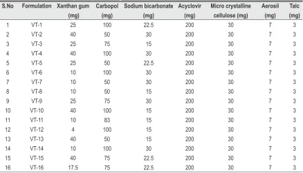
Vaginal tablets of acyclovir were prepared by direct compression method. Accurately weighed quantity of acyclovir was triturated with appropriate quantities of carbopol 934P and xanthan gum (Table 1) in a mortor and pestle. To this sodium bicarbonate, microcrystalline cellulose, aerosil and talc were added, triturated and passed through BSS# 80. The resulting mixture were compressed using 12 mm caplet punches in rotary tablet punching machine (Rimek RSB-4 Mini Press).
Optimization of acyclovir vaginal tablets by D-optimal design
The runs based on D- Optimal design using response surface methodology, were utilized to evaluate the response variables. The formulation variables are 1) Amount of xanthan gum (X1) 2) Amount of carbopol (X2) and 3) Amount of sodium bicarbonate (X3). The responses subjected for the analysis was; 1] Swelling index (Y1) 2]
Time taken for 90% drug release (t90%) in hours (Y2). Each variable was studied at two different levels (-1, +1) and center point (0) which is the midpoint of each factor range. The minimum and maximum range of variables investigated for the variables were 10 mg to 40 mg for Xanthan gum; 50 mg to 100 mg for carbopol 934P and 15 mg to 30 mg for sodium bicarbonate. The responses were subjected to multiple regression analysis to find out the relationship between the factors used and the responses obtained. The effect of formulation variables on the response variables were statistically evaluated by applying one way ANOVA at 0.05 level using a commercially available software package Design Expert 8.04 trial version ( Stat Ease, USA). The optimization of the bioadhesive vaginal tablets was carried out by taking into consideration the amount of polymer as formulation variables whose operating range is mentioned in the Table 1. All experiments were carried out in triplicate.
Compatibility studies
Differential scanning calorimetry
Acyclovir, carbopol 934P, xanthan gum and physical mixture of polymers with drug were subjected for DSC studies using differential scanning calorimeter (Mettler-7, Germany). 5mg of sample was placed in a 50 µl perforated aluminium pan and
0 0
sealed. Heat runs for each sample were set from 5 C to 300 C
8
using nitrogen as purging gas and the samples were analyzed .
Analytical method development of acyclovir
A new, simple, accurate, environmental friendly, cost effective, safe, and sensitive spectrophotometric method for
S.No 1 2 3 4 5 6 7 8 9 10 11 12 13 14 15 16 Formulation VT-1 VT-2 VT-3 VT-4 VT-5 VT-6 VT-7 VT-8 VT-9 VT-10 VT-11 VT-12 VT-13 VT-14 VT-15 VT-16 Xanthan gum (mg) 25 40 25 40 25 10 10 10 25 40 10 4 40 10 40 17.5 Carbopol (mg) 100 50 75 100 50 100 50 50 75 100 83 100 50 100 75 75 Sodium bicarbonate (mg) 22.5 30 15 30 22.5 30 30 15 30 15 15 15 15 30 22.5 22.5 Acyclovir (mg) 200 200 200 200 200 200 200 200 200 200 200 200 200 200 200 200 Micro crystalline cellulose (mg) 30 30 30 30 30 30 30 30 30 30 30 30 30 30 30 30 Aerosil (mg) 7 7 7 7 7 7 7 7 7 7 7 7 7 7 7 7 Talc (mg) 3 3 3 3 3 3 3 3 3 3 3 3 3 3 3 3
estimation of acyclovir using equal volume of 4M urea and 25% sodium acetate as hydrotropic solution was developed.
Preliminary solubility studies of the drug
Solubility of acyclovir was determined by saturation aqueous solubility method in equal volume of 4M urea and 25% sodium acetate in distilled water. An excess amount of drug was added to the 50ml beakers containing equal volume of 4M urea and 25% sodium acetate in distilled water. The
0
beakers were shaken at 27±0.5 C for 24 hours. The solution was then filtered through Whatmann filter paper # 41. Then the filtrate was suitably diluted and analyzed spectrophotometrically against corresponding solvent blank at a wavelength of 255nm (Shimadzu UV-1700).
Preparation of standard stock and calibration curve
The standard stock solution of acyclovir (1mg/ml) was prepared by dissolving 50mg of drug in 50 ml of equal volume of 4M Urea and 25% Sodium acetate. From this stock solution working standard solutions having concentrations 5, 10, 15, 20, 25, 30 and 40 µg/ml was prepared with distilled water. The absorbance of resulting solutions were measured spectrophotometrically at 255nm and a calibration curve was plotted to get the linearity and regression equation.
Analysis of Acyclovir in tablets using 4M Urea and 25% Sodium Acetate
Twenty tablets (commercially available product) were weighed and powdered. Powder equivalent to 200 mg acyclovir was weighed and transferred to a 100 ml volumetric flask containing 80 ml of equal volume of 4M urea and 25% Sodium acetate. The flask was shaken for about 5 min to solubilize the drug. Then volume was made up to the mark with distilled water. Solution was filtered through Whatmann filter paper #41. A part of filtrate was taken and kept at room temperature for 24 hours to check the effect on stability of drug in presence of urea and also to note precipitation. The remaining part of filtrate was appropriately diluted with distilled water and analyzed for drug content measured at a wavelength of 255nm.There was no precipitation in the filtrate for 48 hours.
Validation of the proposed method
The proposed analytical method was validated for parameters such as linearity, percent recovery, precision, robustness,
9,10
LOD and LOQ .
Evaluation of bioadhesive vaginal tablets
Surface pH
One tablet was placed in 10 ml of simulated vaginal fluid (SVF) in small beakers and it was allowed to swell and the pH was measured at time intervals of 1, 2, 3, and 4 up to 8 h by
11
placing the electrode in contact with the surface of the tablet . Average of three determinations was taken and calculated.
Swelling study
One tablet was weighed (W ) and placed in a petri dish 1
containing 5ml of SVF. At interval of 1hr, the tablet was removed from the petri dish and wiped off with a tissue paper. The swollen tablet was then reweighed (W ) and the swelling 2
index were calculated by taking average of three determinations using the formula given below.
Swelling Index = W2 x W1 W1
x 100
Ex-vivo bioadhesion strength
Mucoadhesive strength of the tablet was determined by using
12,13
modified balance method . Fresh sheep vaginal mucosa was obtained from local slaughter house and suitable dimension of the mucosa was fixed to the apparatus using cyanoacrylate adhesive. One end of the tablet is adhered on to the mucosa with little amount of simulated vaginal fluid there by creating adhesive bond between the tablet and the membrane. The other end of the tablet is connected to the pan for the addition of weights. The weights were added slowly to the pan until the tablet gets detached from the mucosal surface, from which mucoadhesive strength of the tablet in grams was determined. The following parameters were also calculated by using the bioadhesion strength.
Force of Adhesion (N) = (Bioadhesion Strength×9.81)/1000
2
Bond Strength (N/m ) = (Force of Adhesion (N))/(Surface Area)
In-vitro dissolution studies
The studies were carried out in USP dissolution apparatus Type II containing 600ml of Simulated Vaginal Fluid pH4.5
0 0
maintained at 37 C± 0.5 C and rotated at 50 rpm. 2ml of solution was withdrawn from the dissolution apparatus at regular predetermined time intervals and same volume of sample was replaced with fresh dissolution medium. The collected samples were diluted suitably and their absorbances were measured spectrophotometrically at 253 nm using
UV-14
visible Spectrophotometer (UV-1601, Shimadzu) .
Kinetic modeling of drug release mechanism
The dissolution data of all formulations were fitted to zero-order, first-zero-order, Hixson-Crowell, Higuchi and Korsemeyer and Peppas models to predict the drug release mechanism.
In-vitro drug Permeation
beaker containing 100ml of SVF and placed on the magnetic stirrer, stirred at 50 rpm by maintaining the temperature at
0
37 C. Samples of 2ml were withdrawn at predetermined intervals and amount of acyclovir was analyzed
15
spectrophotometrically at 253 nm . Average of three determinations was taken and calculated.
In-vitro absorption studies
The dissolution-absorption studies were performed using
16
Continuous Dissolution absorption system . The optimized batch of acyclovir vaginal tablet was used in the absorption studies. The medium consisted of 600ml of SVF of pH 4.5
0
maintained at 37 ± 0.5 C. A freshly sacrificed sheep vaginal mucosa was obtained from the slaughter house and a segment was clamped to the perfusion apparatus. The total volume of the absorption compartment (tube A and tube B of perfusion apparatus) was 30ml of SVF. The drug diffused from the dissolution medium (mucosal side) to the serosal side (absorption compartment). The tablet was placed in the dissolution basket of the designed system and rotated at 50 rpm. Dissolution samples (2 ml) were withdrawn at regular intervals of time and analyzed spectrophotometrically at 253 nm. The transported drug from the absorption compartment was sampled at 3 min later than their corresponding dissolution samples and analyzed spectrophotometrically at 253 nm. The whole experiment was repeated in triplicate (n=3) using fresh dissolution medium as well as fresh vaginal mucosa each time.
Stability studies
Accelerated stability studies were carried out as per ICH guidelines (QIA) where the conditions were maintained 40°±2°C with 75± 5% RH for a period of 6 months. The formulations were closely packed in aluminum foils and stored in stability chamber and evaluated for their physical appearance, drug content and in-vitro drug release studies at intervals of 2 months. The shelf life period of the prepared
17
vaginal tablets is determined by using similarity factor .
RESULTS AND DISCUSSION
Vaginal tablets of acyclovir were prepared by direct compression method. In the present study 16 formulations of acyclovir with varying concentration of Carbopol 934P and Xanthan gum used as bioadhesive agents were prepared, each tablet weighing 400mg.
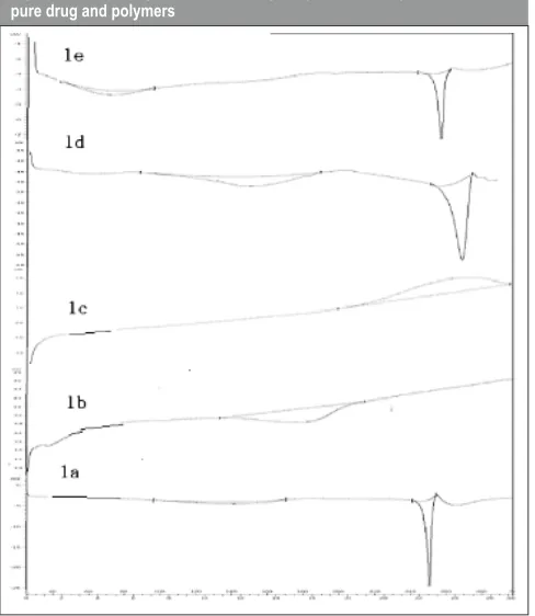
Differential scanning calorimetry
The DSC analysis of pure drug, polymers and the physical mixture were carried out to evaluate any possible interaction between drug and polymer. The DSC thermogram of pure drug acyclovir showed a characteristic endothermic peak at
0
256 C as in figure 1a which is the melting point of acyclovir. DSC thermogram of carbopol 934P revealed a bulge at
0
216.82 C (1b) confirming the amorphous nature of the polymer. DSC thermogram of xanthan gum revealed an
0
exothermic peak at 271 C (1c). Similar sharp endothermic
0
peaks at 256 C (figures 1d, 1e) was observed in physical mixture of acyclovir and polymers. This study confirmed that there was no interaction between the drug and the polymer.
Analytical method development
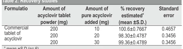
The solubility studies indicated that aqueous solubility of acyclovir was enhanced in hydrotropic solution of equal volume of 4M urea and 25% sodium acetate as compared to solubility in distilled water. The Beer-Lambert's concentration range was found to be 5-40 μg/ml for acyclovir at the wavelength of 255 nm. The solubility of pure acyclovir in distilled water was found to be 10.34mg/ml, whereas in the equal volume of 4M urea and 25% sodium acetate, the solubility was found to be 26.4mg/ml. There was a marked increase in solubility of acyclovir in the hydrotropic solution used. So it was optimized to employ this solution in the analysis of the tablet formulation. A part of the solution was kept at room temperature for 24 hours to check the effect on stability of drug in presence of urea and for precipitation. The study revealed that estimations of acyclovir can be done within 24 hours without any detrimental effect on drug stability. The drug showed good regression value at this wave length. It was evident that there was good correlation between the amounts estimated and the label claim. The estimated label claim was found to be 99.33±0.57mg with a standard error of 0.389. Accuracy and reproducibility of the proposed method were further confirmed by the recovery studies. The results of recovery study revealed that any small change in the drug concentration in the solution could be accurately determined by the proposed method (Table 2). The method
-1
was found to be robust within the Beer's range of 5-40 μg ml . The low values of LOD and LOQ indicated good sensitivity of proposed method. Repeatability results indicated the precision under the same operating conditions over a short
a
mean ±S.D (n= 6)
Amount of acyclovir tablet
powder (mg) 200 200 200
Amount of pure acyclovir
added (mg) 10 20 30
% recovery
a
estimated (mean ±S.D.) 100.6±0.7667 98.30±0.4787 99.36±0.4789
Standard error
0.4657 0.3456 0.3456 Formulatio
Commercial tablet of acyclovir
interval time and inter- assay precision. Intermediate precision study expresses within laboratory variation in different days. In both intra and inter-day precision study for the method co-efficient of variation the values were 0.2014 and 0.2890 that were not more than 1.0% which confirmed the good intermediate precision. The molar absorptivity was
3
found to be 13.67 X 10 , whereas the correlation co-efficient and slope were found to be 0.987 and 0.059 respectively. The
-1
values of LOD were 0.3664 μg ml and LOQ were 0.6789 μg
-1
ml respectively. This developed and validated analytical method for acyclovir was used in the evaluation of drug content of the prepared vaginal tablet formulations.
Evaluation of prepared tablets
Ex- vivo Bioadhesive strength
There is a strong prophylactic and clinical need to develop vaginal products with desired characteristics such as product dispersion throughout the vagina and retention for intended intervals. Retention of a dosage form in vaginal cavity for prolonged intervals is desirable for therapeutic efficacy and minimizes the need of frequent dosing intervals. The bioadhesive strength of the tablets was found to be a function of nature and concentration of polymer. The tablets showed bioadhesive strength between 125 to 390 g. As the polymer concentration increased bioadhesive strength of the prepared tablets increased. Vaginal tablets containing high amount of xanthan gum were found to have increased bioadhesive strength. The bioadhesive strength exhibited by the xanthan
gum and tablets were considered satisfactory for maintaining them in the vaginal cavity.
Force of adhesion and Surface pH
The force of adhesion for all 16 formulations was found to be in the range of 1.22 N for formulation VT 1 and 3.82 N for formulation VT 16. This study demonstrated as the polymer concentration increased the force of adhesion increased. The surface pH of all 16 formulations was determined in SVF pH 4.5. There was no changes in pH and all the formulations possess the pH In the range of 4.52- 4.88.
Swelling Study
The swelling studies for all 16 formulations were performed and it was observed that as the polymer concentration of carbopol 934P increased, there was a marked increase in the swelling index. This was observed in all the formulations. The maximum swelling index was observed to be 156 % in
th
formulation VT 6 at the end of 8 hour and the least swelling index was observed to be 71 % in formulation VT 11 after 8 hours.
In-vitro drug release studies
The simulated dynamic vaginal system used in this study mimics the physic dynamic conditions of the vagina. As evident from the diverse nature of dissolution profiles the influence of polymer seems to be vital in regulating the drug release. In-vitro drug release studies were performed in SVF pH 4.5 for all the prepared formulations by using USP dissolution test apparatus-type II, rotating paddle method. The graphs showing drug release profile for formulations were shown in the figure 2a & 2b. In-vitro dissolution studies showed that the formulation containing carbopol 934P and xanthan gum (VT1, VT3, VT7, and VT8) showed 100% drug release with in a period of 6 hours. In formulations VT4, VT9, VT10, VT12, VT13, VT14, VT15, an increase in concentration of xanthan gum was found to delay the drug release over an extended period of time. Formulations VT2, Vt6 and VT11 sustained the drug release over a period of 8 hours.
In-vitro drug release mechanism
Models with highest correlation coefficient were judged to be most appropriate model for dissolution data. The drug release data when fitted into various kinetic equations, showed Koresmeyer - Peppas release pattern for all formulations
2
except Vt7, that showed zero order with r value of 0.9826,
2
VT15 showed zero order release with r of 0.9784 and VT13
2
showed hixon-crowell release with r value of 0.9879 respectively.
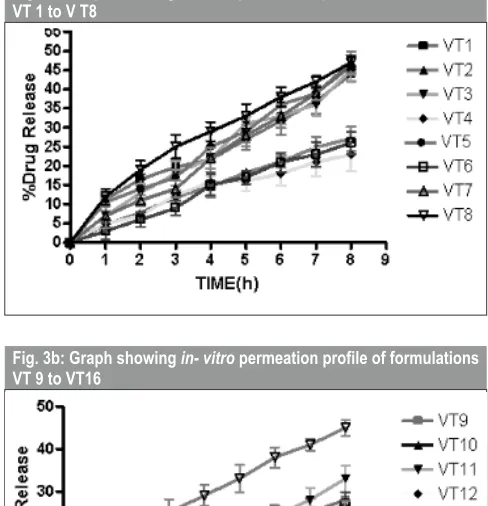
In-vitro drug permeation studies
In-vitro drug permeation was performed using fabricated
apparatus at 37±0.5°C. The graphs showing drug release profile for formulations were shown in the figure 3a & 3b. In-vitro permeation studies revealed that acyclovir vaginal tablet formulations containing increased concentration of Carbopol 934P and less concentration of xanthan gum in the range of 10 - 20mg showed maximum drug permeation of 45%. Tablet formulations containing Xanthan gum in the range of 20 - 40mg showed permeation in the range of 20-25% indicating as the concentration of xanthan gum is increased permeation is delayed.
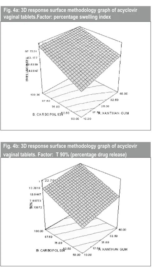
Optimization of vaginal tablet formulations
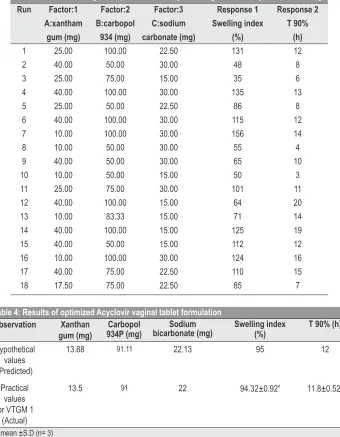
Results of design experiments for acyclovir vaginal tablets by D- Optimal design are shown in table 3. The 3-dimensional response surface graphs for the factors percent swelling index
and T 90 % were plotted shown in figures 4a and 4b respectively. From the numerical optimization results, one of the solutions was selected randomly as the optimized formula and coded as VTGM1 and this optimized formulation was characterized further. The generated optimization study was conducted to study the constraints on the design space and the vulnerability of the experimental model. This is important, since it suggest factors, responses and the goal for each variable with respect to the measured response. The results were shown in the table 4 which confirmed there is a correlation of the predicted results with that of the observed results. It was observed that the response was almost similar to the response predicted by the design expert software. All the further studies were carried out using this optimized batch for swelling and In-vitro studies.
Run 1 2 3 4 5 6 7 8 9 10 11 12 13 14 15 16 17 18 Factor:1 A:xantham gum (mg) 25.00 40.00 25.00 40.00 25.00 40.00 10.00 10.00 40.00 10.00 25.00 40.00 10.00 40.00 40.00 10.00 40.00 17.50 Factor:2 B:carbopol 934 (mg) 100.00 50.00 75.00 100.00 50.00 100.00 100.00 50.00 50.00 50.00 75.00 100.00 83.33 100.00 50.00 100.00 75.00 75.00 Factor:3 C:sodium carbonate (mg) 22.50 30.00 15.00 30.00 22.50 30.00 30.00 30.00 30.00 15.00 30.00 15.00 15.00 15.00 15.00 30.00 22.50 22.50 Response 1 Swelling index (%) 131 48 35 135 86 115 156 55 65 50 101 64 71 125 112 124 110 85 Response 2 T 90% (h) 12 8 6 13 8 12 14 4 10 3 11 20 14 19 12 16 15 7
Table 3: Results of design of experiments for Acyclovir vaginal tablets by D- Optimal design
Hypothetical values (Predicted)
Practical values for VTGM 1
(Actual) Observation Xanthan gum (mg) 13.88 13.5 Carbopol 934P (mg) 91.11 91 Sodium bicarbonate (mg) 22.13 22 Swelling index (%) 95 a 94.32±0.92
T 90% (h)
12
a 11.8±0.52
Table 4: Results of optimized Acyclovir vaginal tablet formulation
Fig. 3a: Graph showing in- vitro permeationprofile of formulations VT 1 to V T8
Fig. 3b: Graph showing in- vitro permeationprofile of formulations VT 9 to VT16
In-vitro drug release studies
The in-vitro drug release data for optimized formulation and
th
the drug release mechanism showed 90% drug release in 12
2
hour with an r value of 0.9931. The curve fits for various release systems for optimized formulation were shown in table 5. The n-value was found to be 0.1392 and the k-value was found to be 56.5185 for the optimized formulation.
Kinetic modeling of drug release
Models with the highest correlation coefficient were judged to be the most appropriate model for the dissolution data. The optimized formulation has shown correlation coefficient
value 0.9931 that showed Peppas would be the most appropriate drug release mechanism where the drug could be released by diffusion process.
Swelling Studies
The swelling studies for optimized formulation showed maximum swelling at the end of the study. The optimized formula showed 95% of swelling in 12 hours. Hence there is a correlation between hypothetical and practical values.
In-vitro absorption studies
The vaginal mucus membrane is the rate-limiting in the proposed absorption system. As the drug released in the dissolution medium, this is available for absorption through the mucus membrane. The concentration between the two sides of the tissue helps to maintain sink conditions. The absorption profile of the optimized formulation showed that the amount of the drug in the absorption medium is 26.7% at
th
the end of 8 hour.
Stability studies
Stability of the tablets is crucial during storage. Stability studies revealed that the tablets kept at elevated temperature
Fig. 2a: Graph showing in- vitro drugrelease profile of formulations VT 1 to VT 8
Fig. 2b: Graph showing in- vitro drugrelease profile of formulations VT 9 to VT 16
Equation
Zero order st 1 order Matrix Peppas Hixon-Crowell
Regression coefficient ( r) 0.6871
0.3213 0.5771 0.9931 0.7932
k 9.9322 0.1861 29.3331 56.5185 0.0493 Kinetic values
0
of 40 C and 75% RH showed maximum stability. The similarity factor value was found to be 62.22, which confirmed that it has an acceptable shelf life up to 3 years.
CONCLUSION
Vaginal tablets of acyclovir were prepared using carbopol 934P and xanthan gum as bioadhesive polymers. The vaginal tablets were found to sustain the release of acyclovir for 12 hrs in simulated vaginal fluid. The present study signifies the utility of vaginal delivery system in retarding the drug release. This may in turn reduce the frequency of dosing, there by improving the patient compliance.
ACKNOWLEDGEMENT
The authors are thankful to Gokula Education Foundation, Bangalore for providing necessary facilities to carry out the research work and Indian Institute of Science, Bangalore for providing DSC facility.
REFERENCES
1. Duchene D, Ponchel G. Bioadhesion, a new pharmacotechnical method for improving therapeutic efficiency. STP Pharma. 1989; 5: 830-8.
2. Park K, Park H. Test methods of bioadhesion, in: Bioadhesive Drug nd
Delivery Systems, CRC Press, Boca Raton, Florida. 2 ed. 1990; 43.
3. Zaneveld LJD, Waller DP, Ahmad N, Quizg J, Kaminski J, Nikurs A, Jonge CD. Properties of new long-lasting vaginal drug delivery system for contraception and anti-microbial agents. J Androl. 2001; 22: 481-90.
4. Kavita V, Sanjay G. The scope and potential of vaginal drug delivery. Pharm sci technol. 2000; 3(10): 359-64.
5. Robinson JR, Bologna WJ. Vaginal and reproductive system treatments using a bioadhesive polymer. J Cont Rel. 1994; 28: 87–94.
th
6. Martindale. The complete drug reference. 34 edition. Pharmaceutical Press: 626-7.
7. http://en.wikipedia.org/wiki/acyclovir. (Accessed on November, 2011).
8. Hirlekar RS, Kadam VJ. Design of buccal drug delivery system for a poorly soluble drug. Asian J Pharm Clin Res. 2009; 2(3): 49-53.
9. Anish VSN, Deveswaran R, Bharath S, Basavaraj BV, Madhavan V. Application of mixed hydrotropic solubilization in spectrophotometric estimation of aceclofenac in tablets. Curr Pharm Res. 2011; 1(3): 223-6.
10. Anish VSN, Deveswaran R, Jhansipriya Marabathuni V, Bharath S, Basavaraj BV Madhavan V. Simultaneous estimation and validation of aceclofenac and paracetamol from bulk and tablets using mixed hydrotropic solubilisation. Asian J Bio Chem Pharm Res. 2011; 1(1): 19-24.
11. Bhat SR, Hani Umme, Ritesh KG, Shivakumar HG. Development and characterization of bioadhesive vaginal films of clotrimazole for vaginal candidiasis. Acta Pharm Sci. 2010; 52: 417-26.
12. Amit G, Ram GS, Ganga S. Development, evaluation and optimization of extended release buccal tablets prepared using progressive hydration technology. Int J Drug Del. 2010; 2: 37-48.
13. Patel VM, Prajapati BG, Patel MM. Formulation, evaluation and comparison of bilayered and multi layered mucoadhesive buccal devices of propranolol hydrochloride. AAPS Pharm Sci Tech. 2007; 8(1): Article 22.
14. Yesim KH, Suleyha H, Dilek MY, Tamer G. Efficacy of a new ketoconazole bioadhesive vaginal tablet on candida albicans. IL Farmaco. 2004; 59:163-7.
15. Gursoy A, Sohtorik I, Uyanik N, Peppas NA. Bioadhesive controlled release systems for vaginal delivery. STP Pharma.1989; 5: 886-92.
16. Kale V, Kasliwal RH, Avari JG. Attempt To design continuous dissolution- absorption system using everted intestine segment for in vitro absorption studies ofslow drug release formulations. Disso Tech.2007; 5:31-6.
17. Wolfgang Grimm. Extension of the international conference on harmonization tripartite guideline for stability testing of new drug substances and products to countries of climatic zones III and IV. Drug Dev Ind Pharm. 1998; 24(4): 313-25.
Fig. 4a: 3D response surface methodology graph of acyclovir vaginal tablets.Factor: percentage swelling index