UNIVERSITY of TRENTO
______________________________________________
Centre for Mind / Brain Sciences – CIMeC Doctoral School in Cognitive and Brain Sciences
XXIII cycle
Interaction between language and analogical reasoning from
the brain imaging perspective
Ph.D. Dissertation of:
Alessia Giovenzana
Advisor: Gianpaolo Basso
Index
______________________________________________
1. Chapter I: Introduction ………...p.5
2. Chapter II: Brain activity during analogical reasoning within different verbal
contexts: words vs. nameable pictures ………..p.11 Introduction……….p.11 Material and methods………...p.13 Results...……….……….p.19 Discussion……….. ….p.26
3. Chapter III: Brain activity during analogical reasoning in language impaired
subjects: the case of developmental dyslexia ……….p.33 Introduction……….p.33 Material and methods………...p.39 Results………..p.44 Discussion……….. ….p.63
4. Chapter IV: Brain activity during analogical reasoning within non-verbal
context……….………...p.75
Introduction……….p.75 Material and methods………...p.76 Results………..p.84 Discussion……….. ….p.93
5. Chapter V: General conclusions …..……….p.99
Chapter I
______________________________________________
Introduction
Analogy (from Greek "ανα-λογos”, "related to") is the cognitive process of transferring information from a particular subject (the analogue or source) to another
particular subject (the target), deriving the relationship between a group of items and
then applying that relationship to help the reasoning about a different group of items.
Traditionally, an analogy is represented as a four term problem: “A:B as C:D”. The
solution requires the ability to retrieve information-concepts from semantic memory,
the ability to form and manipulate mental representations of relations between objects
and events, and to compare the resulting prepositions between pairs (Bunge,
Wendelken, Badre, & Wagner, 2005). Analogical reasoning is considered a core
component of intelligence and cognition (Gentner, 2003): the relational thinking allows
us to concatenate previous/different experiences/concepts and arrive at a new
conclusion, as well as to generalize experience from particulars. The analogical
thinking is the ground/basis of metaphors and gives us the possibility to use concrete
concepts/experience/relations for discussing and explaining new abstract ideas
Functional neuroimaging studies that examined the neural basis of analogical
reasoning documented a clear left hemisphere involvement: relational thinking in
general elicits strong activity in a left-lateralized group of brain regions among which
al., 2005; Cho et al.; Christoff et al., 2001; Kroger et al., 2002; Monti, Parsons, &
Osherson, 2009). As a consequence, particular interest has been devoted to the
functional organization within the prefrontal cortex to investigate if/how different
sub-regions subserve different components of the reasoning process (Bunge, et al.,
2005; Cho, et al.; Hampshire, Thompson, Duncan, & Owen, 2011; Volle, Gilbert, Benoit,
& Burgess).
In addition to the prefrontal cortex, brain areas traditionally related to verbal
language may be involved to a different extent and grading in reasoning (Bunge, et al.,
2005; Christoff, et al., 2001; Green, Kraemer, Fugelsang, Gray, & Dunbar, 2010;
Hampshire, et al., 2011; Luo et al., 2003; Prabhakaran, Smith, Desmond, Glover, &
Gabrieli, 1997; Wharton et al., 2000; Whitney, Grossman, & Kircher, 2009). On the
same track, clinical data suggest that the integrity of the language-dominant
hemisphere is necessary to solve analogical tasks (Baldo, Bunge, Wilson, & Dronkers,
2010; Baldo et al., 2005; Langdon & Warrington, 2000). Langdon and Warrington
documented that patients with left hemisphere lesions were impaired on both verbal
and visuo-spatial tasks (Langdon & Warrington, 2000); Baldo and Colleagues, studying
left-hemisphere stroke patients, suggest a correlation between non-verbal relational
thinking and a lesion of core language regions such as the superior and middle
temporal gyrus (Baldo, et al., 2010; Baldo, et al., 2005). On the other hand, studies with
Alzheimer‟s disease patients suggest that the left prefrontal areas critical for relational
reasoning are those subserving working memory and executive functions, traditionally
not considered core areas for language processing (Waltz et al., 2004; Waltz et al.,
These observations agree with many behavioral evidences of the involvement of
language in reasoning (Baldo, et al., 2005; Carruthers & Bermùdez, 2006;
Hermer-Vazquez, Spelke, & Katsnelson, 1999). Gentner (2003) proposed a more general
theoretical framework for reasoning within which language provides an internal
cognitive tool which fosters high order relational concepts throughout the possibility to
use linguistically shaped relations. The ability to use words that refer to relations (such
as “cause”, “inhibit”, “source”, “advantage”, etc), that Gentner calls “relational
language”, help us to manipulate concepts, relations, and abstract entities. Thus, in
Gentner‟s position, verbal language provides a symbolic system which serves to
develop and learn relational concepts and provides cognitive stability to them. In this
view, the development/acquisition of language, and in particular the relational
language, during childhood contributes to the development of analogy and cognition
because language provides the control over mental processes (Vygotsky, 1962).
Learning specific relational terms provides representational resources that augment
cognitive processes and the possibility of abstraction and generalization (Gentner,
2003; Gentner & Christie, 2010).
Despite the evidences about the strong link between language and thinking, the
neural substrate of verbal language comprise a widespread network of mostly left
lateralized brain areas each subserving different aspects of verbal language (Price,
2000, 2010). It is unclear if all components of verbal language are needed, support or
influence the reasoning itself.
Some behavioral studies suggest a role of covert verbalization during flexible
1999). On the other hand, functional neuroimaging studies demonstrated that the
classical Wernicke-Broca circuit, which subserves overt and covet verbalization by
means of lexical and phonological processing, may be not necessary to perform
reasoning unless verbal language is needed for the decoding of the terms (Monti, et al.,
2009). Interestingly, there area data in the literature which suggest that the access to the
conceptual knowledge, that is the mandatory step for analogical reasoning, may differ
depending on the stimulation modality. For example, Saffran et al. (2003)
demonstrated that pictures and words representing semantic concept evoked
significantly different words associations. Caramazza (1990) suggested that visually
presented objects`, through their perceptual features`, may have a more direct access to
semantic knowledge. Thus, even without entering the question about how conceptual
knowledge is organized in the brain, it is clear that its retrieval and manipulation may
be influenced by the stimulation modality. It is then possible also to argue that the
reasoning may be affected by the stimulus format. While it is known that other factors,
such as the number of visuo-spatial relationships (Christoff, et al., 2001), the relational
complexity (Kroger, et al., 2002), and the associative strength (Bunge, et al., 2005),
strongly influence the activity of the prefrontal cortex during reasoning, to our
knowledge, only one study investigated the modulation induced by the stimulation
context on reasoning using abstract and meaningful pictures. Even in that case the
analysis has been “limited” to the prefrontal cortex subregions (Krawczyk, Michelle
McClelland, & Donovan, 2011).
When I originally planned the research activity for this thesis, the principal aim of
my work was to study the higher cognitive functions, in particular the analogical
reasoning, in a young population of dyslexics in order to understand if the reading
disorder may influence the reasoning in relation to the language load requested by the
stimulus format. In fact, independently from the theoretical approaches to dyslexia and
the cognitive domain explored, up to now the data available in the literature deal
mainly with the nature of the deficit associated with developmental dyslexia. But
evidences also suggest that developmental dyslexics may have a talent for activities
related to the non-verbal domain including complex reasoning. The question I wanted
to investigate was if this observation could be linked to a particular neural brain
organization both at the structural and functional level. For this reason I developed an
fMRI paradigm to investigate brain activity during analogical reasoning performed
either with words or pictures. The hypothesis was that, if a particular talent is present
in dyslexics, they may have shown significant brain reorganization compared to
normal readers, especially with pictures which allow but do not require a mandatory
use of language.
As a first step the fMRI paradigm was applied to study a sample of normal adults.
This study allowed to verify how much reasoning relies on verbal language. In fact,
despite the increasing interest about the neural substrates of reasoning, at present no
fMRI study systematically investigates the contribution of stimulus format/context on
results of this first study will focus on how different verbal context, nameable pictures
versus words, may influence brain activity related to analogical reasoning.
In a second study the same paradigm was applied to investigate brain activity
during analogical reasoning in young normal readers and young dyslexics in order to
tackle the main question that I wanted to investigate.
Finally, considering the results of the first two fMRI studies, a new question arose
about the contribution of language and semantic system to reasoning. Thus, a new
fMRI design was implemented where analogical reasoning had to be performed either
on meaningful or on abstract geometrical pictures. Results form this last study
provided new information about the relation between language and analogical
reasoning that could be derived form the observation of the brain activity elicited by
the two contexts.
Chapter II
______________________________________________
Brain activity during analogical reasoning within different
verbal contexts: words
vs.
nameable pictures
The aim of this study was to explore how brain areas responsive to analogical
reasoning are modulated by the modality of access to semantic information, using
words and nameable pictures to present the terms of the analogy. We assumed that
words require a mandatory phonological and lexical elaboration in order to access the
underlying semantic information. This also may trigger a more verbal strategy to
manipulate the information in order to perform the reasoning. In this respect one may
expect that covert verbalization or inner speech may be always present in a word
context. Instead, when pictures are presented, despite lexical-phonological processing
is possible, and may be somewhat automatically triggered in order to name the picture,
the use of covert verbalization is not mandatory since pictures may have a more direct
access to the semantic system and convey enough information for the reasoning even
without any lexical-phonological support.
We implemented a block fMRI design where analogical reasoning task had to be
performed in word and picture context. We provided a limited time interval during
indicated by the literature to be critical for reasoning, verbal language and
conceptual-semantic competences. In the present study the term “core language areas” will be
used referring to the standard thesis about the localization of language which assert
that human linguistic components are embodied by structures close to the left sylvian
fissure - i.e. the classical Wernicke-Broca circuit (Bookheimer, 2002; Monti, et al., 2009).
On the basis of the literature we expected that core areas for reasoning in
prefrontal cortex as well as areas known to be part of semantic system should not be
modulated by the context of reasoning. Differently, the behavior of core language areas
can be informative respect to the influence of context in analogical reasoning. We
assumed that when reasoning is performed on words the involvement of the
lexical-phonological system is mandatory to access the meaning, but its involvement is not
obvious in a picture context. If the hypothesis of a “privileged access” suggested by
Caramazza et al. (1990) is true, reasoning within a picture context may not engage the
core language regions, especially in the case of a short time available for the response.
Differently, in case a covert verbalization strategy is needed for reasoning despite the
context on which it is performed, we should expect an involvement of verbal language
Material and Methods
Subjects
15 right-handed subjects were recruited for this study (7 males and 8 females,
mean age 27 years, range 22-42). All participants were Italian native speakers with no
history of neurological or psychiatric disorders. Handedness was assessed using the
Edinburgh Inventory (Oldfield, 1971). The study was approved by the Ethics
Committee of the University of Trento and all participants signed an informed consent
form.
Stimuli
The stimuli of picture context were drawn and modified from the Non Verbal
Intelligence Test (Wiederholt, 2004). We prepared 24 stimuli for each task: the
analogical reasoning (AnR) and the semantic judgment (Sem). In the Picture-AnR task,
each stimulus was composed by two pairs of grey scale pictures; in the Picture-Sem
task each stimulus was composed by a triplet of grey scale pictures. In 16 of the
Picture-AnR stimuli, the two picture pairs had the same type of analogical relationship
(True Items) while in the remaining 8 stimuli the type of relationship was different
(False Items). Similarly, in 16 Picture-Sem stimuli the three pictures belonged to the
same category (True items) while 8 did not (False items). Figure 1a and 1b shows an
example of the stimuli used.
The stimuli for word context were prepared on the basis of the picture context
pairs of words; in Word-Sem each stimulus was composed by a triplet of words. In 16
of the Word-AnR stimuli, the two words pairs had the same type of analogical
relationship (True Items) while in 8 stimuli the type of relationship was different (False
Items). Similarly, in 16 Word-Sem stimuli the three words belonged to the same
semantic category (True items) while 8 did not (False Items).
The words were controlled for frequency based on the corpus COLFIS
(http://alphalinguistica.sns.it/BancheDati.htm) and no significant differences were
found between stimuli used for the AnR and the Sem task. Figure 2.1 shows an
example of the stimuli used.
The words and picture stimuli used in both tasks were matched for two main
factors: the ratio of living/non-living items and the semantic distance. Semantic
distance was computed based on Likert ratings (1 = low semantic distance, 7 = high
semantic distance) given by 20 subjects who did not participate to the fMRI
experiment. Each pair of items used in the AnR task and each triplets used in the Sem
task was judged individually. A two-tail t-test for unpaired samples did not reveal any
significant difference in semantic distance between picture and word items within each
task (Picture-AnR: mean 2.97, sd 0.59; Word-AnR: mean 2.86, sd 0.52, p=0.48 n.s.;
Figure 2.1 Examples of the stimuli used
(a) Picture analogical reasoning; (b) Picture semantic judgment; (c) Word analogical reasoning; (d) Word semantic judgment. Transl: (“pecora”=sheep; “lana”=wool; “mucca”=cow; “latte”=milk; “leone”=lion; “giraffa”=giraffe; “elefante”=elephant).
Tasks, fMRI design and procedure
The presentation of the stimuli was performed using a block fMRI design with one
run for each context (Word and Picture). Each run contained 12 blocks, six for each task
(AnR and Sem) presented alternately. Each block contained four stimuli and it was
preceded by specific instructions for task and context lasting 2 s. On AnR task,
participants viewed the two pairs of pictures or words displayed simultaneously on
the screen and they had to indicate whether the analogical relationship between the
two pairs of items was or was not the same. On the Sem task, participants viewed the
indicate whether the three items belonged to the same semantic category. In both tasks,
participants were required to make a yes/no response only during the presentation by
pressing one of two buttons of a response pad with their right hand.
The minimum and maximum stimulus durations were 500 and 5500 ms
respectively. For response times faster than 500 ms, the stimulus disappeared
immediately after the minimum duration. Otherwise, the stimulus disappeared as
soon as the subject gave the response or, in case no response was given, after the
maximum duration. The next stimulus was presented after a blank screen lasting 500
ms. The resulting maximum block duration was 24 s. A variable additional period of
visual fixation was added after the last stimulus of the block to compensate for
responses shorter than 5500 ms and control the blocks onset time. A fixation cross
lasting between 8 and 12 s was presented between blocks. At the beginning and at the
end of the runs a fixation cross was presented for 17.6 s and 17 s respectively. The total
duration of each run was 457.6 s.
Before starting the experiment subjects underwent a training session outside the
scanner with 6 additional trials for each task in order to familiarize with the
experiment.
Imaging data acquisition
Brain images were collected with a 4-Tesla Bruker MedSpec scanner (Bruker Inc.,
Ettlingen, Germany) using an 8-channel head coil. During the scanning sessions, the
motor response was recollected using a fiber optic two button response pad (Cedrus,
on an acrylic screen viewed by the subject through a mirror attached to the head coil
using E-Prime software (Psychological Software Tools, Pittsburgh, PA, USA).
Functional images were acquired using a T2*-weighted gradient-echo echo-planar
sequence (repetition time = 2200 ms, echo time = 33 ms, flip angle = 75°, acquisition
matrix = 64 x 64, slice thickness = 3 mm, inter-slice gap = 0.45 mm, field of view = 192 x
192 mm, number of slices = 37). Each functional run had 208 brain volumes; at the
beginning of each run five dummy scans were acquired.
For the subsequent superimposition of functional statistical parametric maps, a
high-resolution structural 3D T1-weighted image was acquired (MPRAGE sequence,
resolution 1x1x1 mm3, acquisition matrix 256 x 224; number of slices = 176; repetition
time = 2700 ms, echo time = 4.18 ms, inversion time = 1020 ms).
Imaging data analysis
Preprocessing and data analysis were conducted using BrainVoyager QX 1.9
software package (Brain Innovation, Maastricht, The Netherlands).
Functional images from each subject were corrected for slice time acquisition with
cubic spline interpolation. All volumes were realigned using a 3D rigid-body spatial
transformation to the first volume of the first functional run. Temporal filtering
included linear trend removal and a 0.028-Hz (5 cycles in time course) high pass filter
to eliminate low frequency noise. The functional data were co-registered to structural
images and they were spatially smoothed using a Gaussian kernel (full width at half
co-registered functional data were normalized into standard stereotaxic space (Lancaster
et al., 2000).
Statistical analysis was performed using a multi-subject general linear model
random effect analysis in BrainVoyager QX 1.9 software.
A regressor for each set of the four types of trials (W-AnR, AnR, W-Sem and
P-Sem) was created for each functional run and convolved with a standard
hemodynamic response function. Scans acquired during visual fixation were
considered as baseline. The regressors of all subjects were used to implement a
multi-subject GLM random effect analysis. Z-transformation was used for normalization of
signal respect to the baseline. Six motion regressors (3 translation and 3 rotation
parameters) on x, y, z axes were included in the analysis as covariates of no-interest.
Also RT and semantic distance were added as covariates to remove BOLD signal
variation correlated with the response time and/or the semantic distance among items.
The beta maps obtained from the GLM analysis were entered into a 2 x 2 ANOVA
design: [Task (AnR; Sem) x Context (Word; Picture)]. The resulting statistical
parametric maps were corrected for multiple comparisons using the false discovery
rate (FDR) approach with q < 0.005 and excluding all clusters extending less than 0.2
cubic centimeters. The sets of clusters of voxels found to be significantly activated after
this statistical correction were used to define regions of interest (ROI) on which to
perform post-hoc t-tests.
The Talairach Client software (Lancaster, et al., 2000) was used to assign Talairach
Atlas labels for a given x,y,z coordinate, represented by the center of gravity of each
Results
Behavioural in-scanner results
Mean accuracy was 94% and 90% for the Sem and the AnR task respectively
(Picture-AnR: mean 90%, sd 6%; Word-AnR: mean 89%, sd 7%; Picture-Sem: mean
93%, sd 6%; Word-Sem: mean 96%, sd 3%). The ANOVA for repeated measures
revealed that accuracy was significantly higher in Sem task respect to AnR (F[1,14] =
11,06; p<0.004). No effect for context was found (F[1,14] = 0.7; p=0.41) and no
interaction between task and context was present (F[1,14] = 1.33; p=0.27).
The ANOVA for repeated measures revealed that the response time (RT) in Sem
tasks was significantly faster than in AnR tasks (F[1,14] = 268.26; p<0.00001). A
significant effect of context was present with faster RTs for pictures (F[1,14] = 9,7621;
p=0.007). No interaction was found between task and context (F[1,14] = 1.2062; p=0.29).
fMRI results
The fMRI results are summarized in Table 2.1 and 2.2 at the end of the section.
The context effect revealed that the pictures respect to the words were associated
with greater activation in a bilateral set of posterior regions, including primary and
associative visual areas, extending to parietal lobe and to paralimbic regions.
Additional activations were found in the left superior parietal lobule and precuneus
and in the right frontal lobe (inferior frontal and postcentral gyrus).
The words respect to the pictures were associated with greater activation
side and in the left postcentral gyrus. Additional activations were found in the right
cerebellum and the right medial frontal gyrus (Figure 2.2 and Table 2.1).
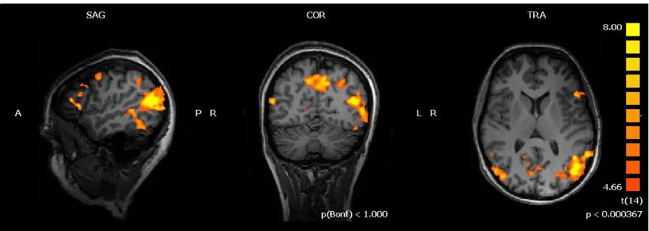
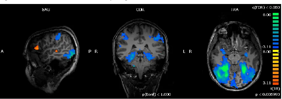
Figure 2.2 Word vs Picture Context
Statistical parametric map of the t-contrast between Word vs Picture context displaying areas responding to words (orange-yellow) and pictures (blue-green) context thresholded at q(FDR) <0.005.
Regions that showed increased activation during solution of analogical reasoning
task were found on parietal, temporal, frontal and paralimbic regions, with a clear
lateralization on the left hemisphere. In particular, analogical reasoning evoked greater
activations in the left fusiform gyrus (BA 37), left parahippocampal gyrus (BA 27),the
posterior part of left middle temporal gyrus (BA 22 and 39), the left middle frontal
gyrus (BA 9 and BA 46) and in the posterior portion of the left inferior frontal gyrus
Figure 2.3 Analogical Reasoning vs Semantic judgment
Statistical parametric map of the t-contrast between Analogical Reasoning vs Semantic judgment task showing areas responding to analogical reasoning thresholded at q(FDR) <0.005.
Notably, no areas displayed an interaction between task and context. Only raising
the statistical threshold (FDR q=0.05) revealed an interaction between the two factors
bilaterally in occipital lobe (Lingual gyrus) and in the parahippocampal gyri.
To asses the influence of context on analogical reasoning, we performed a separate
ROI analysis. We focused the attention on three sets of regions that we found activated:
prefrontal areas found more active in the AnR task, which the literature indicate as
potential core areas for reasoning (BA9, BA46), verbal language areas (posterior part of
IFG - BA 45, BA 44 and the posterior part of superior temporal gyrus – pSTG, BA22)
and regions (left fusiform gyrus - BA37, posterior middle temporal gyrus –pMTG) that
have been linked to conceptual-semantic competences (Bookheimer, 2002; Chao,
Haxby, & Martin, 1999; Martin, 2007).
The specific ROI definition was based both on functional and anatomical criteria.
semantic judgment (NV-AnR + V-AnR vs NV-Sem + V-Sem). Only the BA44 was defined within voxels activated by words vs pictures contrast. The resulting regions of activity were then intersected with anatomical masks using the Talairach Client
software (Lancaster, et al., 2000) in order to include in the analysis only the voxels
belonging to the anatomical regions indicated by the centre of gravity of each cluster
of activation, excluding voxels belonging to adjacent regions. The stereotaxic
coordinates of pMTG were selected on the basis of previous research on picture and
word processing (see Lin et.at. 2011 for a systematic review of functional neuroimaging
studies).
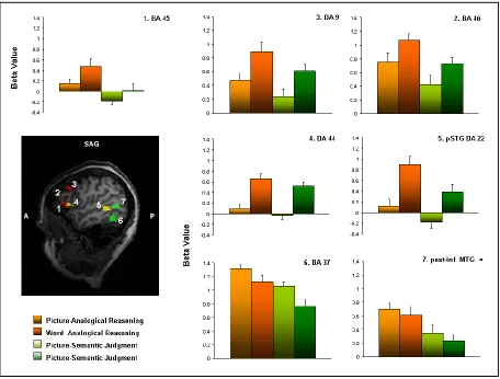
The ROI analysis revealed distinct patterns of activity within these sets of regions
(mean beta values and standard deviations are reported in the graphs of Figure 2.4).
The pre-frontal areas (BA9 and BA46) showed an involvement in both AnR and
Sem task but with significant higher response during the former one (AnR vs Sem: BA9 p=0.02; BA46 p 0=0.0008). Notably, these areas were involved to a similar extent in
word and picture contexts (Words vs Pictures: BA9 p=0.1; BA46 p=0.3). Despite the relative distribution of beta values within linguistic-semantic regions (left fusiform
gyrus -BA37, and the pMTG) was different from that found in prefrontal areas, it
revealed that also in these areas response was higher for analogical reasoning respect
to semantic judgment (AnR vs Sem: BA37 p= 0.0002, pMTG p=0.002), without significative differences between words and pictures contexts (Words vs Pictures: BA37 p=0.25, pMTG p=0.69).
The posterior IFG (BA44) and pSTG (BA22) were influenced by stimulus
context (Words vs Pictures: BA44 p=0.01, pSTG p=0.69). While BA44 did not show differential activation between tasks, BA22 showed a significant higher response in
analogical reasoning (Wors-AnR vs Word-Sem: p= 0.0001).
The only region that showed a selective increased activity for analogical reasoning
not significantly dependent from the context was the anterior part of inferior frontal
gyrus (BA 45)( Words vs Pictures p=0.32; AnR vs Sem p=0.0001) .
Figure 2.4 shows plots of mean normalized beta values for the four types of
Figure 2.4 ROI Analysis
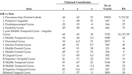
Table 2.1 Context Effect
Talairach Coordinates
Area x y z Voxels Nr of BA Picture vs Word Context
Bil. Occipital & Post. Inf.Temporal Lobe 6 -62 -0.6 111718 18/19/37
L Ant Cingulate Gyrus 0.27 -1.8 37 1200 24
L Precuneus -20 -68 45 1593 7
L Middle Frontal Gyrus -27 22 49 812 8
L Insula -39 -12 4.2 410 13
L Inferior Parietal Lobule -53 -31 35 3713 40
R Inferior Frontal Gyrus 48 -0.79 22 737 9
R Postcentral Gyrus 46 -25 40 2758 2
R Putamen 31 -6 -2.6 830
Word vs Picture Context
L Superior Temporal Gyrus -48 -61 21 1537 39
L Precentral / Inf. Frontal Gyri -51 13 9.3 714 44
L Middle Temporal Gyrus -53 -31 2.1 4147 22
R Superior Temporal Gyrus 49 -33 0.073 1259 22
R Superior Temporal Gyrus 40 -54 29 663 39
R Medial Frontal Gyrus 7.2 36 38 462 8
R Cerebellum 20 -73 -36 3665
Table 2.2 Task Effect
Talairach Coordinates
Area x y z Voxels Nr of BA
AnR vs Sem
L Precuneus-Sup Parietal Lobule -26 -65 27 39918 7/19/30
L Posterior Cingulate -1 -58 27 697 31
L Parahippocampal Gyrus -22 -33 -8 364 27
L Fusiform Gyrus -45 -50 -14 664 37
L post Middle Temporal Gyrus - Angular
Gyrus -42 -65 20 7332 21/37/39
L Middle Temporal Gyrus -54 -44 2.4 1090 22
L Precentral Gyrus -33 -6.5 51 3871 6
L Inferior Frontal Gyrus -49 23 9.7 545 45
L Middle Frontal Gyrus -43 19 20 321 46
L Middle Frontal Gyrus -49 15 33 224 9
R Lingual Gyrus 13 -85 4.2 502 17
R Superior Occipital Gyrus 36 -71 22 519 19
R Middle Temporal Gyrus 35 -67 22 3144 39
R Middle Temporal Gyrus 51 -41 -2.2 298 22
R Superior Temporal Gyrus 49 -8.6 -8.7 247 22
Discussion
In the neuroscience field, converging experimental evidences indicate that a
network of areas within the left hemisphere is critically involved in analogical
reasoning, indicating the prefrontal cortex as the core region. The left hemisphere is
dominant for language in 95% of the normal adult population. Despite there is a
general agreement on the strong relationship between language and reasoning (Baldo,
et al., 2010; Gentner, 2003; Gentner & Christie, 2010), it is not clear how much the
verbal degree of the context influences the activity of this network. In this experiment
we assumed that when reasoning is performed on words the phonological-lexical
system is mandatory to access the meaning, while when reasoning is performed on
meaningful pictures lexical/phonological processing may be triggered, but it may be
not mandatory to the reasoning itself.
In the present investigation, we presented the arguments for analogical reasoning
within either a word or a picture context: our 2x2 design allowed us to verify the
different contribution of the language system to reasoning as a function of the context.
Our results confirmed that the left hemisphere plays a central role in relational
reasoning. In fact, analogical reasoning evoked activity in a clearly left lateralized
circuit including the fusiform gyrus, the parahippocampal region, the posterior part of
the superior temporal gyrus, the middle and inferior frontal gyri. These activations are
consistent with previous neuroimaging studies indicating that analogical reasoning
engages a neural network comprising both anterior and posterior regions in the left
al., 2002; Luo, et al., 2003; Wendelken, Nakhabenko, Donohue, Carter, & Bunge, 2008;
Wharton, et al., 2000).
Interestingly, most of the areas we found more active during analogical reasoning
compared to semantic judgment did not show any context effect. The strong
activations found in the left dorsolateral prefrontal cortex - DLPFC (BA 9 and 46)
confirmed lesion and neuroimaging studies indicating a critical role of these areas for
reasoning (Bunge, et al., 2005; Goel & Dolan, 2001, 2003, 2004; Green, et al., 2010;
Hampshire, et al., 2011; Krawczyk, et al., 2011; Monti, et al., 2009) in particular when it
requires active manipulation and monitoring of information within working memory
(Petrides, 2000; Ramnani & Owen, 2004). Previous studies suggested that, during
analogical reasoning, activity of DLPFC is independent from intra-task features such as
associative strength or number of relations to be considered (Bunge, et al., 2005;
Christoff, et al., 2001; Kroger, et al., 2002; Wharton, et al., 2000). In addition, the results
suggested that the complex processing performed by this region is also independent
from the context, since BA9 and BA46 appeared equally activated in both analogy
tasks. This is in keeping with the idea that frontal cortex is organized according to the
nature of processing required rather than to the modality of the information to be
processed (Owen, 1997; Petrides, 1994; Petrides, Alivisatos, & Evans, 1995).
Also areas traditionally linked to semantic system (Bright, Moss, & Tyler, 2004;
Chao, et al., 1999; Martin, 2007) showed a higher response during analogical reasoning,
independently from the context. The context-independent response of pMTG and
fusiform gyrus confirmed previous neuroimaging data which indicate that these
mainly by the semantic category of the items (Chao, Weisberg, & Martin, 2002; Lin, et
al., 2011; Mahon et al., 2007; Martin, 2007). This observation has been used to support
the idea that the semantic system is organized in the brain in a unitary fashion and can
be accessed independently from the stimulus properties (Bright, et al., 2004;
Caramazza, et al., 1990) and our results are in agreement with this idea.
The pattern of activity of the anterior part of the inferior frontal gyrus (BA 45) not
only did not display any context effect but appeared also to be specific for analogical
reasoning remaining almost silent during the semantic task within both contexts. This
result do not agree with the idea of BA45 being involved in the semantic retrieval per
se (Bookheimer, 2002) while it supports the hypothesis that the anterior IFG
specifically subserves the selection of task-relevant knowledge amidst competing
irrelevant knowledge (Kimberg & Farah, 1993; Thompson-Schill, D'Esposito, Aguirre,
& Farah, 1997; Wagner, Pare-Blagoev, Clark, & Poldrack, 2001). In fact, if BA45
mediates only the semantic retrieval, it might be expected its involvement also during
the semantic judgment condition. The selective activation found during analogy
suggests that it is engaged when it is necessary to control the
search/selection/recovery of semantic properties (Whitney, et al., 2009) relevant for
the reasoning to be performed. On the other hand, it must be taken into consideration
that BA45 is modulated by the semantic distance among items (Bunge, et al., 2005;
Hampshire, et al., 2011). In our analysis we factor out this element introducing the
semantic distance as a confound covariate. Thus, it is possible that part of the activity
within BA45 related to the semantic judgment has been “canceled”. Even if this would
fully excluded, still our data suggest that the activity during analogical reasoning is not
explained by a simple semantic retrieval. Instead, we suggest that its activity must be
related to some additional operation to be performed when semantic knowledge has to
be manipulated to perform relational reasoning, for example selecting the
stimuli-related knowledge relevant in order to find the only one which allows solving the
analogy.
The only strong context effect was evident in the pSTG and IFG (BA 22 and BA
44), brain areas considered critical for lexical and phonological processing (Graves,
Grabowski, Mehta, & Gupta, 2008; Heim, Eickhoff, & Amunts, 2008): strong activation
of these areas was evident whenever the task was performed with words and, notably,
their activity was linked almost only to the word context. Thus, it may be assumed that
the activity in these areas was just due to inner verbalization/speech strongly triggered
by words and not needed when analogical and semantic reasoning was performed on
pictures. Nevertheless, the pSTG displayed an additional modulation being
significantly more active during analogical reasoning. Based on this observation, we
cannot rule out that, when the activation was triggered by words, the processing
performed by this area may have specifically contributed to reasoning. Although part
of the pSTG response has been linked to the phonological access (Graves, et al., 2008),
the exact functional organization of this region and of the adjacent areas of superior
temporal sulcus is not fully understood. It has been suggested that they subserve also
the process of cross-modal binding (Beauchamp & Martin, 2007; Hocking & Price,
Bahlmann, 2009). It is reasonable to hypothesize that this last function may help the
process of analogical reasoning especially when it is performed within a word context.
Overall, these data are consistent with the idea of a sovra-modal system
subserving reasoning. When the system has to work on words, this triggers the
engagement of language structure as an additional step (e.g. lexical-phonological
system). However, it is possible that the triggering of the lexical-phonological system
may influence the performing of the reasoning itself, as demonstrated by the
significantly increased activity in the pSTG during analogical reasoning. In order to
better understand how these areas are coordinated further investigations should be
carried out with electrophysiological technique, such as Transcranical Magnetic
Stimulation (TMS) or Magneto-electroencephalography (MEG). This would allow to
estimate the effect of inhibition and/or facilitation among them and the timing of
activation: if the pSTG plays a specific role in reasoning it could be expected that it will
be active not only in the early phase in relation with phonological access required by
reading but also at a later stage as for BA45. On the other hand, our data suggest also
that analogical reasoning may be performed without the involvement of
lexical-phonological components. When analogical reasoning was performed on pictures, in
fact, we did not observe any clear involvement of BA44 and/or BA22 (pSTG).
Interestingly, we did not use any picture (or word) referring to abstract concepts. It is
possible to argue that visual information conveyed by a picture of a concrete item is
sufficient to perform an analogy among items without passing through any verbal
literature (Bright, et al., 2004; Caramazza, et al., 1990; Saffran, et al., 2003). We suggest
that visually presented object directly access the semantic knowledge under the
top-control of prefrontal regions, while words require also a phonological and lexical
analysis and integration, as documented by the selective activation of BA44 and BA22.
In this respect, the picture of a concrete item may directly activate the semantic system
from which information may flow directly to the working memory and analogical
reasoning apparatus, without requiring a covert verbalization strategy especially if a
limited time is available for the response. The faster RTs in picture context also support
this hypothesis of a more direct elaboration of meaningful pictures.
Results from this study do not allow a final response to the possible role of verbal
strategies, in particular covert verbalization, in analogical reasoning: it is possible that,
provided with more time for the response, the healthy adults may rely also on a
different strategy to ensure the correct solution of analogy, passing through a covert
verbalization of pictures and/or relational terms. In addition, the analogy problems
used in the present experiment were easy and related to concrete objects and
relationships: it could be supposed that increasing the difficulty of the task, for
example introducing abstract meaningless figure or/and multiple simultaneous
relations between stimuli, may prevent the solution of the task based only a direct
access to semantic information.
Chapter III
______________________________________________
Brain activity during analogical reasoning in language impaired
subjects: the case of developmental dyslexia
Developmental dyslexia is a persistent problem that involves a serious difficulty in
identifying written words. This problem affects people of otherwise normal intellectual
capacity and it is characterized by difficulties with accurate and/or fluent word
recognition and by poor spelling and decoding abilities. According to the Diagnostic
and Statistical Manual of Mental Disorders (DSM-IV-TR, 2000) this learning disorder
involves substantially lower reading performance than expected according to the
child‟s chronological age, intelligence, and school grade.
Despite there is a general consensus on considering developmental dyslexia a
disorder with a neurobiological origin (Ramus, 2004), in the last years various theories
of dyslexia have been proposed in order to understand and better define this learning
disability which, in addition to the reading impairment, seems to be associated with
problems in phonology, sensory difficulties in visual, auditory and tactile domains (see
Ramus , 2003 for a review).
The majority of evidences coming from different lines of investigations indicate
particular in the phonological processing. The phonological theory (Snowling, 2000)
postulates that the developmental dyslexia is linked to an impairment in language
domain characterized by deficit in the representation and processing of speech sounds
which causes difficulty in learning and handling the grapheme–phoneme
correspondence.
Both anatomical and functional studies support the idea that developmental
dyslexia is linked to an impairment of language systems. Postmortem, brain
morphometry and diffusion tensor imaging (DTI) studies have documented many
structural differences between dyslexic and control brains within the language
network both in gray and white matter organization (Eckert, 2004). Geschwind and
Galaburda indicated that dyslexics‟ brain showed a peculiar hemispheric asymmetry
due to a smaller left hemisphere associated with a larger right one (Geschwind, 1987).
In addition, Galaburda et al. (1985) observed anomalies of cell migration, such as small
foci of ectopia and microgyria, located in the left perisylvian cortex, associated with an
asymmetry of planum temporale. Recently, areas of decreased fractional anisotropy
have been reported in relation to the perisylvian language network in dyslexic children
(Rimrodt, Peterson, Denckla, Kaufmann, & Cutting, 2010; Steinbrink et al., 2008).
Functional neuroimaging studies suggest a different brain organization not only at
a structural level, but also at a functional one. In particular, dyslexics showed less
activation in the left hemisphere within inferior frontal gyrus, superior temporal
gyrus, occipito-temporal areas, with the additional recruitment in dyslexics of right
frontal regions across reading and phonological tasks (S. E. Shaywitz & Shaywitz, 2005;
Independently from the approaches to dyslexia and the cognitive domains
explored, the majority of the literature focused the attention on the search and
explanation of the nature of the deficit associated to developmental dyslexia, but
evidences also suggest that developmental dyslexia is a more complex picture. In fact,
despite the deficit in various domains, evidences support the idea that the dyslexia
could be linked to a talent in the non-verbal domain that may partly compensate the
language difficulties (Bacon & Handley, 2010; Miles, 1993). Davis (1997) has proposed
that individuals with dyslexia engage in internal monologue using the semantics (or
image of meaning) of words. Since the earliest description of dyslexia at the beginning
of 19th century, it has been suggested that it could be associated with spared or
enhanced visuo-spatial abilities. Orton (1925) suggested that dyslexia may sometimes
be accompanied by spatial talents. Similarly, Geschwind and Galaburda (1987) noted a
high incidence of individuals with dyslexia in professions requiring spatial abilities,
such as art, engineering, or architecture. And there is a growing popular view that
dyslexia is associated with compensatory talents in the visual-spatial arena that allow
individuals with dyslexia to excel in professions that capitalize on such strengths (e.g.,
computer graphics) (West, 1997; Winner et al., 2001).
However, data reported in the literature are not consistent. Winner et al. (2001)
documented that in a wide range of visuo-spatial tasks, dyslexics performed just as
well as or even poorer respect to normal readers; Morris et al. (1998) documented a
relative weakness in a subgroup of dyslexics in non-verbal domains. Conversely, other
evidences support the hypothesis of a non-verbal talent in developmental dyslexia.
impossible versus possible figures (von Karolyi, 2001). Recently, it has been
documented that dyslexia is associated with enhanced abilities in visuo-spatial
processing (von Karolyi, Winner, Gray, & Sherman, 2003) and in the implicit learning
processing in spatial context (Howard, Howard, Japikse, & Eden, 2006).
Trauzelttel-Klosinki et al. (2006) underline that children with developmental dyslexia are faster
and more accurate respect to controls in naming meaningful pictures suggesting a
direct access to the semantic system mediated by pictures. Looking at higher cognitive
functions, the literature offers few and discordant data about the executive functions in
developmental dyslexia. Nevertheless, there are evidences which suggest that in
dyslexics planning and problem solving abilities may be better respect to the normal
readers (Brunswick, Martin, & Marzano, 2010; Reiter, Tucha, & Lange, 2005). It has
been documented that, in visual reasoning, dyslexic participants are more accurate and
adopt different modalities to solve inference problems: dyslexics adopt strategies
involving visuo-spatial representations, while non-dyslexics tend to use abstract verbal
strategies (Bacon & Handley, 2010; Bacon, Handley, & McDonald, 2007).
Altogether the literature data suggest that dyslexics 1) have a cognitive talent for
non-verbal domains; 2) rely on different reasoning strategies and 3) have a different
neural brain organization both at structural and functional level respect to normal
readers.
In this work we try to understand if there is a relationship between these three
evidences through a neuropsychological assessment and a functional MRI
The neuropsychological investigation allows to explore if the non-verbal domain,
and in particular problem solving and reasoning, may be considered a talent of
dyslexics respect to unimpaired readers. It is also possible that dyslexics show only a
relative sparing of these competences respect to their verbal and reading skills, but not
a real superiority respect to normal readers.
In the functional MRI study, we expected that if the non-verbal domain is a real
(or even relative) talent of dyslexics, they may have performed reasoning (specifically
an analogical reasoning task) using different strategies, more related to visual
modalities, which should find their counterpart at a functional level with a different
involvement of brain areas related to reasoning itself.
In the previous study on adult normal readers, we documented that analogical
reasoning is a left hemisphere phenomenon, where the load of language related areas
is modulated by the context within which reasoning is performed. In particular, only
reasoning on word context evoked a greater activity in core areas known to be
involved in lexical-phonological processing, i.e. BA 44 and BA 22 (Graves, et al., 2008;
Heim, et al., 2008).
It could be hypothesized that dyslexics may show brain reorganization secondary
to the reading disability. In the case of deep reorganization, analogical reasoning may
evoke a completely different pattern of activity. For example a possible involvement of
the right hemisphere may be expected since it mediates non-verbal abilities and it has
been proposed to be a possible compensatory system (S. E. Shaywitz & Shaywitz,
2005). A second possibility is that dyslexics may differ from normal readers only when
lateralized, typical of early and middle infancy (Moses et al., 2002) when verbal
strategies are much less used by the child.
The third possibility that we have taken into account is that reading disability
induces a more subtle reorganization where the context within which reasoning is
performed modulates the activity of the areas involved in reasoning. In this case, it
may be supposed that the major differences will be found in language areas within the
word context. On the basis of the results of the previous study, we expected that those
areas which were not modulated by the context, i.e. anterior part of inferior frontal
gyrus (BA45), fusiform gyrus, prefrontal cortex, during the reasoning task would
display a similar pattern of activity in dyslexics as in normal readers.
In the first study it has been argued that pictures of concrete items may directly
access to semantic knowledge and to reasoning-dedicated areas, without requiring the
load of verbal areas. If this is the case, we supposed that this phenomenon may be
Material and Methods
Subjects
The young subjects recruitment was performed following the procedure approved
by the Ethics Committee of the University of Trento. Only children and adolescents
older than 12 years were admitted to the study: in fact there is a general consent,
supported by the physiological development of cognitive functions, that 12 year old
children (or older) have the possibility to give their consent to the participation
understanding the responsibility of this choice (Gill, 2004).
After the preliminary contact with the participants, the investigator had to inform
the family doctors about the possible participation in the experiment and organized a
preliminary visit to the Functional Neuroimaging Lab in order to allow young
participants and their family to understated the aim of the study, ask any additional
information, familiarize with the experimental setting and procedure. In this occasion
the investigator had to ensure that the child was not forced to participate in the
experiment by parents and explained to potential participants that no clinical
diagnostic advantage could be derived by the participation in the research. After this
phase, both parents signed an informed consent form and the subsequent steps (i.e.
neuropsychological evaluation and fMRI experiment) of the study could be
programmed. Participants were allowed to give the consent for only one of the two
Overall, 25 subjects have been recruited for this study: 14 children with a diagnosis
of developmental dyslexia and 11 normal readers without history of neurological or
developmental disorders. All participants were Italian native speakers.
Developmental Dyslexia (DD) Group: 14 young potential participants were recruited (12 males and 2 females; mean aged 15 years old, range 13-19). One potential
participant was excluded from the study because of comorbidity with a relational
disorder. Two of them participated only in the neuropsychological evaluation because
MRI incompatible and absence of parent‟s consent for fMRI procedure. One child was
excluded from fMRI data analysis because of head and legs movement artifacts. Thus,
the final sample for the fMRI experiment was composed by 10 subjects (9 males and 1
females; mean age: 15 years, range: 13-19), while 13 subjects participated in the
neuropsychological evaluation.
Normal Readers (NR) Group: 11 young participants were recruited for the study. After the preliminary visit to the neuroimaging lab, two children were excluded
because of MRI incompatibility or absence of one parent‟s consent for the procedure.
One subject participated only in the neuropsychological evaluation because of MRI
incompatibility.
Overall, 8 young participants (2 male and 6 females; mean aged: 15 years, range
13-19) participated in the MRI experiment. Five of them refused to participate in the
full neuropsychological assessment and were tested only to assess their reading skills.
neuropsychological evaluation. In order to have numerosity homogeneity between
groups, during the analysis the data of two young adults (less than 22 years old) were
added to the NR group. Thus, the NR sample for the fMRI experiment was composed
by 10 subjects (4 male and 6 females; mean aged 16 years, range 14-21).
Neuropsychological assessment
The neuropsychological evaluation focused on non-verbal abilities and higher
executive functions (planning, problem solving). The entire protocol was made of
well-known, standardized tests for the study of intelligence, memory, visual-spatial skills,
reasoning and problem solving.
All the children were administered the Wechsler Intelligence Scales -WISC-III
(Wechsler, 1991) to assess the cognitive abilities.
The reading skills were assessed using the standardized Italian reading tasks for
evaluation of reading abilities (Cornoldi & Colpo, 1995; Sartori, 2007). In particular we
considered the following parameters: reading comprehension, reading speed and
accuracy of words, pseudowords and text.
The planning and problem solving were evaluated with the Raven‟s Progressive
Matrices - RPM (Raven, 1962), the Wisconsin Card Sorting Test -WCST (Stuss et al.,
2000) and Maze and Block Design subtests of WISC-III which are considered, within
the scale, tasks sensible to planning and problem-solving.
Memory was assessed using the Test of Memory and Learning -TOMAL
(Reynolds, 1994) which is a comprehensive battery of 14 memory and learning
which can be combined to obtain a verbal memory index (VMI) and a non-verbal
memory index (NVMI).
Visuo-spatial abilities and visuo-motor integration were evaluated with the
Beery‟s Visuo-Motor integration test (Beery, 1967), which requires copying of
geometrical figures with increasing difficulties, and with the Rey-Osterrieth Complex
Figure test (Osterrieth, 1944) which required the subject to reproduce a complex line
drawing.
Verbal working memory was assessed using the digit span backward and forward
subtests of TOMAL which evaluated the short-term auditory memory. The
visuo-spatial non-verbal working memory was assessed with two subtests of TOMAL
(Memory for Location; Visual Sequencing Memory) and the Coding subtest of WISC
scale which required also the automatization process of the procedure.
fMRI experiment
The fMRI assessment (stimuli, tasks, fMRI design and procedure, data acquisition)
was the same as for the first study previously described (see Chapter II).
Imaging data analysis
Preprocessing and data analysis were conducted using BrainVoyager QX 1.9
software package (Brain Innovation, Maastricht, The Netherlands).
Functional images from each subject were corrected for slice time acquisition with
cubic spline interpolation. All volumes were realigned using a 3D rigid-body spatial
included linear trend removal and a 0.028-Hz (5 cycles in time course) high pass filter
to eliminate low frequency noise. The functional data were co-registered to structural
images and they were spatially smoothed using a Gaussian kernel (full width at half
maximum = 4 mm) and resampled to 2x2x2-mm3 cubic voxels. The structural and
co-registered functional data were normalized into standard stereotaxic space (Talairach
& Tournoux, 1988).
Statistical analysis was performed using a multi-subject general linear model
random effect analysis in BrainVoyager QX 1.9 software. A regressor for each set of the
four types of trials (Picture-AnR, Word-AnR, Picture-Sem and Word-Sem) was created
for each functional run and convolved with a standard hemodynamic response
function. Scans acquired during visual fixation were considered as baseline. The
regressors of all subjects were used to implement a multi-subject GLM random effect
analysis. Z-transformation was used for normalization of signal respect to the baseline.
Six motion regressors (3 translation and 3 rotation parameters) on x, y, z axes were
included in the analysis as covariates of no-interest.
Beta maps were generated for each subject for each of the following contrast:
Word vs Picture; AnR vs Sem; Word-AnR vs Picture-AnR. The beta maps of each subject for each contrast of interest obtained from the GLM analysis were entered into
the ANOVA design to explore the influence of task and context within and between
groups.
The resulting statistical parametric maps were corrected for multiple comparisons
using the false discovery rate (FDR) approach with q < 0.05 and excluding all clusters
The Talairach Client software (Lancaster, et al., 2000) was used to assign Talairach
Atlas labels for a given x,y,z coordinate, represented by the center of gravity of each
cluster of activation.
Results
Neuropsychological evaluation
Only the data of DD group are presented because the NR group in this phase of
study did not reach a significant numerosity (4 subjects).
The scores obtained from the different tests were converted in z-scores (standard
score WISC-III, RPM, TOMAL, WCST, VMI Mean = 100; StandardDev = 15; standard
score for subtests Mean = 10; StandardDev = 2). Table 3.2 and figure 3.1 reports the
performances on the different test used grouped by the cognitive domain examined.
Performances lower than 2 sd below the average were considered impaired;
performances ranging from -1 sd to -2 sd were considered borderline; performances
ranging from -1 to +1 sd were considered in the average; performances ranging from
+1 to +2 sd were considered in the higher average.
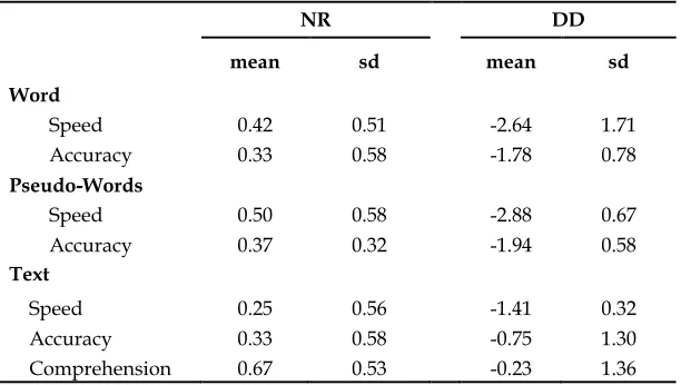
As expected, considering the diagnostic criteria for developmental dyslexia, all
children had general Intelligent Quotient (IQ) within the normal range associated with
a reading impairment in at least two of the tests considered (reading words, pseudo
words, text). The text comprehension was spared: only two dyslexic participants
assessing the reading abilities was within the normal range. The results are reported in
the Table 3.1
Table 3.1 Reading skills
NR DD
mean sd mean sd
Word
Speed 0.42 0.51 -2.64 1.71
Accuracy 0.33 0.58 -1.78 0.78
Pseudo-Words
Speed 0.50 0.58 -2.88 0.67
Accuracy 0.37 0.32 -1.94 0.58
Text
Speed 0.25 0.56 -1.41 0.32
Accuracy 0.33 0.58 -0.75 1.30
Comprehension 0.67 0.53 -0.23 1.36
Concerning the verbal and the non-verbal abilities, all dyslexics showed a
performance within the normal range in both domains, but with significant higher
scores in the non-verbal one (WISC-III: Mean Verbal-IQ = 102, sd=9; Mean
Performance-IQ = 111.5 sd= 10.6, with p =0.01; TOMAL; Mean Verbal Memory Index
=99.8 sd=9.5; Mean Non-Verbal Memory Index =110.5 sd=9.5 with p=0.001): 7 out of 10
children showed a significant (p<0.01) difference between Verbal-IQ and
Performance-IQ on WISC-III and between Verbal and Non-Verbal memory index of TOMAL. The
visuo-spatial skills, evaluated with VMI and Rey‟s Figure, were in the normal range.
values, with the only exception of Raven‟s Progressive Matrices whose score remained
just within the normal limit. Borderline or low performances respect to the normal
range were documented in tasks which implied verbal working memory load and in
the Coding subtest of WISC-III.
The NR group, respect to dyslexics, showed higher performances in verbal
domains and in tasks requiring an automatic processing (Coding) and working
memory.
Notably, in the majority of the reasoning tasks it was possible to observe a trend:
Figure 3.1 Neuropsychological profiles of dyslexics and normal readers. -4 -3 -2 -1 1 2 3 V er b al - IQ V er b al Me m or y P er fo m an ce - IQ N on V er b al Me m or y R ey F ig u re V MI R P M Ma ze s Blo ck D es ig n WC S T D ig its F or w ar d D ig its Ba ck w ar d V is u al S eq u en cin g Me m or y Me m or y fo r L oc atio n C od in g
Verbal Abilities Non Verbal Abilities Problem Solving Working memory
Z -S co re NR DD
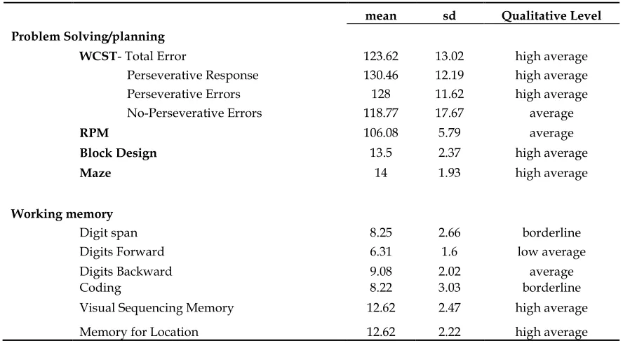
Table 3.2 Summary of neuropsychological results of dyslexics
mean sd Qualitative Level
Verbal Abilities
Verbal IQ - WISC-III 102.46 9.15 average
Informations 10.33 2.5 average
Similarities 10.67 2.55 average
Vocabulary 12.44 3.36 average
Comprehension 11.56 2.46 average
Arithmetic 10.89 1.96 average
Digit Span 8.25 2.66 borderline
Verbal Memory Index-TOMAL 99.85 9.57 average
Memory for Stories 11.18 2.32 average
Word Selective Reminding 11.23 3.35 average
Object Recall 11.69 2.18 average
Digits Forward 6.31 1.6 low average
Paired Recall 9.62 2.99 average
Digits Backward 9.08 2.02 average
Non-Verbal Abilities
Performance IQ - WISC-III 111.46 10.62 average
Picture Completion 12.56 3.09 high average
Picture Arrangement 13.44 3 high average
Block Design 13.5 2.37 high average
Object Assembly 11.88 1.64 average
Maze 14 1.93 high average
Coding 8.22 3.03 borderline
Non-Verbal Memory Index - TOMAL 110.54 11.39 average
Facial Memory 11.54 2.79 average
Abstract Visual Memory 10.85 1.57 average
Visual Sequencing Memory 12.62 2.47 high average
Memory for Location 12.62 2.22 high average
Manual Imitation 10.5 2.2 average
Rey’s Figure 113.27 5.61 average
VMI 108.69 10.87 average
Table 3.2 Summary of neuropsychological results of dyslexics
mean sd Qualitative Level
Problem Solving/planning
WCST- Total Error 123.62 13.02 high average
Perseverative Response 130.46 12.19 high average Perseverative Errors 128 11.62 high average No-Perseverative Errors 118.77 17.67 average
RPM 106.08 5.79 average
Block Design 13.5 2.37 high average
Maze 14 1.93 high average
Working memory
Digit span 8.25 2.66 borderline
Digits Forward 6.31 1.6 low average
Digits Backward 9.08 2.02 average
Coding 8.22 3.03 borderline
Visual Sequencing Memory 12.62 2.47 high average