www.fm.viamedica.pl
Address for correspondence: Michał Szpinda, MD, Department of Normal Anatomy, the Ludwik Rydygier Collegium Medicum in Bydgoszcz, ul. Karłowicza 24, 85–092 Bydgoszcz, Poland, tel: +48 52 585 37 05, fax: +48 52 585 37 53, e-mail [email protected]
The variability and morphometry of the
brachiocephalic trunk in human foetuses
Michał Szpinda, Piotr Flisiński, Gabriela Elminowska-Wenda, Mariusz Flisiński,
Elżbieta Krakowiak-Sarnowska
Department of Normal Anatomy, the Ludwik Rydygier Collegium Medicum in Bydgoszcz, the Nicolaus Copernicus University, Toruń, Poland
[Received 26 January 2005; Revised 25 August 2005; Accepted 2 September 2005]
In prenatal and pediatric cardiovascular surgery knowledge of the various ar-rangements of the aortic arch and its branches as well as the normative data are essential. The variability and morphometric features of the brachiocephalic trunk in 131 human foetuses (65 male, 66 female) ranging from 15 to 34 weeks of gesta-tion were studied by means of anatomical, digital and statistical methods. In all the foetuses examined the left aortic arches were found to have three different arrangements. In 74.05% of cases the usual pattern of the aortic arch with its three main branches were observed. A common origin of the brachiocephalic trunk and left common carotid artery occurred in 20.61% of individuals. In 5.34% of cases the left vertebral artery was an additional vessel and arose from the aortic arch between the left common carotid and subclavian arteries. No significant gen-der differences were found with respect to the brachiocephalic trunk (p ≥ 0.05). The developmental increase in length (r1 = 0.78) and diameter (r2 = 0.83) lated with a linear function but the increase in volume in relation to age corre-sponded to a quadratic function (r3 = 0.73). Our results show the largest increas-es in the brachiocephalic trunk according to the following parameters: the length — between the 4th and 5th, and7th and 8th months, diameter — between the 8th and 9th months and volume — between the 4th and 5th,and 7th and 9th months of gestation (p £ 0.01). The present study constructs a normal range for the mor-phometric features of the foetal brachiocephalic trunk.
Key words: innominate artery, variability, length, diameter, volume, regression analysis, human foetuses
INTRODUCTION
Variations in the branching of the aortic arch are a guide to vascular surgery in ischaemic cerebrovas-cular disease [4, 16]. The number of primary branches may be reduced to 1–2 or increased to 4–6 [1, 3, 14, 19]. Non-invasive techniques such as ultrasonog-raphy, CT and MRI enable pathological changes in the brachiocephalic trunk and carotid arteries to be diagnosed [10]. Duplex-Doppler ultrasonography is
the morphometric features of the brachiocephalic trunk during gestation.
The aim of this study was to examine the follow-ing: the morphometric features (length, diameter and volume) of the brachiocephalic trunk; the influence of sex on the value of the features examined; the developmental trend of the morphometric features of the brachiocephalic trunk.
MATERIAL AND METHODS
The examinations were carried out on 131 foetus-es of both sexfoetus-es (65 male and 66 female) from spon-taneous abortions or stillbirths, cardiovascular abnor-malities having been excluded at necropsy. The ges-tational age ranged from 15 to 34 weeks (Table 1). Foetal age was established by crown-rump (CR) mea-surements. The arterial bed was filled with approxi-mately 15–30 ml of white latex LBS 3060 through a catheter, which was introduced by dorsal access into the thoracic aorta. Specimens were fixed in a 10% neutral formalin solution and branches of the aortic arch were prepared under a stereoscope with Huygens ocular at a magnification of 25–50 times. Afterwards source pictures of aortic arch branching were analysed by Digital Image Analysis System Q 500 MC of Leica (Cambridge). The following mea-surements were made: the length [mm], external di-ameter [mm] and volume [mm3] of the
brachioceph-alic trunk. The developmental growth of the brachio-cephalic trunk was statistically analysed by means
of regression analysis. Gender differences were anal-ysed by means of Student’s t test for two mean in-dependent variables using the PC STAT 1.0 program. The correlation coefficients of the examined features with foetal age (r) were evaluated.
RESULTS
In all the foetuses examined the left aortic arches were found to have three different arrangements (Fig. 1) and to be without gender differences. In 74.05 % of cases the usual pattern of the aortic arch (Figs. 1a, 2) with its three main branches (brachio-cephalic trunk, left common carotid artery and left subclavian artery) was observed. A common origin of the brachiocephalic trunk and left common ca-rotid artery (Fig. 1b) occurred in 20.61% of individu-als. In 5.34% of cases the left vertebral artery was an additional vessel and originated from the aortic arch between the left common carotid and subclavian arteries (Fig. 1c). There were no high or low types of division of the brachiocephalic trunk in the foetuses examined. The statistical analysis of the examined features of the brachiocephalic trunk did not show gender dimorphism (p ≥ 0.05). For this reason the morphometric values obtained are presented with-out regard to sex (Table 2). The morphometric fea-tures of the brachiocephalic trunk indicated the dif-fering developmental dynamic. The largest increase in length of the brachiocephalic trunk was charac-teristically between the 4th and 5th and between the
7th and 8th months of prenatal life (p £ 0.01). The
largest increase in diameter (p £ 0.01) took place between 8th and 9th months. The largest growth in
volume occurred between the 4th and 5th, and 7th and
9th months of gestation (p £ 0.01). The length and
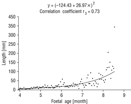
diameter revealed an increase in values with increased foetal age according to the regression line (Figs. 3, 4). The increase in volume in relation to age correspond-ed to the quadratic function (Fig. 5). Positive correla-tion coefficients of these parameters with foetal age were statistically significant (p £ 0.01) and reached the following values: r1 = 0.78 for length, r2 = 0.83
for diameter and r3 = 0.73 for volume.
DISCUSSION
The brachiocephalic trunk develops from the as-cending part of the right dorsal aorta [1, 3, 7, 15]. Regression of the right dorsal aortic root (between the right subclavian artery and the descending aor-ta) and the right ductus arteriosus leaves the normal left aortic arch. The proximal segments of the third pair form the common carotid arteries. Variation in
Table 1. Age and sex of investigated foetuses
Months Weeks Number Male Female
4 15 12 6 6
16 11 5 6
5
18 11 5 6
19 9 5 4
6
22 12 7 5
23 13 6 7
7 25 14 8 6
26 9 4 5
8
29 11 3 8
30 10 7 3
9 33 14 6 8
34 5 3 2
Total 131 65 66
the bicarotid trunk were described by Niżankowski et al. [12] in 0.9% only. In our material the normal pattern of brachiocephalic trunk was observed in 74.05%, while a bicarotid trunk was found in 20.61% of cases. The number of large arteries of the aortic arch might be reduced to one or increased to 6 ves-sels [3, 9, 14, 19], but these anomalies were absent in the material under examination. We noted in 5.34% of individuals the origin of the left vertebral artery from the aortic arch. This pattern occurred in Anson’s investigation [3] in 3.8% of cases (Types III, IV, VII, XIV). In his material the brachiocephalic trunk was absent in 0.5% of cases (Type VI). Anson de-scribed a bi-innominate sequence (Type IX) in 1.2% of cases. In this the normal left and right brachio-cephalic trunks derived from the left aortic arch. In Anson’s material derivation of all the main branches from a single trunk was found in 0.3% of cases. Rob-erts et al. [13] observed a left aortic arch with a ret-ro-oesophageal aberrant right brachiocephalic trunk. Moes and Freedom [11], on the other hand, de-scribed a left aberrant brachiocephalic trunk with
Figure 1. Three different arrangements of aortic arch branching: a. The usual pattern of the aortic arch, b. Reduction to 2 derived
branch-es of the aortic arch, c. Increase to 4 derived branchbranch-es of the aortic arch: A — aortic arch, B — bicarotid trunk, 1 — brachiocephalic trunk, 2 — right common carotid artery, 3 — right subclavian artery, 4 — left common carotid artery, 5 — left subclavian artery, 6 — left vertebral artery.
the origin of the brachiocephalic trunk is closely de-pendent on the type of aortic arch branching. The right brachiocephalic trunk is characteristic for the left aortic arch and this pattern occurs in 99.9% [3, 7]. The normal left brachiocephalic trunk is typical for the right aortic arch [8]. This rare variety (0.1%) re-sults from persistence of the right fourth branchial arch. Savastano et al. [16] presented agenesis of bra-chiocephalic trunk in one case and a hypoplastic right brachiocephalic trunk in two specimens.
Figure 2. The usual pattern of the aortic arch in human foetus
aged 5 months: A — aortic arch, 1 — brachiocephalic trunk, 2 — right common carotid artery, 3 — right subclavian artery, 4 — left common carotid artery, 5 — left subclavian artery, 6 — inter-nal carotid artery, 7 — exterinter-nal carotid artery.
Figure 3. Regression line for the length of the brachiocephalic
trunk in relation to foetal age. Linear function: length = –1.531 + + 1.307 × age; r1 = 0.78.
mirror-image branching. Szpinda [17] presented a new typological variant of the left aortic arch in which a left aberrant hypoplastic brachiocephalic trunk passed behind the trachea and the oesophagus.
Figure 4. Regression line for the diameter of the brachiocephalic
trunk in relation to foetal age. Linear function: diameter = –1.056 + + 0.554 × age; r2 = 0.83.
Table 2. Angiometric analysis of the brachiocephalic trunk for length, diameter and volume
Foetal age [months]
Mean SD Mean SD Mean SD
4 3.38A 0.61 1.23 0.27 3.62G 1.43
5 5.34B 1.37 1.87 0.38 12.98H 5.45
6 6.42 1.41 2.29 0.36 25.57 11.66
7 7.39C 1.54 2.62 0.56 42.13I 24.64
8 9.57D 2.41 2.84E 0.61 60.32J 25.71
9 9.89 2.34 4.45F 0.64 162.27K 87.45
Means lengths marked by the letters A and B, C and D indicate the largest increases (p £ 0.01). Mean diameters marked by the letters E and F indicate the largest in-creases (p £ 0.01). Mean volumes marked by the letters G and H, I, J and K indicate the largest inin-creases (p £ 0.01).
Brachiocephalic trunk
It should be noted that we observed no statisti-cally significant gender differences with respect to either the various arrangements or the values of the morphometric features. The length of the brachio-cephalic trunk depends on the level of its origin and on the level of its bifurcation into subclavian and common carotid arteries. The long type of brachio-cephalic trunk corresponded with a high division and the short type of brachiocephalic trunk is charac-teristic when the division is low, but these types were absent in the material under examination. Our earlier study indicated that during foetal development the termination of the brachiocephalic trunk apparently descended by one vertebra [18].
The angiometric study of the brachiocephalic trunk during gestation showed developmental growth in the length (r1 = 0.78) and diameter (r2 = 0.83)
correlated with a linear function, while the increase in volume in relation to age (r3 = 0.73) corresponded
to a quadratic function. Similar observations concern-ing these common relations between aortic growth and foetal age have been confirmed by other authors [2, 20]. Therefore, in children and adults no system-atic correlation has been found between arterial length and diameter with increased age, the relation being best described as a function of the natural log-arithm of the body weight [6]. Our results show the largest growth of the brachiocephalic trunk concern-ing the followconcern-ing parameters: the length — between the 4th and 5th and the 7th and 8th months, the
diame-ter — between the 8th and 9th months and the
vol-ume — between 4th and 5th and the 7th and 9th months
of prenatal life (p £ 0.01). Our findings indicated that in the age range examined the volume of the
bra-chiocephalic trunk increased approximately 45-fold, from 3.62 ± 1.43 mm3 to 162.27 ± 87.45 mm3. This
result was obtained from the product of the length and the squared diameter, which increased approxi-mately 3-fold and 4-fold, respectively.
The morphometric features of the brachioceph-alic trunk presented in this paper have not previous-ly been discussed in the professional literature. The normative data of the brachiocephalic trunk estab-lished in this work may be helpful in the early diag-nosis of developmental anomalies, especially in pre-natal life in the assessment, with the use of duplex--Doppler ultrasonography, of the condition of foet-uses in pregnant women with hypertension, diabe-tes or twin pregnancy [9, 10, 12]. The angiometric parameters of the brachiocephalic trunk complement current measurement data obtained during invasive (arteriography) and non-invasive (ultrasonography, CT, MRI) techniques.
This study has demonstrated that the normal ranges of the brachiocephalic trunk presented may serve as a basis for comparison of the measurements of the same parameters in foetuses with congenital defects of the heart, the aortic arch and the branch-es of the aortic arch.
REFERENCES
1. Adachi B, Hasebe K (1928) Das Arteriensystem der Japa-ner. Verlag der Kaiserlich-Japanischen Universität, Bd II, Kyoto.
2. Alvarez L, Aranega A, Saucedo R, Contreras JA, Lopez F, Aranega A (1990) Morphometric data concerning the great arterial trunks and their branches. Int J Cardiol, 29: 127–139.
3. Anson BI (1971) Thoracic cavity and its contents. In: Anson BI, McVay Ch (eds.). Surgical anatomy. Vol. 1, W.B. Saunders Company, Philadelphia, London, Tor-onto, pp. 408–412.
4. Azakie A, Mc Elhinney DB, Messina LM, Stoney RJ (1999) Common brachiocephalic trunk: strategies for revascularization. Ann Thorac Surg, 67: 657–660. 5. Fitzgerald SW, Donaldson JS, Poznański AK (1987)
Pe-diatric thoracic aorta: normal measurements deter-mined with CT. Radiology, 165: 667–669.
6. Hofstetter R, Engelhardt W, Prunte K, Rother A, von Bernuth G (1987). Sector echocardiographic determi-nation of the diameter of the large arteries of the heart in children. Z Kardiol, 76: 38–43.
7. Kadir S (1991) Atlas of normal and variant angio-graphic anatomy. W.B. Saunders Company. Philadel-phia, pp. 19–54.
8. Kurata H, Satoh S, Kohno M, Kajiwara H, Mashimo Y, Satoh H (1989) Brachiocephalic arterial aplasia of the right aortic arch with subclavian steal syndrome. Nip-pon Kyobu Geka Gakkai Zasshi, 1: 171–174.
9. Lize I (1970) Abnormal origin of the great vessels from the aortic arch. Folia Morphol, 29: 401–402. Figure 5. Regression line for the volume of the brachiocephalic
trunk in relation to foetal age. Quadratic function: volume = = (–124.43 + 26.97 × age)2; r
10. Midiri M, Finazzo M, Pilato M, Lagalla R, De-Maria M (1999) Right aortic arch with aberrant left innominate artery: MR imaging findings. Eur Radiol, 9: 311–315. 11. Moes CA, Freedom RM (1993) Rare types of aortic arch
anomalies. Pediatr Cardiol, 2: 93–101.
12. Niżankowski C, Rajchel Z, Ziółkowski M (1975) Abnor-mal origin of arteries from the aortic arch in man. Fo-lia Morphol, 34: 109–116.
13. Roberts CS, Othersen HB Jr, Sade RM, Smith CD 3rd, Tagge EP, Crawford FA Jr (1994) Tracheoesophageal compression from aortic arch anomalies: analysis of 30 operatively treated children. J Pediatr Surg, 2: 334–338.
14. Roguin N, Sujov P, Shapir Y, Peleg H, Riss E (1982) Single arterial trunk arising from the aortic arch associated with coarctation of the aorta. Pediatr Radiol, 12: 39–40.
15. Sadler TW (1993) Langman’s medical embryology. Med Tour Press International, Warszawa, pp. 207–219. 16. Savastano S, Feltrin P, Chiesura-Corona M, Mioffa D (1992)
Cerebral ischemia due to congenital malformations of bra-chiocephalic arteries — case reports. Angiology, 1: 76–83. 17. Szpinda M (2005) A new variant of left aberrant bra-chiocephalic trunk in man: case report and literature review. Folia Morphol, 64: 47–50.
18. Szpinda M, Flisiński P, Gościcka D (1999) Skeletopy of the brachiocephalic trunk and the common carotid arteries in human fetuses. Folia Morphol, 58: 127–136. 19. Testut L, Latarjet A (1948) Traite d’Doince, anatomie
humaine. Vol. 7, Paris.