STRUCTURE AND REDOX TRANSFORMATIONS OF IRON(III)
COMPLEXES WITH SOME BIOLOGICALLY IMPORTANT
INDOLE-3-ALKANOIC ACIDS IN AQUEOUS SOLUTIONS
§Krisztina Kovács
1, Alexander A. Kamnev
2*, Alexei G. Shchelochkov
2, Ern
ő
Kuzmann
1,
János Mink
3, Tünde Megyes
3, Attila Vértes
11Research Group for Nuclear Techniques in Structural Chemistry, Hungarian Academy of Sciences; Laboratory of Nuclear
Chemistry, Eötvös Loránd University, H-1518, Budapest 112, Hungary
2Laboratory of Biochemistry of Plant-Bacterial Symbioses, Institute of Biochemistry and Physiology of Plants and Microorganisms,
Russian Academy of Sciences, 410049, Saratov, Russia
3Chemical Research Center of the Hungarian Academy of Sciences, H-1525, Budapest, P.O. Box 77, Hungary
* Corresponding author. E-mail: [email protected]; Fax: +7-8452-970383
Abstract: Interactions of a series of indole-3-alkanoic acids (with n-alkanoic acid side-chains from C1 to C4) with
iron(III) in acidic aqueous solutions have been shown to comprise two parallel processes including complexation and redox transformations giving iron(II) hexaaquo complexes. The structure and composition of the reaction products are discussed, as analysed using a combination of instrumental techniques including 57Fe Mössbauer, vibrational and 1H NMR spectroscopies.
Keywords: indole-3-alkanoic acids, auxin phytohormones, iron(III) complexes, coordination structure, redox
transformations.
INTRODUCTION
Earlier studies have shown [12–16] that both IAA and some other chemically and metabolically related organic substances of biological origin are capable of gradually reducing iron(III) in weakly acidic nitrate-containing aqueous media even under aerobic conditions. This could be of ecological signifi cance since iron(III) has a poor biological availability, which is due to its full hydrolysis and extremely low solubility of ferric hydroxides in a wide pH range, whereas iron(II) species are more soluble and, therefore, more biologically available both for plants and for soil microorganisms. Note also that acidic soils are rather widely spread, comprising about 30% of only arable territories [17]. On the other hand, these processes can result in oxidative degradation of the organic biomolecules involved in plant-microbe interactions in soil [7]. Therefore, knowledge of the chemical processes is of interest both for basic research and in applied fi elds, particularly those related to agricultural and environmental biotechnology.
In the present work, chemical reactions are considered which occur between indole-3-alkanoic acids (with
n-alkanoic acid side chains from Indole-3-acetic acid (IAA) and its close structural analogues, indole-3-carboxylic (ICA), indole-3-propionic (IPA) and indole-3-butyric (IBA) acids, are natural and synthetic phytohormones of the auxin series that regulate plant growth and development [1–3]. IAA (along with some other indolic auxins), being ubiquitous in plants, is also well documented to be synthesized by many soil microorganisms which exude it into soil [4], where it plays an essential role in plant-microbe interactions [5, 6]. Thus, within the soil and/or aquifer environment, auxin molecules can readily be subjected to chemical reactions involving different metal ions including iron [7], which is commonly ubiquitous in soil [8] and is one of the most important micronutrients for virtually all organisms [9]. In the biomedical fi eld, prooxidant activity and cytotoxic effects of IAA and its derivatives upon peroxidase-catalysed oxidation have also been tested for potential novel applications in antitumour therapy [10, 11].
C1 to C4) and iron(III) in acidic aqueous solutions under aerobic conditions, and their products are analysed using a combination of physicochemical instrumental techniques.
RESULTS AND DISCUSSION
In order to follow in-situ redox processes involving iron species in aqueous solutions, 57Fe Mössbauer spectroscopy is a very convenient and informative technique giving, in particular, direct quantitative information on the Fe(II)-to-Fe(III) ratio. As Mössbauer spectra can be obtained for solid matrices only (or, for non-solids, in a solidifi ed state, e.g. rapidly frozen), aqueous solutions can be studied in the frozen state [18]. Note also that rapid freezing (e.g., by inserting small portions of a solution into liquid nitrogen) allows one to obtain a glassy solid that refl ects the structure of the initial § Material presented at the XV-th Conference “Physical Methods in Coordination and Supramolecular Chemistry”,
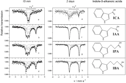
solution. Moreover, in such a frozen solution all processes are virtually ceased, so that by rapidly freezing a successive series of solution aliquots with their subsequent low-temperature Mössbauer spectroscopic measurements, one can obtain „snapshots” of the state of processes that have been „stopped” at certain successive time points [19]. Mössbauer spectra of iron(III)-containing aqueous solutions with different indole-3-alkanoic acids, fi ltered and rapidly frozen 15 min and 2 days after mixing, are shown in Figure 1, a–h. It can be seen that in the solutions which initially contained iron(III) only, already 15 min after mixing some certain amounts of iron(II) are present, which is distinctly evidenced by the appearance of a corresponding component doublet with a large quadrupole splitting (its position is indicated in Figure 1 by a square bracket above the upper spectrum) having varying intensity for different acids (cf. Figure 1, spectra
a to d). The presence of the same iron(II)-related doublet with higher intensities is detected in the spectra of the mixtures obtained after 2 days (cf. Fig. 1, spectra e to h). The Mössbauer parameters of the resulting Fe2+ species (i.e., isomer shifts δ = 1.39 ± 0.01 mm/s and quadrupole splittings Δ = 3.35 ± 0.03 mm/s) are typical of a hexaaquo coordination microenvironment [18].
Fig. 1. Mössbauer spectra of aqueous solutions of 57FeIII nitrate and indole-3-alkanoic acids (their structures are shown in the right-hand panel) fi ltered and rapidly frozen (at T = 80 K) 15 min (a–d) and 2 days (e–h) after mixing the reagents (1:3 molar ratio; fi nal pH ~ 2 to 3). Spectra (a), (e) – indole-3-carboxylic acid (ICA); (b), (f) – indole-3-acetic
acid (IAA); (c), (g) – indole-3-propionic acid (IPA); (d), (h) – indole-3-butyric acid (IBA). The position of the FeII -related doublets is indicated in the upper plots by square brackets.
Comparing the spectral intensities (cf. Figure 1, e–h) it can be seen that after 2 days of contact of the indolic acids with iron(III), in IAA solution there is ferrous iron only, as compared to the Fe–ICA, Fe–IPA or Fe–IBA systems where some remaining ferric iron is still detectable. This indicates a stronger reducing capability of IAA towards iron(III) in the series of indole-3-alkanoic acids, evidently related to the ease of the IAA side-chain decarboxylation [20–23]. Note also that both the relative and absolute intensities of the FeII component in ICA solutions (see Figure 1, spectra a and e) are less than those for the other acids, showing the least reducing capacity of ICA in the series. The other two components of the spectra (see Figure 1, spectra a–h, except spectrum f) represent iron(III) complexes with the corresponding ligands (doublets with δ = 0.52 to 0.55 mm/s and Δ = 0.5 to 0.6 mm/s), that remain in solution after fi ltering out the precipitated complexes, and residual mononuclear Fe3+ ions (evidently partly hydrolysed at weakly acidic pH, which give a very broad single line).
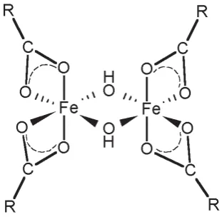
molecular dissolution. In the frozen acetone solutions (at concentrations of each of the complexes 0.1 M and 0.01 M, using 57Fe-enriched samples in the latter case to enhance the intensity of the spectra), the lack of a magnetic structure (due to fast spin-spin relaxation) provides evidence that the iron(III) species have a dimeric structure [16, 18]. This result is in good agreement with the data of elemental analyses, FTIR and FT-Raman spectroscopic results (including those for deuterated samples) for the solid complexes fi ltered out of the solutions, indicating a μ-(OH)2-bridged structure: [L2Fe<(OH)2>FeL2] (where L is the deprotonated IAA, IPA or IBA moiety) [24]. It has to be noted that in the case of ICA, the data of elemental analyses pointed to the possibility of the presence of a mixture of solid products which should be studied in more detail separately.
In the case of FeIII–IAA complex dissolved in methanol, a solution X-ray diffraction study was also performed [24]. Analysis of the data for FeIII–IAA complex as well as, by analogy (considering the closely related Mössbauer, FTIR and FT-Raman spectroscopic results), for the corresponding IPA and IBA complexes, can be interpreted using the general structure represented in Fig. 2. Each iron atom in a complex is surrounded by six oxygen atoms in a slightly distorted octahedral symmetry as follows: four from two deprotonated IAA carboxylate ligands (in the bidentate coordination) and two from the dihydroxo bridge linking the two iron atoms. Nevertheless, it should be noted that under different conditions, a monomeric poorly water-soluble Fe(III)–IAA complex was obtained from aqueous solution which gave different spectroscopic images owing to its different structure [15].
Fig. 2.Schematic representation of solid Fe(III) complexes with IAA, IPA and IBA.
Thus the parameters of the spectra in Figure 1 suggest the existence of two parallel reactions between Fe3+ and the ligands (L); namely, both a redox transformation yielding Feaq2+ ions and Fe3+–L complex formation take place, as shown in the following scheme.
Fe3+ + L Fe
III complex (precipitate and/or dissolved form)
Feaq2+ + [oxidised L product(s)]
While enzymatic oxidation of auxin phytohormone catalysed by plant peroxidases, regarding its mechanism and products, has been under intensive investigation owing to basic interest [21–23] as well as possible biomedical applications (see, e.g. [10, 11] and references therein), chemical oxidation products of auxins are much less studied [25]. Owing to the sophisticated nature of these chemical process involving radical products and/or intermediates [26], this seems to be not an easy and straightforward task.
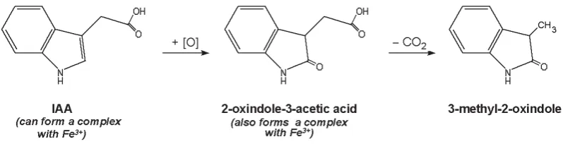
It should be noted that both oxindole-3-acetate and 3-methyl-2-oxindole, which were found to be formed in the course of chemical oxidation of indole-3-acetic acid in the presence of FeIII (see Figure 3), had earlier been reported among products of both its enzymatic [28, 29] and electrochemical oxidation [25] at physiological pH (along with 3-methylene-2-oxindole), i.e. under different conditions and involving different electron transfer modes. Nevertheless, the exact mechanism of chemical oxidation of IAA, in particular, under environmentally relevant conditions has to be elucidated in more detail, which requires further investigations.
Fig. 3. Scheme and some products of chemical oxidation of indole-3-acetic acid in aqueous solution under aerobic conditions in the presence of iron(III).
CONCLUSIONS
Iron(III) ions were shown to be gradually reduced by each of the indole-3-alkanoic acids with n-alkanoic side-chains C1 to C4 in acidic aqueous media under aerobic conditions using Mössbauer spectroscopic measurements in rapidly frozen solutions. The parameters of the Mössbauer spectra indicate that there are two parallel processes, viz
iron(III) complexation and redox transformations. Within the series of the indole-3-alkanoic acids, indole-3-carboxylic acid showed the least reducing capability towards iron(III). After 2 days, indole-3-acetic acid showed virtually a complete reduction of iron(III) to iron(II), whereas iron(III) was still detectable in solutions of the other acids, along with iron(II). Mössbauer parameters of the frozen solutions provide evidence that the resulting iron(II) species is the hexaaquo complex. The solid complexes formed were found to have a dimeric μ-dihydroxo-bridged structure, that was confi rmed using a combination of spectroscopic techniques for indole-3-acetic, indole-3-propionic and indole-3-butyric acids. Among the products of chemical oxidation of indole-3-acetic acid in the presence of iron(III) under aerobic conditions, oxindole-3-acetate and 3-methyl-2-oxindole were detected using vibrational and 1H NMR spectroscopic measurements. The same substances had earlier been reported to be found among the products of enzymatic and electrochemical oxidation in circumneutral media, i.e. under different conditions and involving different electron transfer modes.
EXPERIMENTAL
Mössbauer measurements in aqueous solutions were performed using materials prepared from 57Fe-enriched iron (ca. 90% 57Fe) dissolved in nitric acid at elevated temperature. The stock solution was 0.01 M with regard to iron(III), with pH 0.9. The indole derivatives used (ICA, IAA, IPA, IBA) were dissolved in water adding KOH to the solutions up to pH 6–7. The concentration of the ligands after mixing was 0.03 M. Addition of iron(III) nitrate to an indolic acid in solution (up to the 1:3 metal-to-acid molar ratio) resulted in the colour change of the solutions and the formation of cocoa-brown precipitates indicating complexation of Fe3+ with the indole-3-alkanoic acids. The fi nal pH values of the mixtures were around 2.5 (measured using an OP-211 laboratory pX/mV meter, Radelkis, Hungary).
ACKNOWLEDGEMENTS
The material of this paper was presented as part of the invited lecture at the XV International Conference “Physical Methods in Coordination and Supramolecular Chemistry” (Chişinǎu, Moldova, 27 September – 1 October, 2006). A.A.K. is grateful to the organisers of the Conference and personally to Dr. Marina Fonari and Professor Constantin Turta for their helpful attention and hospitality; partial support for the visit from the Russian Foundation for Basic Research (by RFBR travel grant 06-03-42956) is also acknowledged. This work was supported by The Hungarian Science Foundation (OTKA Grant T043687), NATO (Expert Visit Grants LST.EV.980141 and CBP.NR.NREV.981748; Collaborative Linkage Grant LST.CLG.977664), Russian Academy of Sciences’ Commission (Grant No. 205 under the 6th Competition-Expertise of research projects), as well as under the Agreements on Scientifi c Cooperation between the Russian and Hungarian Academies of Sciences for 2002–2004 and 2005–2007.
REFERENCES
Marumo, S. Auxins. In Takahashi, N. (Ed.),
[1] Chemistry of Plant Hormones, CRC Press, Inc.: Boca Raton, Flo.
(U.S.A.), 1986; Chapter 2, pp. 9-56.
Weyers, J.D.B.; Paterson, N.W.; New Phytol. 2001, 152, 375-407. [2]
Teale, W.D.; Paponov, I.A.; Palme K.; Nat. Rev. Mol. Cell Biol. 2006, 7, 847-859. [3]
Patten, C.L.; Glick, BR.; Can. J. Microbiol. 1996, 42, 207-220. [4]
Lambrecht, M.; Okon, Y.; Vande Broek, A.; Vanderleyden, J.; Trends Microbiol. 2000, 8, 298-300. [5]
Somers, E.; Vanderleyden, J.; Srinivasan, M.; Crit. Rev. Microbiol. 2004, 30, 205-240. [6]
Kamnev, A.A.;
[7] Kovács, K.; Shchelochkov, A.G.; Kulikov, L.A.; Perfi liev, Yu.D.; Kuzmann, E.; Vértes, A.; In
Metal Ions in Biology and Medicine, Vol. 9; Alpoim, M.C.; Morais, P.V.; Santos, M.A.; Cristóvão, A.J.; Centeno, J.A.; Collery, Ph. (Eds.), John Libbey Eurotext: Paris, 2006; pp. 220-225.
Cornell, R.M.; Schwertmann, U. The Fe Oxides: Structure, Properties, Reactions, Occurrences, and Uses, VCH: [8]
New York, 1996. Sigel, A.; Sigel, H., Eds.
[9] Metal Ions in Biological Systems, Vol. 35. Iron Transport and Storage in Microorganisms,
Plants, and Animals. New York: Marcel Dekker, 1998, 824 pp.
Tafazoli, S.; O’Brien, P.J.; Chem. Res. Toxicol. 2004, 17, 1350-1355. [10]
Veitch, N.C.; Phytochemistry, 2004, 65, 249-259. [11]
Kamnev, A.A.; Kuzmann, E. In
[12] Spectroscopy of Biological Molecules: Modern Trends. Annex; Carmona, P.; Navarro, R.; Hernanz, A., Eds. UNED Press: Madrid, 1997, pp. 85-86.
Kamnev, A.A.; Kuzmann, E.; Biochem. Mol. Biol. Int. 1997, 41, 575-581. [13]
Kamnev, A.A.; Kuzmann, E.; Perfi liev, Yu.D.; Vankó, Gy.; Vértes, A.; J. Mol. Struct. 1999, 482-483, 703-711. [14]
Kamnev, A.A.; Shchelochkov, A.G.; Perfi liev, Yu.D.; Tarantilis, P.A.; Polissiou, M.G.; J. Mol. Struct. 2001, 563-[15]
564, 565-572. K
[16] ovács, K.; Kamnev, A.A.; Kuzmann, E.; Homonnay, Z.; Szilágyi, P.Á.; Sharma, V.K.; Vértes, A.; J. Radioanal. Nucl. Chem. 2005, 266, 513-517.
Von Uexkull, H.R.; Mutert, E.; Plant Soil, 1995, 171, 1-15. [17]
Vértes, A.; Nagy, D.L., Eds. Mössbauer Spectroscopy of Frozen Solutions. Akad. Kiadó: Budapest, 1990. [18]
Krebs, C.; Price, J.C.; Baldwin, J.; Saleh, L.; Green, M.T.; Bollinger, J.M., Jr.; Inorg. Chem. 2005, 44, 742-757. [19]
Savitsky, P.A.; Gazaryan, I.G.; Tishkov, V.I.; Lagrimini, L.M.; Ruzgas, T.; Gorton, L.; Biochem. J. 1999, 340, [20]
579-583.
Gazaryan, I. G.; Lagrimini, L.M.; Ashby, G. A.; Thorneley, R.N.F.; Biochem. J. 1996, 313, 841-847. [21]
Harrod, J.F.; Guerin, C.; Inorg. Chim. Acta 1979, 37, 141-144. [22]
Hinman, R.L.; Lang, J.; Biochemistry (USA), 1965, 4, 144-158. [23]
Kovács, K.; Kamnev, A.A.; Mink, J.; Németh, Cs.; Kuzmann, E.; Megyes, T.; Grósz, T.; Medzihradszky-[24]
Schweiger, H.; Vértes, A.; Struct. Chem. 2006, 17, 105-120. Hu, T.; Dryhurst, G.; J. Electroanal. Chem. 1997, 432, 7-18. [25]
Candeias, L.P.; Folkes, L.K.; Dennis, M.F.; Patel, K.B.; Everett, S.A.; Stratford, M.R.L.; Wardman, P.; J. Phys. [26]
Chem. 1994, 98, 10131-10137.
Shchelochkov, A.G.; Kamnev, A.A.; Tarantilis, P.A.; Polissiou, M.G.; In
[27] Metal Ions in Biology and Medicine,
Vol. 7. Khassanova, L.; Collery, Ph.; Maymard, I.; Khassanova, Z.; Etienne, J.-C., Eds., John Libbey Eurotext: Paris, 2002, pp. 37-40.
Gazaryan, I.G.; Chubar, T.A.; Mareeva, E.A.; Lagrimini, L.M.; Van Huystee, R.B.; Thorneley, R.N.F.; [28]
Phytochemistry 1999, 51, 175-186.