COMPARATIVE ESEM ANALYSIS OF Er,Cr:YSGG TREATED DENTIN SURFACE vs CONDITIONER
36 TREATED DENTIN SURFACE
Rashmi Issar
1, Shashi Ranjan
1Department of Conservative Dentistry & Endodontics, Patna Dental College & Hospital, Patna, Bihar
2Department of Oral & Maxillofacial Pathology, Dr.B.R.Ambedkar Institute
3Dental Surgeon, Health & Family Welfare, Govt. Of Meghalaya
4Department of Conservative Dentistry & Endodontics, Patna Dental College & Hospital, Patna, Bihar. 5,6Dept of Oral and Maxillofacial Pathology, Sree
A R T I C L E I N F O
INTRODUCTION
Michael Buonocore with his pioneering work in 1955 laid the foundation for the first meaningful proof of intra
by using 85% phosphoric acid to etch enamel.
conditioning of dentin were to remove the intrinsic weakness of the smear layer to permit bonding to underlying dentin, demineralize the superficial dentin matrix & uncover both intertubular and peritubular dentin to permit resin infiltration into surface.2 When the etchant first contacts the smear layer it begins to dissolve hydroxyapatite crystal within the surface of the intertubular dentin and along the surface of the outermost peritubular dentin and penetrates primarily along the tubules (Selvig, 1968).3
International Journal of Current Advanced Research
ISSN: O: 2319-6475, ISSN: P: 2319-6505,
Available Online at www.journalijcar.org
Volume 8; Issue 11 (A); November 2019
DOI: http://dx.doi.org/10.24327/ijcar.2019
Copyright©2019 Rashmi Issar et al. This is an open access article distributed under the Creative Commons Attribution License, which permits unrestricted use, distribution, and reproduction in any medium, provided the original work is properly cited.
Article History:
Received 10th August, 2019 Received in revised form 2nd September, 2019
Accepted 26th October, 2019
Published online 28th November, 2019
Key words:
Dentin, acid etching, laser etching, ESEM
ESEM ANALYSIS OF Er,Cr:YSGG TREATED DENTIN SURFACE vs CONDITIONER
36 TREATED DENTIN SURFACE
Ranjan
2, Deirimika Lakiang
3, Pankaj Singh
4, Masthan
and Aravindha Babu N
6Department of Conservative Dentistry & Endodontics, Patna Dental College & Hospital, Patna, Bihar
Department of Oral & Maxillofacial Pathology, Dr.B.R.Ambedkar Institute of Dental Sciences & Hospital, Patna, Bihar Dental Surgeon, Health & Family Welfare, Govt. Of Meghalaya
Department of Conservative Dentistry & Endodontics, Patna Dental College & Hospital, Patna, Bihar. Dept of Oral and Maxillofacial Pathology, Sree Balaji Dental College & Hospital, Chennai, Tamil Nadu
A B S T R A C T
Introduction: To achieve an ideal substrate for dentin bonding the surface needs a prior treatment either through acid etching or application of hard tissue lasers. The advent of lasers with its various advantages and multiple applications has lead to a wide acceptance of this modality as an option for etching of dental hard tissues.
Background: The present study aims to compare surface characteristics after etching by 37% phosphoric acid and Er,Cr:YSGG on the dentinal surfaces of extracted tooth specimens.
Methods: The sectioned dentin samples of forty posterior non diseased extracted teeth were reduced to a thickness of 1.0 ± 0.5mm. The 40 prepared and mounted specimens were then divided in 3 groups GrD1, GrD2 and GrD3. The GrD1 served as control group while GrD2 was treated by Conditioner 36(36% phosphoric acid) and GrD3 by Er,Cr:YSGG laser. Then an ESEM comparative analysis was done for smear layer removal and the diameter of dentinal tubules.
Results: Chi Square test was performed for removal of smear layer and GSA image analyser was used to measure tubule diameter. The difference between group GrD2 and GrD3 for smear layer removal was statistically insignificant (p>.05). The ‘t’ value for student‘t’ test between groups GrD2 and GrD3 was 18.10 for the comparison of dentinal tubule diameter which was a highly significant result( p< .001).
Conclusion: The smear layer removal of lased dentinal surfaces and acid treated surfaces showed no statistical differences. The mean diameter of dentinal tubules after acid etching was 2.78 µm and lased surfaces were 1.09 µm on an average. This was a statistically significant result which will have great influence on hypersensitivity but further in vivo studies are needed to prove this.
Michael Buonocore with his pioneering work in 1955 laid the foundation for the first meaningful proof of intra-oral adhesion by using 85% phosphoric acid to etch enamel.1 Goals of acid were to remove the intrinsic weakness of the smear layer to permit bonding to underlying dentin, demineralize the superficial dentin matrix & uncover both intertubular and peritubular dentin to permit resin infiltration t contacts the smear layer it begins to dissolve hydroxyapatite crystal within the surface of the intertubular dentin and along the surface of the outermost peritubular dentin and penetrates primarily along the tubules
Effects of acid etching are typically limited to .1
superficial region of intertubular dentin and to a depth of 5µm along the walls of peritubular dentin.
Bonding to dentin is thought to basically rely on a micromechanical entanglement of hydrophilic resins into this demineralised microporous dentin thus forming a reticular intertwined hybrid tissue composed of collagen, re mineral particles and resin(Nakabayashi
Meerbek et al 1993a,1993b).
others(1984) the drawbacks of acid etching were damage to tooth structure, technique sensitivity, accidental spillage to adjacent tooth and hybridization deficit.
As a possible alternative to acid conditioning the use of therapy has shown a promising front. Er,Cr:YSGG (Erbium,
International Journal of Current Advanced Research
6505, Impact Factor: 6.614
www.journalijcar.org
2019; Page No.20409-20413
//dx.doi.org/10.24327/ijcar.2019.20413.3987
This is an open access article distributed under the Creative Commons Attribution License, which permits unrestricted use, distribution, and reproduction in any medium, provided the original work is properly cited.
ESEM ANALYSIS OF Er,Cr:YSGG TREATED DENTIN SURFACE vs CONDITIONER
Masthan K.M.K
5Department of Conservative Dentistry & Endodontics, Patna Dental College & Hospital, Patna, Bihar
of Dental Sciences & Hospital, Patna, Bihar
Department of Conservative Dentistry & Endodontics, Patna Dental College & Hospital, Patna, Bihar. Balaji Dental College & Hospital, Chennai, Tamil Nadu
To achieve an ideal substrate for dentin bonding the surface needs a prior treatment either through acid etching or application of hard tissue lasers. The advent of lasers with its various advantages and multiple applications has lead to a wide acceptance of this modality as an option for etching of dental hard tissues.
The present study aims to compare surface characteristics after etching by 37% phosphoric acid and Er,Cr:YSGG on the dentinal surfaces of extracted tooth
The sectioned dentin samples of forty posterior non diseased extracted teeth were reduced to a thickness of 1.0 ± 0.5mm. The 40 prepared and mounted specimens were nd GrD3. The GrD1 served as control group while GrD2 was treated by Conditioner 36(36% phosphoric acid) and GrD3 by Er,Cr:YSGG laser. Then an ESEM comparative analysis was done for smear layer removal and the
re test was performed for removal of smear layer and GSA image analyser was used to measure tubule diameter. The difference between group GrD2 and GrD3 for smear layer removal was statistically insignificant (p>.05). The ‘t’ value for een groups GrD2 and GrD3 was 18.10 for the comparison of dentinal tubule diameter which was a highly significant result( p< .001).
The smear layer removal of lased dentinal surfaces and acid treated surfaces showed no statistical differences. The mean diameter of dentinal tubules after acid etching was 2.78 µm and lased surfaces were 1.09 µm on an average. This was a statistically gnificant result which will have great influence on hypersensitivity but further in vivo
Effects of acid etching are typically limited to .1-.5µm of the superficial region of intertubular dentin and to a depth of 5µm
the walls of peritubular dentin.4
Bonding to dentin is thought to basically rely on a micromechanical entanglement of hydrophilic resins into this demineralised microporous dentin thus forming a reticular intertwined hybrid tissue composed of collagen, residual mineral particles and resin(Nakabayashi et al 1982, Van 1993a,1993b).5As quoted by Liberman & others(1984) the drawbacks of acid etching were damage to tooth structure, technique sensitivity, accidental spillage to
hybridization deficit.
As a possible alternative to acid conditioning the use of laser has shown a promising front. Er,Cr:YSGG (Erbium,
Research Article
The hydrokinetic laser system was investigated in 1 Eversole & Rizoiu.The interest in the Er+3 laser is based on the wavelength it can emit i.e. 1.54 µm and 2.7µ
former coincides nicely with the absorption minimum of
optical silica fibres, allowing long range optical
communication incorporating optical amplifiers. The later wavelength coincides nicely with the peak of water absorption. As water is contained in every biological tissue, efficient interaction and dense optical energy deposition is guaranteed. The effects of Er lasers on dentin were summarized as a (a) Corrugated wavy profile due to less demineralization of peritubular dentin due to lesser water content when compared to intertubular dentin. Protruding dentinal tubules are seen under SEM (b) Opened dentinal tubules with no widen absence of smear layer (c) increased Ca:P ratio making the surface acid resistant (d) increased surface regularities with increased power settings.7
The increased roughness of the laser etched surface in laser etching conventional etching and more control on the area to be etched(600 µm tip lases a surface area of 1.8×10
with reduced technique sensitivity are few of the advantages of laser etching over conventional etching .8
The present study is a comparative ESEM analysis of Er,Cr:YSGG tested dentin surface vs acid etched dentin surface.
MATERIALS AND METHODS
Forty posterior extracted non-diseased human teeth stored in normal saline after cleaning were taken as sample. Horizontal sections of dentin surfaces were prepared thr
of the sample teeth by double sided diamond disk at slow speed. Each specimen was polished by 400 grit SiC paper for 30 sec to produce a smear layer. The thickness of each specimen was 1.0 mm ±0.5mm and was measured by a bur gauge of .1mm sensitivity. The dentin specimens were subdivided into GrD1 (4 samples) acting as control, GrD2 (18 samples) acid etched and GrD3 (18 samples) lased.
An acrylic resin platform was made and the specimens were placed in it stabilized by elastomeric impres
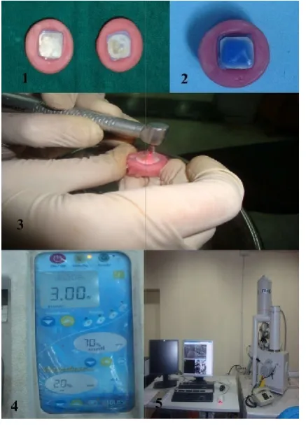
during the etching procedure. GrD2 was acid etched using 36% phosphoric acid gel (Conditioner 36, Dentsply). The application time of acid etchant was 15 sec. The specimens were then rinsed with water for 30 sec and dried by oil free compressed air for 15 sec. (Fig. 1-3)
Er,Cr:YSGG laser (Biolase) of 2.78 µm wavelength with the following settings was used-power -3W, air
20%. (Fig. 4) The beam for was aligned perpendicular at 1mm distance and moved in a sweeping fashion over the s
for 15 sec. The specimens were then dried with an oil free air source for 15 sec.
The specimens were then observed under ESEM (Fig. 5) at x1000 & ×5000 magnification. Parameters studied for dentin specimens were 1) removal of smear layer 2) diamet dentinal tubule opening measured by GSA image analyser. The collected data values were subjected to the Chi square & SND test.
The hydrokinetic laser system was investigated in 1995 by laser is based on the wavelength it can emit i.e. 1.54 µm and 2.7µ-2.9µm. The former coincides nicely with the absorption minimum of
optical silica fibres, allowing long range optical
rating optical amplifiers. The later wavelength coincides nicely with the peak of water absorption. As water is contained in every biological tissue, efficient interaction and dense optical energy deposition is guaranteed.
n were summarized as a (a) Corrugated wavy profile due to less demineralization of peritubular dentin due to lesser water content when compared to intertubular dentin. Protruding dentinal tubules are seen under SEM (b) Opened dentinal tubules with no widening(c) absence of smear layer (c) increased Ca:P ratio making the surface acid resistant (d) increased surface regularities with
The increased roughness of the laser etched surface in laser more control on the area to be etched(600 µm tip lases a surface area of 1.8×10⁻² cm2 ), with reduced technique sensitivity are few of the advantages of
The present study is a comparative ESEM analysis of Er,Cr:YSGG tested dentin surface vs acid etched dentin
diseased human teeth stored in normal saline after cleaning were taken as sample. Horizontal sections of dentin surfaces were prepared through middle third of the sample teeth by double sided diamond disk at slow speed. Each specimen was polished by 400 grit SiC paper for 30 sec to produce a smear layer. The thickness of each specimen was 1.0 mm ±0.5mm and was measured by a bur sensitivity. The dentin specimens were subdivided into GrD1 (4 samples) acting as control, GrD2 (18 samples) acid etched and GrD3 (18 samples) lased.
An acrylic resin platform was made and the specimens were placed in it stabilized by elastomeric impression material during the etching procedure. GrD2 was acid etched using 36% phosphoric acid gel (Conditioner 36, Dentsply). The application time of acid etchant was 15 sec. The specimens were then rinsed with water for 30 sec and dried by oil free
Er,Cr:YSGG laser (Biolase) of 2.78 µm wavelength with the 3W, air- 70 %, water- 20%. (Fig. 4) The beam for was aligned perpendicular at 1mm distance and moved in a sweeping fashion over the specimen for 15 sec. The specimens were then dried with an oil free air
The specimens were then observed under ESEM (Fig. 5) at x1000 & ×5000 magnification. Parameters studied for dentin specimens were 1) removal of smear layer 2) diameter of dentinal tubule opening measured by GSA image analyser. The collected data values were subjected to the Chi square &
Figure 1 Dentin specimens mounted on acrylic platforms
Figure 2 Acid etching of dentin specimen
Figure 3 Laser etching of dentin specimen (perpendicular angulation of the
beam to specimen is maintained)
Figure 4 Wattage, water and air settings for laser etching on Biolase
Figure 5 EDAX equipment for ESEM analysis
RESULTS
In the present study Chi- square & Student ‘t’
statistical comparison between GrD2 & D3 for smear layer removal and diameter of dentinal tubules after surface treatment.
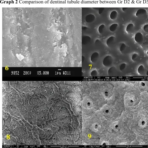
Group D2 showed twelve (66.7%) specimen free of smear layer and six specimens revealed presence of smear layer.(Figure 7-9) Fourteen (77.8%) of Group D3 specimens showed absence of smear layer where as four specimens showed presence of smear layer. (Graph 1)
Chi-square test reveals that comparison of removal of smear layer in the two groups viz D2 & D3, do not differ significantly (p>.05). Comparison between the groups reveals non- significant results (chi-square=0.28, p>0.05).
Absence of smear layer in two groups combined is 26 out of 36(72.2%). SND test shows that z=1.89, p=0.061: indicating significance 10% level.
Group D2 & D3 showed dentinal tubule opening in all the samples. For comparison of SEM impression between Groups D2 & D3, the mean diameter of tubules was tested for significance of their difference. Student ‘t’ test was applied and the ‘t’ value with 16 df came out to be 18.10 which is highly significant (p<0.001). (Graph 2).The mean tubule diameter in group D2 was 2.78 µm & in group D3 was 1.09 µm & the comparison between the groups showed a highly significant result.( Figure 7-9)
Dentin specimens mounted on acrylic platforms Acid etching of dentin specimen
of dentin specimen (perpendicular angulation of the beam to specimen is maintained)
Wattage, water and air settings for laser etching on Biolase EDAX equipment for ESEM analysis
square & Student ‘t’ were used for statistical comparison between GrD2 & D3 for smear layer removal and diameter of dentinal tubules after surface
Group D2 showed twelve (66.7%) specimen free of smear layer and six specimens revealed presence of smear 9) Fourteen (77.8%) of Group D3 specimens showed absence of smear layer where as four specimens showed presence of smear layer. (Graph 1)
square test reveals that comparison of removal of smear layer in the two groups viz D2 & D3, do not differ nificantly (p>.05). Comparison between the groups reveals
square=0.28, p>0.05).
Absence of smear layer in two groups combined is 26 out of 36(72.2%). SND test shows that z=1.89, p=0.061: indicating
International Journal of Current Advanced Research Vol 8, Issue 11(A), pp 20409-20413, November 2019
Graph 1 Comparison of absence & presence of smear layer between GrD2 & GrD3
Graph 2 Comparison of dentinal tubule diameter between Gr D2 & Gr D3
Figure 6 Gr D1 (dentin control)Note the smear layer covered surface
Figure 7 Gr D2(acid etched dentin surface) Note the open dentinal tubules
showing funnel shaped configuration
Figure 8 Gr D3 (lased dentin surface seen at 1300X magnification) Note the
DISCUSSION
The use of laser therapy has shown a promising front from the
current research. Currently, laser etching is proving to become
an alternative to acid etching of enamel. Er,CR:YSGG laser etching does not involve either vibration or heat; also, the easy handling of the apparatus makes this treatment highly attractive for routine clinical use.9
In the present study the specimens of enamel & dentin were polished by 400 grit silicon carbide paper to create a smear layer of around 1-2 µm thickness which is the same as that created by regular grit bur in a clinical situation.10
In the study gel form of the etchant was used however there was still spillage of acid over the tooth specimen seen.8 In comparison laser showed more control over the area to be etched.
3 W energy density used in this study avoided any cavity formation.11 According to T. Dostalova et al at 3 W power settings it is possible to etch the tooth surface without removing the enamel & dentin, the border is well defined & roughness is clearly visible. 70% air & 20% water (Figure 4) was used as it reduces the thermal effect of laser & increases the cleaning & cooling of the substrate.12 In a study by D.C. Atrrill et al, no water coolant was used in one of the subgroups which was irradiated by Er:YAG laser. They concluded that the cracks were wider & more prominent in this group. So, laser etching should be accompanied by water if excessive iatrogenic damage to tissues is to be prevented.13
Cooling with water spray is highly recommended so as to avoid over heating & consequent pulpal injury, microfissures, mineralization defects, carbonization & dehydration of the tissues. (S. Botta.2009).14 Meister et al found that in dehydrated moist dentin the quantity of ablated volumes of dentin was much lower than dentin containing water. Thus the exogenous has a greater effect than the endogenous water on ablation potential of Er,Cr:YSGG laser (P. Ekworapoj, 2007).
15
In the present study the beam was aligned perpendicular (Figure 3) to all the specimens for maximum cutting effectiveness of the laser beam. 7, 8 In a study by Ferreira L.
regions irradiated at different angles showed most of the surfaces having a scratched appearance with interspersed open
dentinal tubules & areas covered by melted surfaces.16
Almost all the specimens showed the absence of smear layer in the present study with no statistically significant difference between acid etched & laser etched surface. (Figure 7-9) This
concurs well with previous studies by M. Hossain et al (2001),
Piyamart et al (2007), Arlene et al (2008) 11, 15, 17.
In the present study SEM observation showed that laser irradiation produces recrystallized dentin surface (Fig. 8-9) which was also reported in studies by M. Hossain et al, S. Lin et al11, 18.
ESEM observation of the lased dentin specimens revealed rough surface topography. The features included absence of smear layer, open dentinal tubules, microroughness, and crater-like appearance.(Figure 8-9) The scaly or flaky surfaces are believed to be associated with the microexplosion effect which which was also observed by L. Ceballos et al & Visuri et al.19, 20 This lased dentin surface possessed an advantage enlarged surface area for adhesion. The scaly surface appearance of laser ablated dentin, along with the cuff-like appearance of peritubular dentin described by Aoki et al., was also evident in the SEM images from the current study(Fig 8-9). 21
In the present study the SEM micrographs of the irradiated dentin showed the depletion of the intertubular dentin more than peritubular dentin. The peritubular dentin still remained, indicating more resistance to laser energy due to higher mineral content and lack of collagen as an organic matrix. Collagen matrix is rich in water content, and laser energy is likely to be absorbed more by the interubular dentin than the
peritubular dentin as described by M. Hossain et al &
Ekworapoj et al11
Another difference between acid etchant and laser actions related to dentin is their effect on the structure of dentin tubules. When an acid etchant is applied, the peritubular dentin is preferentially etched, resulting in funnel-shaped openings to
the tubules.(Figure 7) According to A. Secilmis this structure
may contribute, with polymerization shrinkage, to pull the tags away from the walls thus leading to microleakage.
On the other hand, laser irradiation produces no demineralization of peritubular dentin, and the dentinal tubules remain open, parallel, with no widening. (Figure 8-9) This might probably contribute to the reduced microleakge after
laser etching.7
Dentinal tubules were not visible on the surface of the control group (Figure 6) as they were completely covered by smear layer.
Lased specimens showed open dentinal tubules of approximately 1 µm diameter (measured by GSA image analyser of SEM phtotos) (Fig. 8-9), while acid etched dentin specimens displayed patent dentinal tubules of around 2.8 µm in diameter (Fig. 7). A. Boyde & Scott DB showed the tubule diameter in the middle third of coronal dentin normally ranges from .8 -1.2 µm thus the inference that Er, Cr:YSGG laser had no effect on tubule diameter can be drawn. 22 The same results were observed by M. Hossain et al.23According to Pashley et al, 1996 the tubular dentin permeability is responsible for dentin sensitivity & pulpal irritation while the intradentin permeability is the one which is important for resin infiltration of the matrix to create a hybrid layer. Widely open dentinal tubules as seen after acid etching permits access of bacteria, it’s by- products & toxic chemicals like acids to the pulp. This is a major cause for postoperative pain, sensitivity & pulpal damage after bonding of tooth coloured restorations. As Er,Cr:YSGG might have no effect on dentinal tubule diameter & thus it may lead to reduction in hypersensitivity clinically.
CONCLUSION
Based on the clinical efficiency observed in relation to reduced chair side time, more control over lasing and less technique sensitivity and the ESEM results this study is in favour of laser
etching. However further clinical studies with regard to substrate composition after lasing and bond strength evaluation are needed to validate the effectiveness of Er,Cr:YSGG for the purpose of etching.
References
1. Simomsen R.. Present status of the acid etch technique. Clinical applications of the acid etch technique, Page 13-18
2. Pashley D., The effects of acid etching on pulpodentin complex.Operative dentistry, 1992, 17, 229-242.
3. Sasaki H.,D.C. Lobo, Moriyama Y., Watanabe S.,
Villaverde A., Tanaka S., Moriyama E., Brugnera A. . Tensile bond strength and SEM analysis of enamel etched with Er:YAG laser and phohsphoric acid: a comparative study In vitro. Brazilian Dental Journal, 2008 Vol. 19, No. 1
4. Apel C., Meister J., Gotz H., Duschner H., Gutknecht N.. Structural changes in human dental enamel after subablative Erbium laser irradiation and its potential use for caries prevention. Caries Research, 2005;39:65-70 5. Freitas P., Rapozo M., Eduardo C., Featherstone J. . In
vitro evaluation of Er,Cr:YSGG laser treated enamel demineralization. Lasers in Med Sci, 2008
6. Parker S. Introduction, history of lasers and laser light production.BDJ, 2007 Vol. 202, No 1:21-31
7. Secilmis A., Altintas S., Usumez A., Berk G.
Evaluation of mineral content of dentin prepared by Er,Cr:YSGG. Laser in Med. Sci., 2007
8. Usumez S., Orhan M., Usumez A. . Laser etching of enamel for direct bonding with an Er,Cr:YSGG
hydrokinetic laser system. Am J Dentofacial
Orthop,2002;122:649-656
9. Özer T., Basara G., Berk N. Laser etching of enamel for orthodontic bonding. Am J Dentofacial Orthop, 2008, 134:193-7
10. Goes M., Coelho M., Simnoides. Morphological effect of type, concentration & etching time of acid solutions on enamel & dentin surfaces. Braz. Dent. J., 1998, 9(1) 11. Hossain M., Nakumura Y., Yamada Y., Suzuki N.,
Murakami Y., Matsumoto K. Analysis of surface roughness of enamel and dentin after Er,Cr:YSGG laser irradiation.Journal of Clinical Laser Medicine & Surgery, 2001, Vol. 19, Number 6,pg 297-303
12. Olivi G., Angiero F., Benedicent S. Use of the Er:YAG on human enamel tissues. Influence of the air water spray on the laser tissue interaction:SEM evaluations. Lasers Med Sci Jun, 2009
13. Attrill D., Farrar S., King T., Dickinson M. Er:Yag laser etching of dental enamel as an alternative to acid etching. Lasers Med Sci, 2000, 15; 154-161
14. Botta S., Ana P., Zezell D., Powers J., Matos A. Adhesion after Er,Cr:YSGG laser application at three different irradiation condition. Lasers in Med. Sci., 2008 15. Ekworapoj P., Sharanbir K. Sidhu, John F. McCabe .Effect of different power parameters of Er,Cr:YSGG laser on human dentin. Lasers in Med. Sci., 2007 16. Ferreira L., Apel C., Francci C., Simoes A.Influence of
etching time on bond in dentin irradiated with Er lasers. Lasers Med Sci Aug., 2009
17. Tachibana A.,Marques M., Soler J., Matos A..
International Journal of Current Advanced Research Vol 8, Issue 11(A), pp 20409-20413, November 2019
bonding of a self etching adhesive system. Lasers Med Sci, 2008, Oct.,23;435-441
18. Lin S., Caputo A., Eversole L.,Rizoiu I. Topographical characteristics and shear bond strength of tooth surfaces cut with a laser-powered hydrokinetic system. Journal of Prosthetic Dentistry, 1999; 82: 451-5
19. Cellabos L., Toledano M., R. Osorio R., Tay F.. Bonding to Er:YAG laser treated dentin. J. Dent Res, 2002, 81(2):119-122
20. Visuri S., Walsh J., Wigdor H. Erbium laser ablation of dental hard tissue:Effect of water cooling. Laser in Surgery & Medicine,1996, 18: pg. 294-300
21. Dunn W., Davis J., Bush A.Shear bond strength and SEM evaluation of composite bonded to Er:YAG laser-prepared dentin and enamel. Dental Materials Vol. 21, Issue 7, 2005, July, Pg. 616-624.
22. Ramos R., Chimello D., Chinelatti M. Effect of Er:YAG laser on bond strength to dentin of a self etching primer & two single bottle adhesive systems. Lasers Med Sci,2002, 31:164-170
23. Hossain M, Yamada Y., Nakamura Y. A study on surface roughness & microleakage test in cavities prepared by Er:YAG laser irradiation & etched bur cavities. Lasers Med Sci, 2003 18:25-31
How to cite this article:
Rashmi Issar et al (2019) 'Comparative Esem Analysis of Er,Cr:YSGG Treated Dentin Surface vs Conditioner 36 Treated Dentin Surface', International Journal of Current Advanced Research, 08(11), pp. 20409-20413.
DOI: http://dx.doi.org/10.24327/ijcar.2019.20413.3987