an example of a missed post-traumatic
unilateral facet subluxation –
a case report
Jeffrey R Tuling,
BSc, DC*
William H Hsu,
BSc, DC, DACBR, FCCR(C)**
Les lésions unilatérales des facettes, dont les
subluxations, surviennent dans 4 à 16 p. 100 des cas de blessure à la colonne cervicale (les subluxations unilatérales des facettes forment un sous-groupe particulier de cas). Ces lésions se produisent dans les accidents d’automobile sous l’action des forces de flexion et d’écartement ou de la flexion de la tête déjà en rotation.
Comme, dans la plupart des cas, les subluxations unilatérales des facettes ne présentent pas de signes ni de symptômes neurologiques, les diagnostics tardifs sont relativement fréquents. Aussi est-il impératif de poser le bon diagnostic pour donner au patient des soins plus efficaces et, indirectement, lui éviter des séquelles. Pour ce faire, le clinicien ou la clinicienne peut avoir recours à l’examen radiologique.
Une évaluation appropriée des troubles associés au coup de fouet cervical repose sur une prise adéquate de radiographies. Les signes radiologiques de subluxation cervicale sont peu visibles sur les clichés ordinaires; par contre, les clichés pris en oblique facilitent de beaucoup l’établissement du diagnostic. Ils se révèlent donc d’une grande utilité en clinique.
Voici le cas d’une femme de 29 ans dont la subluxation unilatérale d’une facette est passée inaperçue; il sera question, dans le présent article, des observations et des caractéristiques radiologiques, ainsi que le l’utilité clinique des clichés pris en oblique chez les patients souffrant de troubles associés au coup de fouet cervical.
(JACC 1999; 43(3):168–175)
M O T S C L É S : subluxation ou dislocation unilatérale des
facettes, post-traumatisme, incidence en oblique,
radiographie, troubles associés au coup de fouet cervical. Unilateral facet injuries occur in 4-16% of patients with
cervical spine injuries, (of which unilateral facet subluxations occur as a subgroup in this population). These injuries arise in motor vehicle accidents because of flexion-distraction forces, or flexion of an already rotated head.
Due to the common presentation of a lack of
neurological signs and symptoms, delayed diagnosis of unilateral facet subluxations are common. As a result, it is imperative that the proper diagnosis is made so that patient care can be made more effective with ultimately less sequelae. For the clinician, diagnosis can be aided by the use of radiographic analysis.
Adequate radiographic analysis is an important tool to accurately assess whiplash associated disorders. Radiographic findings of facet subluxations are subtle on routine views. Utilizing oblique views, the diagnoses are usually evident. Therefore, oblique views are very useful in the clinical setting.
This case report will describe a 29-year-old female with a missed unilateral facet subluxation, radiographic findings and characteristics, and the clinical utility of oblique views in patients suffering from whiplash associated disorders.
(JCCA 1999; 43(3):168–175)
K E YW O R D S: unilateral facet subluxation/dislocation,
Introduction
Unilateral facet injuries are significant since they occur in 4 to 16% of patients with cervical spine injury presenting to hospital clinics.1,2,3 Facet injuries can present in a range
from subtle facet subluxations to overt facet dislocations. A facet joint subluxation does not imply full dislocation. A dislocation is defined as a displacement of a bone in rela-tion to the apposing bone at the joint, resulting in a com-plete loss of continuity of the joint surfaces.4 Whereas a
subluxation is a partial loss of contiguity of the joint sur-faces. Although joint surfaces are incongruous, a signifi-cant proportion remains apposed.4
Unilateral facet injuries occur in motor vehicle acci-dents as a result of simultaneous rotation of the head with an accompanying distraction-flexion force or flexion of an already rotated head.1,2,3,5,6,7,8 These movements create
forces that prestress the capsules of the zygapophyseal joints, rendering the joints more susceptible to injury.9
As a result, many who suffer from unilateral facet inju-ries can manifest the associated symptoms commonly found in whiplash-associated disorders due to cervical spine and/or ligamentous injury.10 Two weeks of delay in
treatment due to incorrect diagnoses in unilateral facet in-juries occurs in upwards of 40% of cases.1,11 Frequently,
these patients have no neurologic deficit or have an iso-lated radiculopathy that may be overlooked in a cursory exam.8 Additionally, unilateral facet injuries are
com-monly missed on initial films due to the subtlety of radio-graphic signs.12 It is only through the persistent complaints
from the patient that the correct diagnosis is confirmed.1,11
Beyer and Cabanela8 summarize three reasons for delay
in diagnosis of unilateral facet injuries. First, there is inad-equate radiographic evaluation. Second, associated inju-ries may lead the concern of the physician away from the cervical spine. Third, there is usually a lack of serious symptoms associated with unilateral facet dislocation or fracture-dislocation.8
The case of a 29-year-old female patient with neck pain resulting from a motor vehicle accident is presented.
Case report
A 29-year-old female bank manager presented to our clinic with recent reaggravation of right neck pain of six days duration. The complaint was sudden in onset without recent trauma. However, she was involved in a motor vehicle accident that occurred seven months prior to this presentation.
Seven months before, the patient was in the front pas-senger seat of a car on an expressway. Road conditions were wet due to snow. The patient was wearing a three-point seat belt but did not have airbag protection. The car went out of control, slid into a ditch, rolled several times, and landed upright. She had immediate pain in her neck and right hand. She also noticed swelling of the right wrist and forearm. The patient felt that she hit her head but could not recall the specific details of the accident. She felt that she did not lose consciousness.
She was transferred via an ambulance to the local hospi-tal where she was assessed. Radiographs of her head, neck and hand were taken and reported as normal. She was then given a neck brace and discharged. The next day she saw her physician whereupon she was prescribed Advil and physiotherapy, which included two weeks of wearing a soft cervical collar and four months of electrotherapy.
The presenting complaint was described as a dull ache, with an intensity of 2/10, occurring with neck movement. The pain was aggravated by rotating her head to the right and became more intense with increased neck activity. The relieving factors included massage. There were no other associated symptoms such as weaknesses or paraesthesiae in the upper limbs.
Gross active range of motion of the cervical spine on right rotation was limited by 20° by mild pain in the upper right trapezius fibres. Passive and active right lateral flexion produced pain in the upper right trapezius at the end range of motion. Left lateral flexion elicited a pulling sensation on the right side. Forward flexion produced mild pain while all other ranges were full and pain free. Resisted ranges were full and pain free except for right lateral
* Clinical Sciences Resident II, Department of Graduate Studies and Research, Canadian Memorial Chiropractic College, 1931 Bayview Avenue, Toronto, Ontario M4G 3E6. Tel: (416) 482-2340. e-mail [email protected]
** Assistant Clinical Professor, Department of Clinical Education, Canadian Memorial Chiropractic College. Address reprints and all correspondence to J. Tuling.
flexion which reproduced her presenting complaint. Neurological examination revealed intact upper and lower limb sensation, full motor strength, and 2+ reflexes. Spurling’s (lateral flexion with light overpressure), Jackson’s (rotation with light overpressure) and maximal foraminal compression (lateral flexion, extension and ro-tation with slight overpressure) on the right reproduced the patient’s symptoms with pain into the upper right trapezius fibres. Facet rub produced pain at the right C4–C5 level. Valsalva’s and Doorbell’s were negative. Motion palpa-tion revealed mild restricpalpa-tions at C6-T1 on the right.
Based upon the presentation and clinical findings at the time of this work-up, this patient was tentatively diagnosed as having a Whiplash Associated Disorder (WAD) Type II according to the parameters set out by the Quebec Task Force13 and facet irritation and associated restriction of the
cervical spine.
Radiographs taken at the time of the accident were ob-tained from the hospital for reassessment by our radiology department (Figures 1, 2). These included APOM, AP, lateral and oblique views of the cervical spine. On close inspection of the oblique films, a misalignment of the right C5–C6 facet joint was noted with the C5 inferior facet perched on the C6 superior facet. Only one-third of the facet was still in contact. A post-traumatic facet subluxa-tion of C5 on C6 on the right had been missed.
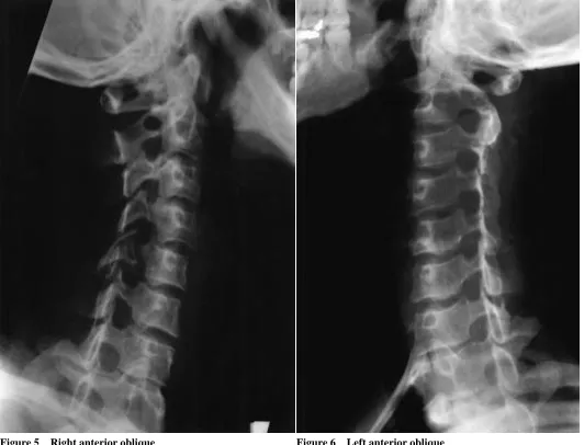
Additional films were obtained at our clinic to assess for any progression (Figures 3, 4, 5 and 6). They confirmed the partial dislocation. Additionally, a 4 mm antero-listhesis of C4 on C5 was detected which was not present on the initial films. This would indicate disruption likely involving the supraspinous, interspinous and facet capsular ligaments. Based upon these findings, the diagno-sis was changed to a Whiplash Associated Disorder (WAD) Type IV because of the facet subluxation, as well as the ligamentous tear at C4–C5. The patient was referred to a rehabilitation clinic for further assessment and was lost to follow-up.
Discussion
The Quebec Task Force13 recommendations for clinical
practice states that patients who present with a Whiplash Associated Disorder (WAD) of Grade II and III, should obtain a three-view baseline radiologic examination of the cervical spine. These baseline radiologic films include the antero-posterior, lateral and open mouth views. These
rou-tine views are usually adequate, and is substantiated by numerous investigators.14,15,16,17,18,19 In patients having a
Grade II or III WAD, flexion/extension views may occa-sionally be indicated.13 If there is any clinical suspicion of
more serious injuries, then other appropriate radiographs should be obtained.
The three-view baseline studies may be incomplete in that significant ligamentous instability may exist in the absence of an obvious vertebral column fracture or dislo-cation.14,15,16 Certain investigators have found a 23% to
26% false-negative rate for their lateral views, and as a
result state that the lateral view is not adequate to screen for cervical spine injury.10,20 Doris et al.20 state that if the
three views prove positive for fracture or dislocation, then no further radiographic screening is necessary. However, if the three views are negative, then the patient undergoes the five-view standard series which includes the oblique views.20 The oblique views are obtained to search for
pos-sible posterior laminar fracture, unilateral facet disloca-tion, and facet subluxation.20
Additionally, extended radiographic evaluation of cases involving cervical spine trauma should also be considered. Further sequelae such as delayed instabil-ity can occur at a later date which can be detected through radiographic studies.
The radiographic characteristics (Table 1) can individu-ally or collectively aid in the diagnosis of a unilateral facet subluxation. Some of these findings are not immediately obvious, but should routinely be sought in cases of cervical spine trauma.
A lateral cervical radiograph yields the most valuable information in regards to unilateral facet subluxations. If anterolisthesis is present on the lateral view, it is regarded as an auxiliary sign of facet subluxation/dislocation. The body of the dislocated vertebra may be anteriorly dis-placed less than or equal to one-half the sagittal diameter of the inferior vertebral body.1,7,21 Fanning or widening of
the interspinous distances may also be visualized on the lateral view and suggest tearing of the interspinous
liga-Right anterior oblique Left anterior oblique
ment and facet capsule. Loss of cervical lordosis may sug-gest hyperflexion injuries likely resulting from torn nuchal and interspinous ligaments with disruption of the zyg-apophyseal capsules.22 Increased height and asymmetry
of the intervertebral discs posteriorly may also indicate a flexion injury.22
Rotational injuries are more apparent if a double con-tour of the posterior margins of the vertebral bodies is seen.22 Another radiographic sign indicating possible facet
subluxation is displacement of the interfacetal joint.3 If the
rotational component is severe, then full dislocation arises and produces the “bow tie sign” which is seen on the lat-eral view. The bow tie sign arises from the combination of the anteriorly displaced articular pillar relative to its former opposing pillar.3,5 Abrupt changes of the
inter-laminar space at the level of the injury also correlate well with rotational-flexion trauma producing facet subluxa-tion/dislocation.3
When viewing the oblique radiographs, the cervical facets should overlap each other and appear similar to
Figure 3 Lateral radiograph of the cervical spine obtained at our clinic at a later date shows a horizontally oriented C5 articular pillar with a 4mm anterolisthesis of C4 on C5.
the traditional rooftop shingles of a house. If there is an interruption of this pattern, then it is indicative of the “shingles on the roof” sign. A disruption of the “shingles on the roof” appearance will confirm the presence of a unilateral facet dislocation23 or facet subluxation on the
oblique view. It must also be emphasized that there are associated fractures affecting the zyagapophyseal joints or laminae in 35% of patients with unilateral facet sub-luxations/dislocations.2
On the anteroposterior view, the spinous processes may
be displaced to the side of the facet injury superior to the facet that was subluxated/dislocated.7,24 Tracheal or
laryn-geal deviation is also indicative of ligamentous disruption in the cervical spine and rotational injury.22
In unilateral facet dislocation/subluxation, computer-ized tomography (CT) is useful to detect and assess associ-ated fractures of the neural arch, disc herniations, prevertebral hematomas, and the relationship of any bony fragments to the spinal cord.25 Axial CT can be utilized to
identify the “naked facet” sign,26 if there is an absence of
Figure 5 Right anterior oblique Figure 6 Left anterior oblique
Table 1
Suggestive Radiographic Findings of Unilateral Facet Subluxations
A Lateral Radiograph 1. Anterolisthesis
2. Disc (increase in posterior disc height) 3. Vertebral body (anterior wedge deformity) 4. Double posterior contour
5. Rotation (abrupt narrowing of the distance between articular pillar and spinolaminar line)
6. “Bowtie” sign
B Oblique Radiographs (Definitive diagnostic film) 1. “Shingles on roof” sign
C Anteroposterior Radiograph 1 Deviated spinous processes
one facet surface due to a unilateral facet subluxation/ dislocation.27 Magnetic resonance imaging (MRI) can be
utilized for the detection of soft tissue injuries in acute and chronic cases. MRI can reveal lesions involving the liga-ments, discs, spinal cord, muscles and prevertebral tis-sues.25,28,29
The priority in treating a subluxation or full unilateral facet dislocation is preservation of the continuity and func-tion of the spinal cord. Other goals of treatment are the relief of neurological compression, spinal stabilization and speedy restoration of a patient’s neurological condition. Thus, medical referral is necessary for subluxations or full unilateral facet dislocations.
There are two schools of thought regarding the appro-priate treatment of facet subluxations/dislocations. One feels that nonoperative treatment is adequate, the other feels that operative treatment is necessary. In Beatson’s21
study, dislocations could be left in unreduced positions with little or no symptoms. Cotler et al.30 found that closed
reduction was safe and effective. This is further supported by Braakman et al.,1 who found that spontaneous
stabilization would occur regardless of whether reduction was carried out or not. Rorabeck et al.11 found that
unre-duced dislocations would eventually develop pain;
sponta-neous fusion occurred in only 20% of patients in the study. Beyer et al.8 found that open reduction provided fewer
complaints of stiffness and pain, and resolution happened more frequently. O’Brien31 found that there was a late
instability rate of 17% after closed treatment of these inju-ries.
Conclusion
The radiographs of this patient’s head, neck and hand were read and interpreted as normal by radiologists at the hospi-tal. Seven months after the accident, this patient continued to experience symptoms related to the cervical spine. Cer-tain pathologies such as ligamentous disruptions may be missed and may not be evident soon after trauma resulting in a delayed instability. Therefore, follow-up x-rays are recommended, particularily if the patient is nonresponsive to treatment.
An adequate radiographic series is crucial for an accu-rate diagnosis of whiplash associated disorders. As dem-onstrated in our case report, radiographic findings of facet subluxation are subtle on routine radiographs but more evident on oblique films.
This case report emphasizes the importance of review-ing any previous radiographs, regardless of their source, by the treating chiropractor. The facet subluxation was missed and the delayed ligamentous laxity was not evident until the follow-up radiographic study. Delayed diagnosis of facet subluxation is common due to its frequent lack of neurological symptoms and signs. However it represents disruption of the stabilizing ligaments of the cervical spine, and it impacts upon the prognosis of the patient’s condition. Thus, early proper diagnosis and early manage-ment are crucial.
Despite its limitations, a plain film radiographic study remains a crucial component of a proper assessment of patients with related injury to the neck in motor vehicle accidents. It is only through this awareness and course of action that whiplash patients can be appropriately diag-nosed and properly treated.
Acknowledgement
References
1 Braakman R, Vinken PJ. Unilateral facet interlocking in the lower cervical spine. J Bone Joint Surgery 1967; 49B:249–257.
2 Scher AT. Unilateral locked facet in cervical spine injuries. AJR 1977; 129:45–48.
3 Young JWR, Resnik CS, DeCandido P, Mirvis SE. The laminar space in the diagnosis of rotational flexion injuries of the cervical spine. AJR 1989; 152:103–107.
4 Rogers LF. Radiology of Skeletal Trauma. New York: Churchill Livingstone, 1982:38.
5 Taveras JM, Ferrucci JT. Radiology. New York: Lipincott-Raven Publishers, 1996:7.
6 Mirvis SE, Young JWR. Imaging in Trauma and Critical Care. Baltimore: Williams and Wilkins, 1992:312. 7 Harris JH, Edeiken MB. The Radiology of Acute
Cervical Spine Trauma 2nd ed. Williams and Wilkins, 1987:119–121.
8 Beyer CA, Cabanela ME. Unilateral facet dislocations and fracture-dislocations of the cervical spine: A review. Orthopedics 1992; 15(3):311–315.
9 Dvorak J, Panjabi MM, Gerber M, Wichman W. CT-functional diagnostics of the rotatory instability of upper cervical spine. An experimental study on cadavers. Spine 1987b; 12:197–205.
10 Bachulis BL, Long WB, Hynes GD, Johnson MC. Clinical indications for cervcial spine radiographs in the
traumatized patient. Am J Sur 1987; 153:473–478. 11 Rorabeck CH, Rock MG, Hawkins RJ, Bourne RB.
Unilateral facet dislocation of the cervical spine: an analysis of the results of treatment in 26 patients. Spine 1987; 12:23–27.
12 Andreshak JL, Dekutoski MB. Management of unilateral facet dislocations: a review of the literature. Orthopaedics 1997; 20:917–926.
13 Spitzer WO, Skovron ML, Salmi LR, Cassidy JD, Duranceau J, Suissa S, Zeiss E. Scientific monograph of the Quebec Task Force on whiplash-associated disorders: Redefining “Whiplash” and its management. Spine 1995; 20(8s):supplement..
14 Allen BL Jr, Ferguson RL, Lehmann TR, O’Brien RP. A mechanistic classification of closed, indirect fractures and dislocations of the lower cervical spine. Spine 1982; 7:1–27.
15 Clark CR, Ingram CM, El-Khoury GY, Ehara S. Radiographic evaluation of cervical spine injuries. Spine 1988; 13:742–747.
16 White AA, Southwick WO, Panjabi MM. Clinical instability in the lower cervical spine: a review of past and current concepts. Spine 1976; 1:15–27.
17 Ross SE, Schwab CW, David ET, Delong WG, Born CT. Clearing the cervical spine: initial radiologic evaluation. J Trauma 1987; 27(9):1055–1060.
18 Cotler HB. Fractures and dislocations of the subaxial cervical spine. Spine: State of the art reviews 1991; 5(2):203–216.
19 Cybulski GR, Douglas RA, Meyer PR, Rovin RA. Complications in three-column cervical spine injuries requiring anterior-posterior stabilization. Spine 1992; 17(3):253–256.
20 Doris PE, Wilson RA. The next logical step in the emergency radiographic evaluation of cervcial spine trauma: The five-view trauma series. J Emergency Medicine 1985; 3(5);371–385.
21 Beatson TR. Fractures and dislocations of the cervical spine. J Bone Joint Surgery 1963; 45B:21–35. 22 Clark WM, Gehweiler JA, Laib R. Twelve significant
signs of cervical spine trauma. Skeletal Radiology 1979; 3:201–205.
23 Harris JH. The radiology of acute cervical spine trauma. Baltimore, Williams and Wilkins, 1978: 25–26, 61–70. 24 Scher AT. Anterior cervical subluxation: an unstable
position. Am J Roentgenology 1979; 133:275–280. 25 Murphy MD, Batitzky S, Bramble JM. Diagnostic imaging
of spinal trauma. Radiology Clinics North America 1989; 27:855–872.
26 Yochum TR, Rowe LJ. Essentials of Skeletal Radiology, 2nd ed. Baltimore: Williams and Wilkins, 1996:680–692. 27 Kornberg M. The computed tomographic appearance of a
unilateral Jumped cervical facet (the “False” facet joint sign). Spine 1986; 11:1038–1040.
28 Harris JH, Yeakley JW. Hyperextension dislocation of the cervical spine. Ligament injuries demonstrated by magnetic resonance imaging. J Bone Joint Surgery 1992; 74B:567–570.
29 Davis SJ, Teresi LM, Bradley WJ, Ziemba MA, Bloze AE. The widened disc space – a sign of cervical spiune hyperextension injury. Radiology 1991; 180:245–251. 30 Cotler HB, Miller LS, DeLucia FA, Cotler JM, Davne SH.
Closed reduction of cervical spine dislocations. Clin Orthop. 1987; 214:185–199.