University of Pennsylvania
ScholarlyCommons
Publicly Accessible Penn Dissertations
1-1-2016
Development of Shear-Thinning and Self-Healing
Hydrogels Through Guest-Host Interactions for
Biomedical Applications
Christopher Blake Rodell
University of Pennsylvania, [email protected]
Follow this and additional works at:http://repository.upenn.edu/edissertations
Part of theBiomedical Commons
Recommended Citation
Rodell, Christopher Blake, "Development of Shear-Thinning and Self-Healing Hydrogels Through Guest-Host Interactions for Biomedical Applications" (2016).Publicly Accessible Penn Dissertations. 1978.
Development of Shear-Thinning and Self-Healing Hydrogels Through
Guest-Host Interactions for Biomedical Applications
Abstract
Hydrogels have emerged as an invaluable class of materials for biomedical applications, owing in part to their utility as structural, bioinstructive, and cell-laden implants that mimic many aspects of native tissues. Despite their many positive attributes, conventional hydrogels face numerous challenges toward translational
therapies, including difficulty in delivery (i.e., invasive implantation) as well as limited control over biophysical properties (i.e., porosity, degradation, and strength). To address these challenges, the overall goal of this dissertation was the development of a class of supramolecular hydrogels that can be implanted in vivo by simple injection and that have tunable properties — either innate to the system or achieved through
additional modifications. Toward this, we developed guest-host (GH) hydrogels that undergo supramolecular assembly through complexation of hyaluronic acid (HA) separately modified by adamantane (Ad-HA, guest) and β-cyclodextrin (CD-HA, host).
Modular modifications were made to GH hydrogels to enable tuning of biophysical properties, including the incorporation of matrix-metalloproteinase cleavable peptides between HA and Ad to form enzymatically degradable assemblies. Additionally, dual-crosslinking (DC) of methacrylated CD-HA (CD-MeHA) and thiolated Ad-HA (Ad-HA-SH) by Michael addition subsequent to GH assembly was explored to stiffen hydrogels in vivo following injection. Finally, injectable and tough double network (DN) hydrogels were fabricated, where GH hydrogels were formed in the presence of an interpenetrating covalent network
(methacrylated HA, MeHA) crosslinked by Michael addition with a dithiol under cytocompatible conditions.
Both GH and DC hydrogels were further explored in vivo, with application to attenuate the maladaptive left ventricular (LV) remodeling that occurs following myocardial infarction (MI) that can result in heart failure. DC hydrogels reduced stress within the infarct region, prevented early ventricular expansion and thereby ameliorated progressive LV remodeling. Moreover, the preservation of myocardial geometry reduced incidence and severity of ischemic mitral regurgitation — an undesirable and devastating consequence of LV remodeling. Overall, the body of work represents a novel approach to engineer biomaterials with unique properties toward biomedical therapies.
Degree Type
Dissertation
Degree Name
Doctor of Philosophy (PhD)
Graduate Group
Bioengineering
First Advisor
Keywords
Cyclodextrin, Heart Failure, Hyaluronic Acid, Hydrogel, Injectable, Supramolecular
Subject Categories
DEVELOPMENT OF SHEAR-THINNING AND SELF-HEALING HYDROGELS THROUGH GUEST-HOST INTERACTIONS FOR BIOMEDICAL
APPLICATIONS
Christopher B. Rodell
A DISSERTATION
in Bioengineering
Presented to the Faculties of the University of Pennsylvania
In Partial Fulfillment of the Requirements for the
Degree of Doctor of Philosophy
2016
_____________________
Supervisor of Dissertation
Dr. Jason A. Burdick
Professor of Bioengineering
_____________________
Graduate Group Chairperson
Dr. Jason A. Burdick
Professor of Bioengineering
Dissertation Committee
DEDICATION
“Don’t downgrade your dreams just to fit your reality. Upgrade your convictions to match your destiny” – Stuart Scott
This one’s for everyone who stood by me, especially my parents and Michele,
ACKNOWLEDGEMENT
The past five years have been an amazing time for growth—both
professional and personal—and I am forever indebted to the multitude of wonderful
people who I have been blessed to share this time with. I would like begin by
acknowledging my thesis committee, including Drs. Andrew Tsourkas, Robert
Gorman, and Russel Composto. In their own ways, each member of my committee
has welcomed me into the community at Penn, furthered my thoughts, and taught
me much—earning my most sincere respect and thanks.
Foremost, I would like to thank my advisor, Jason Burdick, for his continuing
support and guidance. Jason is an exceptional mentor, exuding a positive
personality and excitement for science that are coupled with sharp wit, humble
demeanor, and personable character. Most importantly, he remains truly invested
in his lab members and easily accessible at any time. He has been there to guide
my growth, discuss my decisions, and share in my excitement (and frustrations)
throughout the past years. His support in my life, both professional and personal,
will no doubt continue over the years and always be truly appreciated.
In addition to the boss, I am thankful to the JAB Lab members, past and
present. It became clear to me very early on that this is a diverse and talented
group (likely assembled by some evil genius) where it’s a great strength that every
person is unique—having their own interests, talents, and personalities. It’s
because of these differences that I’ve learned something from all of them and am
thankful. In particular, I owe special thanks to the postdocs, especially, William
Gramlich, Danielle Soranno, Steven Caliari, and Chris Highley, who have been
Ryan “The Cat” Wade and Brendan Purcell, who were my bay mates, mentors,
and friends; and to Adam Kaminski and Neville Dusaj who were outstanding
undergraduate researchers with bright futures ahead.
I would be remiss to forget how I arrived here at Penn. Along my journey,
there were countless mentors and teachers who have forever influenced my life.
Among the great scouters of all time, the Boda Family, Dave Willoughby, and Phil
Carson have indelibly influenced by personal growth. Moreover, I was lucky to
have many great teachers throughout my life, including but not limited to Mrs.
Cathy Nix, Dr. Bob, Dr. Steve Clark, Dr. David Rice, and my BS/MS mentor Dr.
Michael Moore who have each taken special care to develop not only my mind and
career, but also shape my personality and passions.
Of course, no influence is greater than that of family, and mine has been an
unwavering source of support and love throughout my life. I would like to thank
them all, especially my parents (Bob & Debbie), my brothers (Eric and Rob), my
sister (Lynne) and their respective families. I am blessed to have a second
family-to-be in the Rotellas, especially Frank and Gloria. To all of my family, I am thankful
for their acceptance and support, despite the grad school schedule which has kept
me from always being there. Most of all, there is one person who gives me
something to look forward to—be it a weekend of studying, our upcoming wedding,
or a lifetime together: Michele. Through the struggles of distance, late night trips
to lab, and never-ending exams and deadlines we have remained a team, making
each other stronger with each passing challenge — I look forward to the next and
ABSTRACT
DEVELOPMENT OF SHEAR-THINNING AND SELF-HEALING HYDROGELS THROUGH GUEST-HOST INTERACTIONS FOR BIOMEDICAL
APPLICATIONS Christopher B. Rodell
Jason A. Burdick, Ph.D.
Hydrogels have emerged as an invaluable class of materials for biomedical
applications, owing in part to their utility as structural, bioinstructive, and cell-laden
implants that mimic many aspects of native tissues. Despite their many positive
attributes, conventional hydrogels face numerous challenges toward translational
therapies, including difficulty in delivery (i.e., invasive implantation) as well as
limited control over biophysical properties (i.e., porosity, degradation, and
strength). To address these challenges, the overall goal of this dissertation was
the development of a class of supramolecular hydrogels that can be implanted in
vivo by simple injection and that have tunable properties — either innate to the
system or achieved through additional modifications. Toward this, we developed
guest-host (GH) hydrogels that undergo supramolecular assembly through
complexation of hyaluronic acid (HA) separately modified by adamantane (Ad-HA,
guest) and β-cyclodextrin (CD-HA, host).
Modular modifications were made to GH hydrogels to enable tuning of
cleavable peptides between HA and Ad to form enzymatically degradable
assemblies. Additionally, dual-crosslinking (DC) of methacrylated CD-HA
(CD-MeHA) and thiolated Ad-HA (Ad-HA-SH) by Michael addition subsequent to GH
assembly was explored to stiffen hydrogels in vivo following injection. Finally,
injectable and tough double network (DN) hydrogels were fabricated, where GH
hydrogels were formed in the presence of an interpenetrating covalent network
(methacrylated HA, MeHA) crosslinked by Michael addition with a dithiol under
cytocompatible conditions.
Both GH and DC hydrogels were further explored in vivo, with application
to attenuate the maladaptive left ventricular (LV) remodeling that occurs following
myocardial infarction (MI) that can result in heart failure. DC hydrogels reduced
stress within the infarct region, prevented early ventricular expansion and thereby
ameliorated progressive LV remodeling. Moreover, the preservation of myocardial
geometry reduced incidence and severity of ischemic mitral regurgitation — an
undesirable and devastating consequence of LV remodeling. Overall, the body of
work represents a novel approach to engineer biomaterials with unique properties
TABLE OF CONTENTS
DEDICATION ...ii
ACKNOWLEDGEMENT ... iii
ABSTRACT ... v
TABLE OF CONTENTS ... vii
LIST OF TABLES ...xv
LIST OF FIGURES ... xvi
CHAPTER 1 ... 1
Introduction: Material Design for Biomedical Applications ... 1
1.1 Introduction ... 1
1.2 From Molecular to Macroscopic ... 4
1.2.1 From Molecular Organization to Nanostructure ... 4
1.2.2 Building at the Mesoscale ... 6
1.2.3 Macroscopic Materials and their Sub-Assemblies ... 7
1.3 From Static to Dynamic ... 9
1.3.1 Incorporating Degradation into Biomaterials ... 10
1.3.2 Stimuli-Responsive Biomaterials ... 13
1.4 From Bioinert to Biocomplex ... 16
1.4.1 Materials to Deliver Therapeutics ... 18
1.4.2 Materials to Present Matrix Cues ... 20
1.4.3 Materials for Tissue Integration ... 22
1.5 Moving into the Future ... 23
CHAPTER 2 ... 36
Specific Aims and Research Overview ... 36
2.1 Introduction and Specific Aims ... 36
Specific Aim 1 ... 38
Specific Aim 2 ... 38
Specific Aim 3 ... 39
Specific Aim 4 ... 40
2.2. Research Overview ... 41
2.3 References ... 43
CHAPTER 3 ... 44
Supramolecular Guest-Host Interactions for the Preparation of Biomedical Materials ... 44
3.1 Introduction ... 44
3.2 The Structure of Self-Assembly ... 46
3.2.1 Direct Molecular Recognition ... 46
3.2.2 Polymeric Assembly ... 50
3.3 Diverse Biomedical Applications ... 53
3.3.1 Injectable Hydrogels ... 53
3.3.2 Therapeutic Delivery ... 58
3.3.3 Cell-Material Interactions ... 62
3.4 Conclusions and Future Directions ... 67
3.5 References ... 69
CHAPTER 4 ... 80
4.1 Introduction ... 80
4.2 Methods ... 83
4.2.1 General Materials ... 83
4.2.2 HA-TBA Preparation ... 83
4.2.3 Ad-HA Synthesis ... 84
4.2.4 β-CD-HDA Synthesis ... 84
4.2.5 CD-HA Synthesis ... 85
4.2.6 Determination of Apparent Binding Affinity ... 86
4.2.6 Hydrogel Formation ... 87
4.2.7 Rheological Characterization ... 87
4.2.8 Fluorescent Labeling ... 88
4.2.9 Hydrogel Erosion and BSA Release ... 88
4.3 Results and Discussion ... 89
4.3.1 Chemical Modification of HA ... 89
4.3.2 Inclusion Ability of Guest and Host Macromers ... 91
4.3.3 Hydrogel Formation ... 93
4.3.4 Control of Network Properties ... 95
4.3.5 Shear-thinning and Recovery ... 102
4.3.6 Hydrogel Erosion and Biomolecule Release ... 105
4.4 Conclusion ... 109
4.5 References ... 110
CHAPTER 5 ... 116
5.1 Introduction ... 116
5.2 Experimental Methods and Materials ... 119
5.2.2 Hydrogel Formation ... 120
5.2.3 Microscopic Characterization ... 121
5.2.4 Atomic Force Microscopy ... 121
5.2.5 Diffusive Microparticle Tracking ... 122
5.3 Results and Discussion ... 123
5.3.1 Material Preparation and Polymer Co-Localization ... 123
5.3.2 Control of Heterogeneity ... 128
5.3.3 Micromechanical Characterization ... 130
5.4 Conclusions ... 135
5.5 References ... 136
CHAPTER 6 ... 141
Selective Proteolytic Degradation of Guest-Host Assembled, Injectable Hyaluronic Acid Hydrogels ... 141
6.1 Introduction ... 141
6.2 Materials and Methods ... 144
6.2.1 General Materials and Methods ... 144
6.2.2 Peptide Synthesis ... 145
6.2.3 Peptide Degradation ... 146
6.2.4 Peptide Modification of Hyaluronic Acid ... 146
6.2.5 CD-HA Synthesis ... 147
6.2.6 Hydrogel Formation ... 148
6.2.7 Rheological Characterization ... 148
6.2.8 In Vitro Hydrogel Erosion ... 149
6.3 Results and Discussion ... 150
6.3.1 Modulation of Peptide Proteolysis ... 150
6.3.2 Chemical Modification of Hyaluronic Acid ... 153
6.3.3 Hydrogel Formation and Mechanical Properties ... 155
6.3.4 Control of Proteolytic Hydrogel Degradation ... 158
6.4 Conclusion ... 164
6.5 References ... 165
CHAPTER 7 ... 171
Shear-Thinning Supramolecular Hydrogels with Secondary Autonomous Covalent Crosslinking to Modulate Viscoelastic Properties In Vivo ... 171
7.1 Introduction ... 171
7.2. Materials and Methods ... 175
7.2.1 General Materials and Methods ... 175
7.2.2 HA-TBA Preparation ... 175
7.2.3 Ad-HA Synthesis ... 175
7.2.4 β-CD-HDA Synthesis ... 177
7.2.5 CD-HA Synthesis ... 178
7.2.6 HA-SH Synthesis ... 179
7.2.7 MeHA Synthesis ... 181
7.2.8 AHA Synthesis ... 182
7.2.9 VS-HA Synthesis ... 183
7.2.10 MaHA Synthesis ... 184
7.2.11 Ad-HA-SH Synthesis ... 185
7.2.12 CD-MeHA Synthesis ... 186
7.2.14 Hydrogel Formation ... 189
7.2.15 Mechanical Characterization ... 189
7.2.16 MRI Imaging and Analysis ... 190
7.2.17 In Vitro Degradation ... 191
7.2.18 In Vivo Degradation ... 191
7.2.19 Myocardial Infarct Model ... 191
7.2.20 Echocardiographic and Hemodynamic Assessment ... 193
7.2.21 Histological Analysis and Immunohistochemistry ... 193
7.2.22 Statistical Analysis ... 194
7.3 Results and Discussion ... 195
7.3.1 Guest-Host Hydrogels for Shear-Thinning Delivery ... 195
7.3.2 Dual-Crosslinking Hydrogels with Controlled Michael Addition Kinetics... 200
7.3.3 Therapeutic Potential: Mechanical Stabilization of Myocardial Infarct ... 209
7.4. Conclusion ... 216
7.5 References ... 217
CHAPTER 8 ... 223
Injectable Shear-Thinning Hydrogels for Minimally Invasive Delivery to Infarcted Myocardium to Limit Left-Ventricular Remodeling ... 223
8.1 Introduction ... 223
8.2 Methods ... 227
8.2.1 Hydrogel Synthesis and Preparation ... 227
8.2.2 In vitro hydrogel evaluation ... 228
8.2.4 Ovine infarct model ... 232
8.2.5 Echocardiography. ... 233
8.2.6 Magnetic resonance imaging and analysis ... 234
8.2.7 Post-mortem analysis ... 238
8.2.8 Percutaneous intramyocardial injection ... 238
8.2.9 Statistical analysis ... 239
8.3 Results ... 239
8.3.1 Development of Injectable Hydrogels with Controlled Biophysical Properties ... 239
8.3.2 Finite Element Assessment of Myofiber Stress and LV Wall Deformation ... 242
8.3.3 Assessment of Myocardial Thickness ... 244
8.3.4 LV Dilation and Functional Assessment ... 247
8.3.5 Mitral Regurgitation Severity. ... 248
8.3.6 Geometric Remodeling of the Mitral Valve and Subvalvular Apparatus. ... 250
8.3.5 Percutaneous Hydrogel Delivery ... 252
8.4Discussion ... 254
8.5 Conclusion ... 258
8.6 References ... 260
CHAPTER 9 ... 266
Injectable and Cytocompatible Tough Double-Network Hydrogels Through Tandem Supramolecular and Covalent Crosslinking... 266
9.1 Introduction ... 266
9.2.1 Material Synthesis ... 267
9.2.2 Hydrogel Formation ... 270
9.2.3 Mechanical Testing: Shear Rheology. ... 271
9.2.4 Mechanical Testing: Compressive Dynamic Mechanical Analysis 271 9.2.5 Mechanical Testing: Tensile Analysis ... 272
9.2.6 Cell Encapsulation and Viability ... 272
9.2.7 Injection ... 273
9.2.8 Statistical Analysis ... 274
9.3 Results and Discussion ... 274
9.3.1 Hydrogel Design and Network Properties. ... 274
9.3.2 Double Network Mechanical Characterization ... 277
9.3.3 Supramolecular Self-Healing ... 282
9.3.4 Double Network Cytocompatibility ... 285
9.3.5 Double Network Injectability ... 290
9.4 Conclusions ... 292
9.5 References ... 293
CHAPTER 10 ... 297
Summary, Limitations, and Future Directions ... 297
10.1 Overview ... 297
10.2 Specific Conclusions, Limitations, and Future Directions ... 298
10.3 Conclusions ... 309
LIST OF TABLES
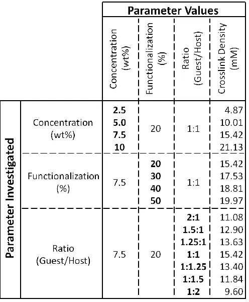
Table 4.1. Hydrogel compositions examined. ... 95
Table 7.1. Volumes of guest-host hydrogels post-injection. ... 199
Table 7.2. Complete geometric, functional, and histological outcomes of infarct
model. ... 215
Table 8.1. Ellipsoid dimensions of a 0.3 mL hydrogel injection, based on MRI
data. ... 231
Table 8.2. Animal usage and assessment of gross cardiac geometry. ... 234
Table 8.3. Direct measurement of myocardial thickness at 8 weeks post-MI. .. 246
Table 8.4. MRI assessment of myocardial thickness over time. ... 246
LIST OF FIGURES
Figure 1.1. The biomaterials toolbox. ... 5
Figure 1.2. Dynamic and responsive materials. ... 10
Figure 1.3. Biologically complex materials. ... 18
Figure 3.1. Molecular guest-host assembly. ... 49
Figure 3.2. Schematic representation of guest-host polymeric assemblies. ... 52
Figure 3.3. Guest-host hydrogel injection. ... 54
Figure 3.4. Guest-host interactions in drug delivery systems. ... 62
Figure 3.5. In situ hydrogel formation. ... 65
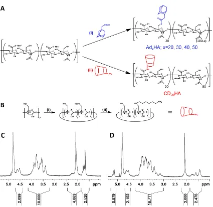
Figure 4.1. Synthesis of guest (Adx-HA) and host (CD20-HA) macromers. ... 90
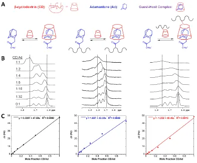
Figure 4.2. Binding affinity determination. ... 92
Figure 4.3. Overview of hydrogel formation. ... 93
Figure 4.4. Moduli dependence on guest-host mole fraction. ... 94
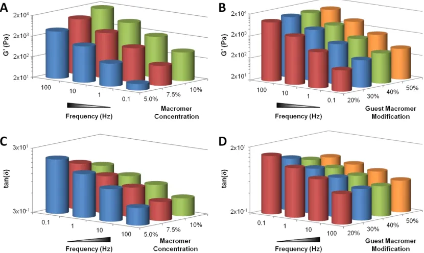
Figure 4.5. Representative frequency dependence. ... 96
Figure 4.6. Frequency dependence of material properties. ... 98
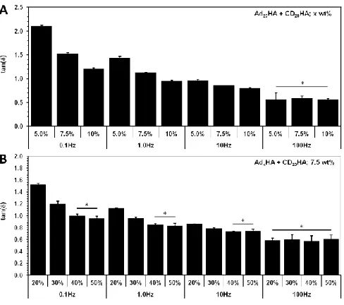
Figure 4.7. Statistical comparisons of frequency dependence. ... 99
Figure 4.8. Statistical comparison of loss tangent. ... 99
Figure 4.9. Representative loss tangent and determination of relaxation time. 101 Figure 4.10. Bulk relaxation behavior of hydrogels. ... 101
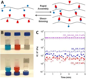
Figure 4.11. Guest-host hydrogel flow and recovery characteristics. ... 104
Figure 5.1. Polymer structure and self-assembly... 124
Figure 5.2. Binary associations and heterogeneity in guest-host hydrogels. .... 126
Figure 5.3. Temporal evolution of hydrogel porosity and component co-localization. ... 127
Figure 5.4. Three-dimensional representation of structure. ... 128
Figure 5.5. Control over hydrogel porosity. ... 130
Figure 5.6. Examination of guest-host hydrogel heterogeneity via atomic force microscopy (AFM). ... 132
Figure 5.7. Microrheological examination of guest-host hydrogels. ... 133
Figure 6.1. Overview of material design. ... 144
Figure 6.2. Proteolytic degradation characterization. ... 153
Figure 6.4. Rheological characterization of guest-host assembled hydrogels. . 156
Figure 6.5. Rheological characterization of hydrogel mechanics and flow. ... 158
Figure 7.1. Synthesis of Ad-HA. ... 176
Figure 7.2. Synthesis of CD-HA. ... 178
Figure 7.3. Synthesis of HA-SH. ... 180
Figure 7.4. Synthesis of MeHA. ... 181
Figure 7.5. Synthesis of AHA... 182
Figure 7.6. Synthesis of VS-HA. ... 183
Figure 7.7. Synthesis of MaHA. ... 184
Figure 7.8. Synthesis of Ad-HA-SH. ... 185
Figure 7.9. Synthesis of CD-MeHA. ... 187
Figure 7.11. Guest-host hydrogel formation enables shear-thinning injection and
hydrogel retention. ... 196
Figure 7.12. Rheological examination of guest-host hydrogel dynamics. ... 197
Figure 7.13. MRI analysis of hydrogel retention. ... 199
Figure 7.14. Michael-Addition hydrogel formation, tuning of crosslinking kinetics. ... 201
Figure 7.15. Real-time rheological observation of Michael-addition crosslinking with variation in the Michael-acceptor reactivity... 202
Figure 7.16. Dual-Crosslinking hydrogel formation, retention, and altered biophysical properties. ... 204
Figure 7.17. Rheological examination of hydrogel properties prior and subsequent to disulfide reduction. ... 205
Figure 7.18. MRI reconstruction of hydrogel injection. ... 205
Figure 7.19. Histological confirmation of hydrogel injections ... 206
Figure 7.20. In vivo degradation behavior modulated by dual-crosslinking. ... 207
Figure 7.21. Macroscopic images of subcutaneous hydrogel injections ... 209
Figure 7.22. Epicardial GH injection in the infarct border zone. ... 210
Figure 7.23. Histological, geometric, and functional outcomes after hydrogel injection. ... 212
Figure 7.24. Immunohistochemical (IHC) staining. ... 213
Figure 7.25. Response of pressure-volume (PV) relationships to varying load. 214 Figure 8.1. Schematic long-axis representation of LV remodeling... 226
Figure 8.3. Reconstruction of hydrogel geometry based on MRI data of injections
embedded in myocardial wall. ... 231
Figure 8.4. MRI based determination of myocardial thickness over time. ... 237
Figure 8.5. MI model and therapeutic groups. ... 240
Figure 8.6. Rheological examination at physiological conditions. ... 241
Figure 8.7. Material retention in vivo. ... 242
Figure 8.8. Finite element analysis of hydrogel injection. ... 243
Figure 8.9. Myocardial wall thickness. ... 245
Figure 8.10. Histological examination 8 weeks post-MI. ... 245
Figure 8.11. MRI assessment of cardiac geometry and function. ... 247
Figure 8.12. Assessment of mitral regurgitation severity. ... 249
Figure 8.13. Valve-associated Geometric Changes due to LV remodeling. ... 251
Figure 8.14. Percutaneous hydrogel injection... 253
Figure 9.1. Methacrylate modifications. ... 269
Figure 9.2. Guest-host polymer modifications. ... 269
Figure 9.3. Methacrylated guest-host polymer modifications. ... 270
Figure 9.4. Properties of single network hydrogels and schematics of hydrogels
examined. ... 276
Figure 9.5. Dependence of double network properties on structure and
composition. ... 279
Figure 9.6. Dependence of compressive elastic moduli on strain rate. ... 280
Figure 9.7. Dependence of compressive hydrogel properties on covalent
Figure 9.9. Dependence of tensile hydrogel properties on network structure. .. 281
Figure 9.10. Supramolecular self-healing at macroscopic and molecular scales.
... 283
Figure 9.11. Evaluation of network energy throughout loading and unloading
cycles. ... 284
Figure 9.12. Evaluation of network recovery from cyclic compressive loading. 284
Figure 9.13. Development of cytocompatible crosslinking conditions. ... 285
Figure 9.14. Mechanical characterization of cell laden double network hydrogels.
... 286
Figure 9.15. Cell encapsulation, distribution, and long-term viability. ... 288
Figure 9.16. Cell distribution within single and double network hydrogels. ... 289
Figure 9.17. Temporal characterization of material properties. ... 289
CHAPTER 1
Introduction: Material Design for Biomedical Applications
Adapted from: Rodell CB, Tibbett MW, Burdick JA, Anseth KS. Progress in Material
Design for Biomedical Applications. Proceedings of the National Academy of
Sciences (PNAS)112, 14444-14451 (2015).
1.1 Introduction
Biomaterials have been used to augment tissue function and treat diseases
and injuries for thousands of years – whether selecting coral or wood for dental
implants or fabric for sutures, implant materials historically originated by evaluating
potential materials in our surroundings that could be used for a specific biomedical
application. Many times this selection process simply involved consideration of the
mechanical properties of the material to restore basic function at the implant site;
typically, the materials themselves were never originally designed to interface with
living tissues. Today, this is no longer the case, as we now have an advanced
toolbox of synthetic and processing techniques to rationally create, design, and
process materials with specific properties in mind. These advancements have
come hand in hand with the integration of theory with experiments, materials
chemistry and biology with engineering, and basic science with application. As
science is often the stealth technology that enables breakthroughs in medical
devices that improve health care and save lives.
In fact, the last few decades of research have led to the emergence of
numerous biomaterial options, along with an increasing sophistication in the ability
to tune and manipulate complex physical and biological properties. Such advances
in biomaterial science have not only driven and enabled new medical products, but
have served as new tools for investigation of important biological questions. The
modern biomaterial evolution initiated with the design of materials – including hard
materials like metals and ceramics – that focused on outcomes such as
mechanical properties and biocompatibility. This approach led to the clinical
implementation of numerous materials for biomedical applications, such as joint
replacement, pacemakers, and orthodontics. The contemporary age of
biomaterials has advanced with a further focus on functionality, where materials
are now smarter and responsive to their environment; they incorporate bioactive
signals, and they have multifunctional design. These strategies are leading to
progress and improvements in fields ranging from medical devices, to drug
delivery, to regenerative medicine.
As one example, vascular stents have been widely used to open blocked
vessels and restore blood flow to ischemic tissues, and the design of these stents
has significantly evolved with time. With the development of Nitinol®, a metal alloy
of nickel and titanium with unique shape memory and superelastic properties, stent
design has improved to be implanted with simpler, minimally-invasive procedures
transitioned from passive mechanical devices to those that actively regulate the
biological interface by integrating biodegradable polymer coatings that locally elute
drugs to limit restenosis and resulting stent failure. These advances enhanced both
the functionality and efficacy of stent technology for clinical use. Similarly, the
coating of traditional metal orthopaedic implants with bioactive ceramics improved
clinical outcomes by facilitating osseointegration with bony tissue, and after the
discovery of bone morphogenetic proteins and their recombinant production,
spinal fusion surgeries benefited from material delivery systems that enabled their
local presentation (e.g., INFUSE®). Collectively, these examples demonstrate how
material design can be used to present biological signals that result in new medical
devices and implants with superior clinical performance. In fact, a recent report
estimated the 2012 global biomaterial market at $44.0 billion and forecasted a 15%
compounded annual growth rate between 2012-2017, reaching $88.4 billion by
20172.
This perspective focuses primarily on recent developments in polymers and
soft materials, due to the large technological growth in these systems since the
1990s. This review is organized to highlight some of the major advances and
modern thinking in biomaterial design, such as the ability to manipulate and control
biomaterial properties at multiple length-scales, introduce dynamic behavior into
biomaterials, and capture biocomplexity and additive functionalities. We then
conclude with a forward-looking perspective about the current challenges and
1.2 From Molecular to Macroscopic
Biomaterials fabrication has evolved across all size scales—from molecular to
macroscopic—to impart biochemical and biophysical cues into cell culture
platforms for regenerative medicine, to achieve optimal outcomes in drug delivery
systems, and to improve in vivo success of medical implants. Our increased
understanding of native tissue architecture and cell-material interactions, as well
as the development of processing methods and chemical syntheses has driven the
design of new materials. This section will highlight advances that have been made
in the development of a toolbox of synthetic approaches and fabrication techniques
that impart defined structures over a range of biologically relevant length scales.
1.2.1 From Molecular Organization to Nanostructure
An increased understanding of biological structures, with a focus on their
biochemical composition and organization, has provided insight into the manner
by which molecular structure and chemistry impart properties into biological
systems. Covalent bonds endow stability (e.g., peptide bonds) while secondary
structures confer material resilience (e.g., resilin3, elastin4). Peptide coupling,
recombinant protein synthesis, and evolution via phage display have become
invaluable tools to recapitulate similar functionalities in synthetic biomaterial
analogues. Likewise, synthetic approaches (e.g., bio-orthogonal chemistry) have
evolved to enable the fabrication and functionalization of biomaterials (e.g.,
hydrogels) that capture aspects of native biological structures5. Collectively, these
including post-modification of cell culture matrices and to crosslink implantable
materials.
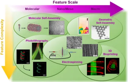
Figure 1.1. The biomaterials toolbox. The toolbox of biomaterials processing techniques that enable formation of highly controlled structures with biochemical and biomechanical features that vary across many size scales, as well as levels of complexity. These include nanoscale molecular self-assembly6, photolithography7,8, electrospinning9,10, geometric
self-assembly11,12, and 3D bioprinting13.
Covalent chemistries have dominated the biomaterials field since its
conception. However, the emergence of supramolecular chemistry has begun to
enhance our understanding of biology and capacity for creating precise,
physiologically structured materials. Nobel Laureate Jean-Marie Lehn insightfully
described supramolecular interactions as “chemistry beyond the molecule,” 14
since they enable dynamic macromolecular interactions, as well as the
the body, supramolecular presentation of bio-signals is exemplified by native
extracellular matrix (ECM) interactions, including receptor-ECM interactions and
heparin-binding proteins. As such, biomolecule presentation through
supramolecular interactions has emerged as a means of controllable delivery16,
including through cyclodextrin-mediated sequestration of small molecules17 or
biomimetic electrostatic protein-matrix interactions18. Beyond the capacity for
single molecule-matrix interactions, the general ECM structure itself is largely the
result of self-assembly (e.g., fibrillar structure of collagen) and can be
recapitulated, in part, by well-designed synthetic analogues. These higher order
motifs are exemplified by self-assembling nanostructures from peptide
amphiphiles6 (Figure 1.1, top left), though many alternative means of biologically
inspired supramolecular materials have been explored and their implications
toward cell behavior recently reviewed19.
1.2.2 Building at the Mesoscale
While self-assembly processes based on molecular design have achieved vast
success in recapitulating certain aspects of the biological nanostructure, they face
notable challenges. Among these is relative homogeneity at larger scales
(resulting from thermodynamically controlled assembly) and physiologically low
mechanical properties (owing to the underlying weak intermolecular forces). In
order to address these aspects at the nano and mesoscale, more active
processing methods have been utilized to impart defined structure. Notably,
become a dominant technique to mimic the nanofibrous nature of ECM20. The
functional importance of such microstructural organization cannot be discounted,
as it enables mechanical anisotropy21 and therefore holds great promise for
formation of biomedical implants including vascular grafts22 and orthopedic
connective tissues23. Toward formation of porous architectures, other processes
such as phase separation, leaching, and directional freezing have also emerged
as versatile methods to process biomaterials that permit cell and tissue
infiltration24,25.
The aforementioned methods allow realization of bi-continuous structures at
the nano- and micro-scale, yet they often display limited capacity toward
generating complex topographical, mechanical, or biomolecular presentation. For
modulation of these aspects, post-processing of larger scaffolds, such as by
light-mediated reactions (Figure 1.1, bottom left), has become instrumental toward
spatiotemporal control of biochemical signals on hydrogel surfaces26 or within 3D
hydrogels through either focused single photon7 or multiphoton27 irradiation
methods. Building on these advances, selective photopolymerization28, addition
reactions29, and degradation mechanisms30,31 have enabled extension of
photopatterning methodologies toward 2D and 3D presentation of spatially or
temporally varying mechanical properties.
1.2.3 Macroscopic Materials and their Sub-Assemblies
Ultimately, material design for biomedical applications must achieve the
properties suitable for in vivo implantation, preferably with necessary tissue
interfacing to achieve functionality. Methods reminiscent of industrial processes,
such as injection molding, have been employed to achieve macrostructure control
in biomaterials. These approaches have enabled recreation of complex structures
at the macroscale with utility toward application in craniofacial32 and meniscal33
implants. In some cases, the biological interaction with these materials has been
mediated by biomolecule presentation within the scaffold, such as sequestration
of heparin and, correspondingly, endogenous BMP-2 to enhance bone formation34.
Toward their utility in tissue engineering applications, material assemblies often
require advanced structural flexibility in order to recapitulate the inhomogeneity of
tissue structures (e.g., spatiotemporal presentation of cells and matrix). To achieve
this, appropriate molecular-, nano-, and meso-scale signals may be engineered
into macroscale structures through either modification of bulk hydrogels
(top-down) or directed component assembly (bottom-up) approaches. A powerful
means of achieving controlled signal presentation within a homogenous scaffold is
photolithography (vide supra), which embodies the top-down methodology.
Alternatively, two primary means of bottom-up approaches have emerged to
create tissue-scale structures with non-homogenous cell and material
compositions. First, pioneering work by the Whitesides group12 has demonstrated
means by which materials may be pre-cast into microgel components with the
desired composition and allowed to passively self-assemble (Figure 1.1, top right)
through hydrophobic or capillary forces35. Owing to the thermodynamic control of
large length scales. Toward increasing the complexity of allowed structures, such
tools as field-driven input and direct serial manipulation have also been
employed11. As an alternative to this self-assembled approach, techniques like 3D
printing (Figure 1.1, bottom right)—the direct spatially controlled deposition of
materials, with or without included cells or signals — have emerged to introduce
material structure at the macro-scale. Within only the past few years, these
methods have been extended to include processes such as: sacrificial printing to
enable perfusion and viability within a secondarily cast hydrogel36, layer-by-layer
printing of pluronics or other thermogels37, and methods to directly write complex
structures in 3D13. Looking forward, it is expected that further inclusion of smaller
scale sub-assemblies, such as nanostructured materials and microscale
patterning will aid in furthering success of these approaches.
1.3 From Static to Dynamic
Beyond control of material structure from the molecular to the macro-scale,
biomaterials are also evolving from a traditional, pre-engineered static design to
those that have dynamic properties. Historically, biomaterials were intended to
provide consistent functions, such as mechanical support (e.g., orthopaedic
implants) or optical properties (e.g., contact lenses). This approach has led to the
successful design of numerous clinically-used biomaterials; yet, advances in
material design and polymer chemistry have recently allowed us to incorporate
materials that are degradable, to eliminate permanent implantation or a second
surgery for implant retrieval, to those that have stimuli-responsive properties,
where various chemical and biological signals can trigger changes in biomaterial
properties or release drugs on-demand.
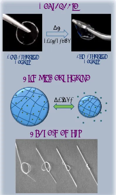
Figure 1.2. Dynamic and responsive materials. Dynamic biomaterials based on polymer degradation38, stimuli-responsive properties (e.g., local changes in temperature
or pH) for the release of therapeutics, or temperature induced shape-memory changes (e.g., self-tying suture)39.
1.3.1 Incorporating Degradation into Biomaterials
Biodegradable materials are those that transition from an initial, stable structure
into soluble products that can be resorbed and processed by the body. Examples High Crosslink
Density
Dt
degrada on
Low Crosslink Density
Degradable
Shape-memory Stimuli-Responsive
of such system have been around for numerous years, with biodegradable sutures
perhaps being the most common40. Original resorbable sutures consisted of
materials such as catgut that degraded via inherent biological mechanisms, but
these were later engineered from synthetic and hydrolytically degradable polymers
(e.g., poly(α-hydroxy esters)). Other examples of biodegradable materials used in
the clinic include biodegradable films that limit undesired adhesions after surgical
procedures and degradable fixation devices (e.g., screws and plates) in
orthopaedics41. Important considerations in the design of biodegradable materials
are the rate of degradation and ensuring that the degradation products are
non-toxic when released.
Biodegradable materials have been applied widely to biomedical applications
to provide temporal control over material presentation, including towards the
engineering of tissues or the release of drugs and growth factors42. For tissue
engineering, the material may temporarily provide a 3D structure or ‘scaffold’ for
the growing tissue, whereas degradable materials for drug delivery are engineered
to protect and then release molecules at desired rates. Hydrogels are one such
class of biomaterials that have been designed with degradable linkers, for example
through the introduction of hydrolytically or enzymatically cleavable bonds into the
crosslinks. Degradable hydrogels have been synthesized from a range of
materials, including synthetic polymers (e.g., poly(ethylene glycol)43, poly(vinyl
alcohol)44, and poly(propylene fumarates)45) and biologically-derived polymers
(e.g., hyaluronic acid46) (Figure 1.2). Towards tissue engineering or wound healing
delivered or recruited cells to secrete their extracellular matrix, but not persist too
long so as to impede tissue formation. For example, hydrogels have been
optimized for cartilage tissue engineering by tuning the degradation rate to control
matrix production and distribution by encapsulated chondrocytes47. Likewise, for
delivery of entrapped biomolecules, hydrogel degradation is primarily used to alter
the diffusion and kinetics of molecule release, which subsequently controls their
spatiotemporal presentation to local cells and tissues48. Often times, these
biological signals are designed to act as morphogens and influence tissue
formation and healing49.
As a complement to hydrolysis, which often occurs at pre-engineered rates
throughout the bulk of a material, biomaterials have also been engineered to
degrade via proteases, more similar to how tissues are remodeled in the body. In
pioneering studies by Hubbell and colleagues, peptides were incorporated into
hydrogel crosslinks that cleave through cell-produced proteases50,51, such as
matrix metalloproteinases (MMPs), elastases, and plasmin52-54. With this
protease-mediated degradation and the addition of cell-adhesive signals, these biomimetic
hydrogels were remodeled by cells55 and could be tuned for specific applications,
such as the regeneration of bone and vascular structures50,54,56,57. In some
examples, only growth factors were embedded into the matrices and their release
occurred in a “cell-demanded” fashion58. This approach can also be harnessed to
control the delivery of molecules to treat diseases where protease activity is
here, the drug release rate and dose are controlled through a feedback mechanism
(i.e., elevated protease activity releases more drug more quickly).
Although these examples have focused on hydrogels, there are many other
instances where degradation is used to control the dynamic properties of
biomaterials. As one highlight, drug delivery reservoirs have been incorporated
into synthetic devices, where they are covered by a thin biomaterial film (e.g.,
poly(lactic-co-glycolic acid) (PLGA))62. Subsequent drug release is mediated
through degradation of the films, where the timing is dependent on the
biodegradable polymer design. Release profiles can be pulsatile, and efficacy has
been shown for the delivery of chemotherapeutics from device for targeting
tumors63. Furthermore, stents have been designed to incorporate various drugs
through biodegradable coatings and reservoirs (vide supra), where drugs (e.g.,
paclitaxel) are released through polymer degradation at concentrations and rates
that can locally influence tissue response (e.g., suppress unwanted scarring or
restenosis)64.
1.3.2 Stimuli-Responsive Biomaterials
Beyond degradation, biomaterials have been designed to respond to a range
of environmental stimuli that may involve signals such as changes in temperature,
ionic strength, light exposure, mechanical stress, magnetic fields, or pH65. These
stimuli may initiate from the local biomaterial environment (e.g., after implantation)
or be introduced as an external “trigger” (i.e., active systems) (Figure 1.2).
ligand-receptor binding67, and nanometer-scale protein motions68, where material
properties and therapeutic release are altered based on the biological
environment. Important examples in this area are the release of insulin in response
to glucose catalysis69 or biochemically-triggered growth factor release70, where the
disease stimuli controls drug delivery. Materials are also designed so that the
presence of specific proteins can disassemble nanoparticles, opening up
disease-triggered therapeutics and diagnostics71. Hydrogels with pH responsive swelling
changes provide advantages for the oral delivery of therapeutics, where
biomaterials are stable in the stomach and then release drugs in the intestines72.
As active systems, biomaterials are being designed with dynamic properties
that introduce temporal signals to cells, towards the engineering of tissues, the
expansion of stem cells, or to understand complex cellular processes. One
common dynamic hydrogel system includes those fabricated from poly(N-isopropyl
acrylamide), which transition from a swollen to a collapsed hydrogel when
processed through its lower critical solution temperature (LCST). Changes in
volume, mechanics, and optical transparency occur when the material transitions
through its LCST, and these changes have been exploited to release cells and cell
sheets for tissue engineering73. Another stimulus of particular interest is light, due
to the allowed spatiotemporal control. Anseth and colleagues introduced light as a
trigger for the cleavage of crosslinks (e.g., containing o-nitro benzyl groups) in
hydrogels30 for the release of tethered signals or to probe how dynamic mechanical
material mechanics at a user-defined time75. Beyond light, dynamic properties may
be introduced in ionically crosslinked gels, by the addition of multivalent cations76
or in physically associated DNA-based gels through the introduction of
complementary DNA77. All of these systems have been used to probe cell behavior
in response to dynamic environments.
Actively controlling biomaterials once implanted in the body is more
challenging, particularly to introduce the stimuli to materials that are implanted
deep within tissues. Light penetration can be attenuated at many depths and
wavelengths; however, there are numerous examples where light has been used
to either form materials78 or alter their properties when implanted79. Ultrasound is
another trigger that can be introduced to disrupt polymer structure and release
therapeutics80,81. As described with the biodegradable reservoirs above, a similar
system has been developed with electrochemically activated microchips with
release through the dissolution of a gold membrane82. While this system is
non-polymeric, it constitutes an important example of stimuli-responsive properties for
controlled release in implanted materials.
As a sub-set of responsive biomaterials, shape-memory materials exhibit
changes in geometry based on triggers such as temperature or light83,84. In brief,
these materials are fixed into a temporary shape (usually under stress) and then
transition into a relaxed permanent shape following an external or environmental
trigger. (Figure 1.2) Such a dynamic process may lead to the next generation of
minimally-invasive implantable constructs, capable of altering their geometry once
developed many years ago and have found commercial application, but the last
few decades have led to an increase in the number of polymeric systems available
for biomedical applications, along with those that under multiple transitions that
allow for sequential geometric changes. These polymers can be processed from a
range of covalently and physically crosslinked polymers and copolymers, including
from biodegradable polymers83,84, and have the potential to be designed for
degradation, elution of drugs, or even signaling to local cells for improved wound
healing.
1.4 From Bioinert to Biocomplex
Building upon advances in dynamic and responsive biomaterials, another
recent direction in soft biomaterials is the design of systems that engage with,
respond to, and integrate into the biological landscape. Such systems extend
beyond passive biological function (bioinert), and researchers seek to engineer
materials that actively interface with biologically complex environments
(biocomplex). Discoveries in the biological sciences have revealed how
information is processed and exchanged in the body, exposing new routes toward
engineering material-tissue interactions. For example, the language of the genetic
code presents novel therapeutics; the critical role of the ECM informs tissue
engineering and regenerative medicine; the genetic basis of many diseases (e.g.,
cancer, Marfan syndrome) transforms the way patients are treated; the
therapy; and the discovery of the microbiome restructures the way we think about
bacteria.
Toward fueling advances in medicine, these basic scientific discoveries are
essential in the design of future biomaterials. For example, biocomplex materials
have the potential to perceive malignant dysfunction and respond by releasing
therapeutics to restore homeostasis; alternative systems could mimic critical
aspects of the ECM to direct tissue morphogenesis ex vivo. Often, the biggest
challenge is reducing the biological complexity into essential elements (e.g., rate
limiting steps, critical signaling factors) that enable a synthetic material to perform
a desired task. In this manner, biomaterials scientists are leveraging biologic
understanding to design materials that are structurally simple, yet functionally
complex in order to communicate with, react to, and synergize with biology to
address clinical needs. This section articulates the concept of biocomplex
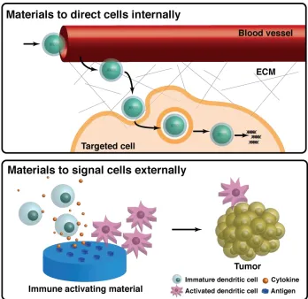
Figure 1.3. Biologically complex materials. Biocomplex biomaterials interact with and direct cells both internally and externally. For example, advanced drug delivery vehicles introduce exogenous nucleic acid content to up- or down-regulate protein expression by transporting sensitive biomolecules through the circulation, actively targeting specific cells, and releasing the therapeutics into the cytoplasm. In addition, biocomplex materials are designed to present external signals to cells, either those that are delivered with the matrix or those that are recruited exogenously. These biomaterial niches can be loaded with multiple cues, presented in concert or sequentially, to communicate, recruit or signal to cells locally. For example, immune activating materials cooperate with native biological signaling to recruit naïve immune cells to a site in the body, activate them with target antigens, and equip them to target specific cells or tissues, such as malignant tumor cells.
1.4.1 Materials to Deliver Therapeutics
Bioinert micro- and nanocarriers that achieve long circulation times in the blood
have transformed parenteral administration of small-molecule drugs 85,86. Potent
scale; however, because of their nature, they present unique challenges for
delivery. For example, translation of these therapeutics requires carriers that not
only circulate for extended periods of time, but also shield the sensitive molecular
cargo from degradation in the bloodstream, target specific cells or tissues, and
release cargo at the appropriate site of action. Additionally, the design of
biocomplex nanocarriers that address these challenges must be balanced by the
need for structural simplicity that enables reproducible manufacturing.
Some of the most clinically relevant biotherapeutics, whose efficacy hinges on
the design of biocomplex delivery systems, are nucleic acids (NAs). NA-based
therapies, such as ribonucleic acid (RNA) interference (e.g., microRNA (miRNA)
or short interfering RNA (siRNA)) draw inspiration from native mechanisms and
regulation in the transcription and translation of genetic material into protein. RNA
interference is a native avenue for post-transcriptional silencing of gene
expression, whereby miRNA or siRNA selectively prevent protein synthesis87. In
addition, exogenous messenger RNA (mRNA) can induce the production of
specific proteins to upregulate protein expression 88. Since NA-based therapies
alter intracellular machinery, their efficiency relies on cytoplasmic delivery.
Moreover, these biomolecules are particularly sensitive to in vivo conditions,
exhibiting very short half-lives before biochemical decomposition is observed.
Therefore, successful translation requires a delivery vehicle that offers protection
from clearance or nuclease degradation, site-specific targeting, passage across
Toward this end, biocomplex polymeric and lipid-based nanoparticle
formulations have been developed to deliver NAs intravenously, some of which
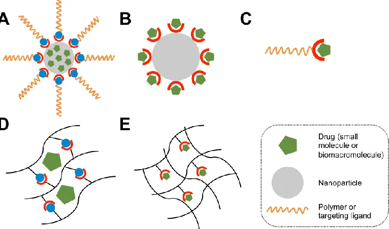
have progressed to clinical trials (Figure 1.3)91. Specific advances have focused
on stable nucleic-acid lipid particles (SNALPs) and ionizable lipids that package
NAs and increase transport across the cell membrane89,90. The majority of NA
nanocarriers unintentionally accumulate in the liver, and to overcome this issue,
lipid structures have been recently developed that allow for selective passive
targeting of heart, lung, and vascular endothelial tissues92. Targeting to tumor cells
has been achieved by functionalizing the surface of delivery vehicles with ligands
that bind specifically to proteins that are selectively expressed on the surface of
tumor cells 93. A major challenge that remains in the clinical use of NA therapeutics
is their escape from endosome or lysosomes into the cytoplasm. To address this
issue, Sahay et al. identified NPC1 as a critical protein in the trafficking of lipid
nanoparticles that can be exploited in the design of materials that better escape
the endosome94.
1.4.2 Materials to Present Matrix Cues
The fields of tissue engineering and regenerative medicine rely on the
proliferation and maintenance of human cells outside of the body. Seminal culture
scaffolds have been designed to permit cell survival and proliferation, but are
inherently passive. While these bioinert scaffolds provided a route to maintain and
culture cells, recent improvements in scaffold design integrate biofunctional
of signals from the extracellular environment that synergize with the genetic code
to instruct cell function, such as proliferation, phenotype, and differentiation. As
discussed, ECM cues include both biophysical and biochemical signals that vary
on multiple length and time scales. Additional signals are introduced by
neighboring cells via cell-cell contacts or secreted cytokines and growth factors.
Biocomplex 3D culture matrices seek to recapitulate critical ECM cues and
cell-cell signaling events through spatiotemporal control over matrix mechanics and
ligand presentation 96. Toward this goal, both static and temporally controlled
presentation of adhesive ligands has been exploited to bias chondrogenic
differentiation of hMSCs30,97. In addition, dynamic control over substrate modulus
has been leveraged to reveal new mechanism of ‘mechanical memory’, bias
differentiation of mesenchymal stem cells, and mature cardiomyocytes28,98-100.
In another approach to communicate with cells, researchers have developed
biocomplex materials that exploit the language of the immune system to treat and
detect disease (Figure 1.3). These strategies utilize an understanding of how the
immune systems senses a foreign substance, arms itself for attack, and carries
out the attack. Mooney and coworkers demonstrated implantable devices that
interact with the immune system to suppress tumor growth101; specifically,
chemotactic factors were released locally to recruit dendritic cells, that were then
activated by local presentation of tumor antigens, which then instructed the
immune system to target cancer cells. An alternative approach employs implanted
materials to cooperate with the immune system to prime a pre-metastatic niche for
detection of cancer. Further, a suite of synthetic vaccines have been developed
similarly that communicate with immune cells to increase immunogenicity and,
ultimately, vaccine efficacy102.
1.4.3 Materials for Tissue Integration
Beyond intracellular and extracellular signaling, biocomplex materials have
been designed to orchestrate multicellular events. For example, Miller et al. used
3D printing of sacrificial sugar networks embedded within hydrogels to fabricate
vascularized neo-tissues36. Culver et al. employed laser writing of adhesive
peptides to instruct multicellular organization for the fabrication of vascular
networks within hydrogels103. Further, gradient biomaterials that present
biochemical ligands in a spatially defined fashion have been used to recapitulate
osteochondral and osteotendinous interfaces104,105.
Despite these in vitro advances, a major hurdle in the clinical utility of
implantable biomaterials (including joint replacements, smart drug delivery
materials, and cell carriers) is non-specific protein adsorption and the
accompanying foreign body response (FBR)106. No implanted material is truly
bioinert; proteins rapidly adsorb to the surface of biomaterials in the body with
random orientations and configurations106,107. Early, this proteinaceous layer
facilitates neutrophil adhesion and activation106,107. With time, macrophages fuse
to form foreign body giant cells that attack the implant surface while recruiting
fibroblasts, which deposit ECM and form a dense, fibrotic capsule that isolates the
has developed, the community has begun to present an array of biocomplex
materials that mitigate the FBR and assist integration with resident tissue. For
example, Jiang and colleagues developed Zwitterionic hydrogels that demonstrate
ultra-low protein fouling109, and the surface chemistry and nanotopography of
implant surfaces has been designed to limit macrophage activation106. Ratner and
colleagues further showed that implant porosity can be exploited to tune the FBR
and tissue integration110. While these advances demonstrate that biocomplex
materials can assist in the organization of multiscale tissues, clinical translation
remains hindered by an incomplete understanding of which critical signals to
present and integrate within biocomplex scaffolds.
1.5 Moving into the Future
There are thousands of different types of medical devices, diagnostic kits, and
pharmaceutical formulations that exist today as a result of advances in biomaterial
science and engineering. The polymers and soft biomaterials employed are
diverse in their origin, classification, and properties, and many products integrate
multiple components that are carefully selected for their performance and function.
Yet, as we look towards the future, the design of soft biomaterials is unifying
around concepts that include: hierarchy, complexity, dynamics and adaptation, as
well as healing111, and to realize this potential, better experimental methods
and modeling tools are needed so that we can understand how to synthesize and
synthesis of polymers with controlled molecular weights, defined sequences, and
integrated biological functionality, biomaterial systems depend on how these
structural elements assemble and interact at complex biological interfaces, as
this hierarchy ultimately dictates performance and function.
The biomaterials that are native to our body (e.g., the ECM) are profound in
their ability to remodel, adapt, and store and retrieve information; this is critical
during processes such as development, growth, and wound healing. While
biomaterials do not need to mimic all aspects of the complexities of a living
organism112, understanding the fundamentals of these processes unlocks future
opportunities in rational design of biomaterials. Contemporary research topics
include the development of healable materials, drug delivery systems that interact
with and deliver their contents in response to signals from cells, active materials
that promote healing of tissues that could not otherwise occur, medical devices
that integrate seamlessly with tissues at the implant site, and stealth nanosystems
that serve as sentinels to monitor and treat disease. Many of these developments
occur and will continue to revolve around multidisciplinary institutes and
environments that eliminate barriers and bring together chemists, biologists,
engineers, and clinicians, that bridge the academic-industrial divide, and engage
researchers on a global scale.
Key to all of these advances will be synthetic tools that allow control of
biomaterials from the molecular to the macro, for patterning and dynamically
revealing biological functionalities, and for engineering biocomplex materials with
delivered in the right context, locality, and time. Importantly, all of this must be
achieved in a manner that allows manufacturing at large scales and that
overcomes any regulatory challenges with the translation of new materials.
Biological complexity demands better tools to characterize changes in material
properties in situ, from molecular level features of degradation to structural
changes and functional properties. The body is a dynamic environment, so
biomaterials constantly experience changes that alter performance, and this
highlights the profound need for methods to allow tracking of biomaterials in
physiologically complex niches and/or improved in vitro assays that allow better
prediction of in vivo performance. Finally, to facilitate the discovery process,
methods to screen and model the broad and diverse experimental space is critical.
Clearly, rational material design will remain an important and leading approach of
the community, but combinatorial and high-throughput strategies that are
complemented by biological assays will allow mining of huge data sets to evolve
1.6 References
1. U.S. NSaTC. Materials Genome Initiative for Global Competitiveness. Executive Office of the President, National Science and Technology Council (2011).
2. marketsandmarkets.com (2013) Biomaterials Market [By Products (Polymers, Metals, Ceramics, Natural Biomaterials) & Applications (Cardiovascular, Orthopedic, Dental, Plastic Surgery, Wound Healing, Tissue Engineering, Ophthalmology, Neurology Disorders)]–Global Forecasts to 2017. Report Code BT 1556. Available at www.marketsandmarkets.com/Market-Reports/biomaterials-393.html. Accessed August 15, 2015.
3. Qin, G., Hu, X., Cebe, P. & Kaplan, D. L. Mechanism of resilin elasticity. Nat
Commun3, 1003 (2012).
4. Debelle, L. & Alix, A. J. P. The structures of elastins and their function.
Biochimie81, 981-994 (1999).
5. Azagarsamy, M. A. & Anseth, K. S. Bioorthogonal Click Chemistry: An Indispensable Tool to Create Multifaceted Cell Culture Scaffolds. ACS
Macro Lett.2, 5-9 (2013).
6. Hartgerink, J. D., Beniash, E. & Stupp, S. I. Self-assembly and mineralization of peptide-amphiphile nanofibers. Science 294, 1684-1688 (2001).
7. DeForest, C. A. & Anseth, K. S. Photoreversible Patterning of Biomolecules within Click-Based Hydrogels. Angew. Chem. Int. Ed. 51, 1816-1819 (2012).
8. Lee, S.-H., Moon, J. J. & West, J. L. Three-dimensional micropatterning of bioactive hydrogels via two-photon laser scanning photolithography for guided 3D cell migration. Biomaterials29, 2962-2968 (2008).
9. Wade, R. J., Bassin, E. J., Rodell, C. B. & Burdick, J. A. Protease-degradable electrospun fibrous hydrogels. Nat. Commun.6 (2015).