1 JNSD 2018; Vol.4, No.1: 1-7
Original Article
Open Access
www.jnsd.tums.ac.ir
Association of vitamin D receptor gene polymorphisms with
acute myeloid leukemia: a case-control study
Rahim Asghari
a, Ali Esfahani
a, Mohammad Asghari
b, Sakineh Shab-Bidar
c, Morteza
Bonyadi
d, Tahereh Mohammadian
d, Kamran Ali-Moghaddam
d, Shahrbano Rostami
e,
Zohreh Ghoreishi
faHematology and Oncology Research Center, Tabriz University of Medical Sciences, Tabriz, Iran bRoad Traffic Injury Research Center, Tabriz University of Medical Sciences and Department of
Statistics and Epidemiology, Faculty of Health, Tabriz University of Medical Sciences, Tabriz, Iran cDepartment of Community Nutrition, School of Nutritional Sciences and Dietetics, Tehran University of Medical Sciences, Tehran, Iran
dCenter of Excellence for Biodiversity, Faculty of Natural Sciences, University of Tabriz, Tabriz, Iran eHematology- Oncology and Stem Cell Transplantation Research Center Research Center, Tehran
University of Medical Sciences, Tehran, Iran
fNutrition Research Center, Tabriz University of Medical Sciences, Tabriz Iran bDoctorate of Pharmacy, Mashhad University of Medical Sciences, Mashhad, Iran
c Psychiatry and Behavioral Sciences Research Center, Ibn-e-Sina Hospital, Mashhad University of
Medical Sciences, Mashhad, Iran
A B S T R A C T
Article History
Received:
29/10/2017
Revised:
25/12/2017
Accepted:
18/01/2018
Objective: Vitamin D receptor (VDR) gene polymorphism has a role in susceptibility to risk of cancers. The aim of this study was to investigate the association of VDR gene polymorphisms with acute myeloid leukemia (AML).
Methods: In this case-control study, patients diagnosed with AML and healthy adult subjects were selected. Four single nucleotide gene polymorphisms of VDR gene (BsmI, TaqI, FokI, and ApaI) were determined using polymerase chain reaction-restriction fragment length polymorphism (PCR-RFLP) and the odds of having AML was determined by unadjusted and adjusted logistic regression analysis.
Results: One hundred and thirty-three AML patients and 300 healthy people were included in the study. There were significant associations between the polymorphisms of FokI, and ApaI on the one hand and increased risk of AML (P= .021, and P<.001) on the other. The odds of the disease in patients with FF genotype were 2.5 times higher than patients with ff genotype and the odds of the disease in individuals with AA genotype was 5.6 times higher than the reference category of aa. In contrast, BsmI polymorphism had a protective effect, such that for those with BB and Bb genotypes there were 91% and 86% lower odds for getting AML than bb genotype, respectively (P<.001).
Conclusion: This study shows that there is a significant association between VDR gene polymorphisms and odds of getting AML. Further studies on different ethnic groups in populations with due consideration of environmental factors interacting with genotypes are highly recommended.
Citation: Rahim Asghari, Ali Esfahani, Mohammad Asghari, Sakineh Shab-Bidar, Morteza Bonyadi, Tahereh Mohammadian, Kamran Ali-Moghaddam, Shahrbano Rostami, Zohreh Ghoreish. Association of vitamin D receptor gene polymorphisms with acute myeloid leukemia: a case-control study. J Nutr Sci & Diet 2018; 4(1): 1-7.
Keywords: Vitamin D receptor, Polymorphism, Acute myeloid leukemia
Introduction
Vitamin D controls various biological processes in the body such as skeletal
_________________________
Corresponding author:
Vitamin D receptor gene and acute myeloid leukemia
2 JNSD 2018; Vol.4, No.1: 1-7
metabolism, immunity response and cell proliferation and duplication. Epidemiological and laboratory research has indicated that vitamin D deficiency is associated with many common diseases and disorders including rickets, skeletal disorders, diabetes, cardiovascular disorders, autoimmunity disorders and cancer [1].
The active form of vitamin D, 1, 25-dihydroxy vitamin D or cholecalciferol, acts by adjoining to its nuclear receptor in target cells (vitamin D receptor/VDR). The vitamin D receptor belongs to the transcriptional regulatory factors family. In order to have a suitable interaction with DNA, VDR undergoes heterodimerization with the retinoid-X- receptor/RXR and connects with the responsive elements of vitamin D in the promotor region of the target cells [2]. The responsive element of vitamin D is detected in most of the genes involved in cell proliferation and duplication, apoptosis, and invasion and metastasis of cancer cells. Thus, it seems that VDR and gene polymorphism have a major role in the risk of cancer.
Results of studies on the relationship between VDR gene polymorphisms and cancers are controversial. In fact, in studies on different regions and ethnic groups, inconsistent results have been reported. In the past years, the association between common VDR gene polymorphisms (FokI, BsmI, TaqI, ApaI and Cdx2) and the risk of different solid tumors in the skin, prostate, breast, colon, ovary, bladder, kidney and brain have been assessed [3]. However, according to research findings, this association has not been fully understood for adult acute Leukemia. The objective of this study was to determine and compare the frequency of VDR gene polymorphism in patients diagnosed with acute myeloid leukemia (AML) with healthy individuals in order to assess the association of polymorphisms with the risk of cancer for the first time.
Subjects and methods
Participants
In this cross-sectional study, patients were recruited from among individuals referring to the Blood and Cancer Section of Shahid Ghazi Hospital, Tabriz University of Medical Sciences, Tabriz and Shariaty Hospital, Tehran University of Medical Sciences, Tehran, Iran. The case group included patients with the following inclusion criteria: diagnosis with AML, older than 18 years, willing to participate in the study, and no other
malignancies. The control group consisted of subjects older than 18 years, willing to participate in the study and not having a first degree relative diagnosed with cancer. Written informed consent was obtained from all the individuals participating in the study. The research project was approved by the Ethics Committee of Tabriz University of Medical Sciences (Ethical Code: 5/4/1720).
Sample size
Primary data for determining the sample size was obtained from Taieb et al. study (OR=1.94) [4]. Based on the G power software (3.2.1 version), with a 95% confidence interval and 80% power, the sample size was calculated to be 386 subjects in either group. Assuming the probability of a 10% missing of the subjects, the sample size was increased to 425 subjects. The subjects were recruited by convenience sampling with a 1 to 3 ratio rate (because of the infrequent prevalence of AML) for the case and control groups.
Extracting DNA from blood samples
A 2-ml blood sample was obtained from the left vein of each subject and stored in vials containing EDTA as an anticoagulant. For extracting DNA, the DNGTM-Plus kit was used (Sinaclon corporations, catalogue number: DN8118C). During the extraction period, the samples were stored at -20ºC.
Assessing VDR gene polymorphisms
Rahim Asghari
, et al.
3 JNSD 2018; Vol.4, No.1: 1-7
BsmI (rs1544410) and FokI (rs10735810) Polymorphisms
The BsmI polymorphism sequence was duplicated using forward primer (5´-AGTGTGCAGGCGATTCGTAG-3´) and return primer (5´-ATAGGCAGAACCATCTCTCAG-3´), as described in the Avila et al.’s study [5]. The FokI polymorphism sequence was also duplicated using forward primer (5′-ATG GAA ACA CCT TGC TTC TCC CTC-3′) and return primer (5′-GAT GCC AGC TGG CCC TGG CAC TG-3′), as described in the Hutchinson et al’s study [6].
The PCR condition of both BsmI and FokI polymorphisms consisted of 34 cycles ─ one cycle, primary denaturation at 94ºC for 5 minutes, denaturation at 94ºC for 30 seconds, annealing at 58ºC for 30 seconds, extension at 72ºC for 30 seconds, and a final extension at 72ºC for 5 minutes. Eventually, the duplicated products of enzymatic digestion created the genotypes BB (1 band containing 191 base pairs), bb (2 bands containing 117 and 74 base pairs), Bb (3 bands containing 191, 117 and 74 base pairs) for the Bsm1 polymorphism and the genotypes FF (1 band containing 272 base pairs), ff (2 bands containing 208 and 64 base pairs) and Ff (3 bands containing 272, 208 and 64 base pairs) for the Fok1 polymorphism.
Apa1 (rs7975232) and Taq1 (rs731236) Polymorphisms
The Apa1 polymorphism sequence was duplicated using forward primer (5′- CAGAGCATGGACAGGGAGC -3′) and
backward primer (5′-
AGGAGAGGCAGCGGTACTG -3´), as described in the Chang et al.’s study [7]. The Taq1 polymorphism sequence was duplicated using
forward primer
(5′-CAGAGCATGGACAGGGAGC-3′) and
backward primer
(5′-AGGAGAGGCAGCGGTACTG-3´), as described in the Hutchinson et al.’s study [6]. Both polymorphism sequences were duplicated at 34 cycles; primary denaturation at 94ºC for 5 minutes, denaturation at 94ºC for 30 seconds, annealing at 60ºC for 30 seconds, extension at 72ºC for 30 seconds and for 32 cycles and a final extension at 72ºC for 5 minutes. Eventually, the duplicated products of enzymatic digestion created the genotypes AA (1 band containing 454 base pairs), aa (2 bands containing 241 and 213 base pairs), Aa (3 bands containing 454, 241 and
213 base pairs) for the Apa1 polymorphism and the genotypes TT (a band containing 454 base pairs), tt (2 bands containing 159 and 295 base pairs) and Tt (3 bands containing 454, 159 and 295 base pairs) for the Taq1 polymorphism.
Statistical analysis
Statistical analysis was performed using the SPSS software (version 22). The qualitative data were expressed as frequencies and percentages and quantitative data as means and standard deviations. For assessing the relationship between VDR gene polymorphisms and risk of AML, logistic regression was performed. All the tests were two-sided with an 80% statistical power. A p-value < 0.05 was considered to show statistical significance.
Results
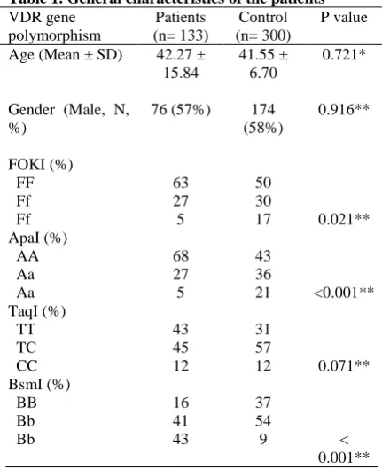
One hundred and thirty-three patients with AML and 300 healthy subjects as the control group completed the study. The mean age of the patients (57% males) and the control group (58%) was 42.27 ± 15.84 and 41.55 ± 6.70 years, respectively. There was no significant difference between the two groups as regards age (P = 0.71) or gender distribution (P = 0.916) (Table 1). On the whole, 18% of the patients were suffering from AML-M3 and the rest were afflicted with other types of AML. In general, FokI
Table 1. General characteristics of the patients
P value Control
(n= 300) Patients
(n= 133) VDR gene
polymorphism
0.721* 41.55 ±
6.70 42.27 ±
15.84 Age (Mean ± SD)
0.916** 174
(58%) 76 (57%)
Gender (Male, N, %)
FOKI (%)
50 63
FF
30 27
Ff
0.021** 17
5 Ff
ApaI (%)
43 68
AA
36 27
Aa
<0.001** 21
5 Aa
TaqI (%)
31 43
TT
57 45
TC
0.071** 12
12 CC
BsmI (%)
37 16
BB
54 41
Bb
< 0.001** 9
43 Bb
Vitamin D receptor gene and acute myeloid leukemia
4 JNSD 2018; Vol.4, No.1: 1-7
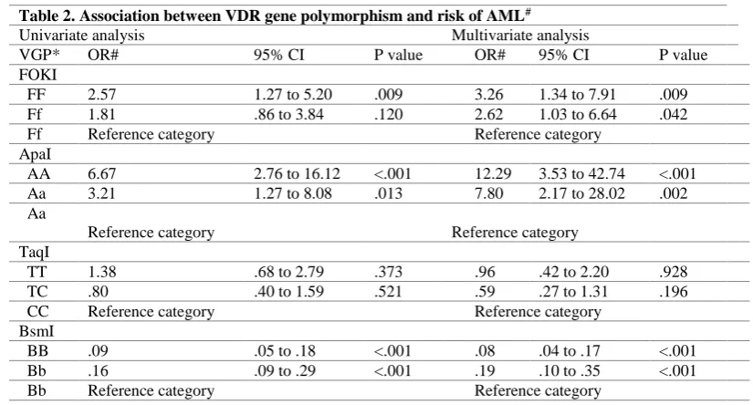
polymorphism was significantly associated with AML (P = 0.021), such that the odds of the disease in patients with FF genotype were 2.5 times higher than those with ff genotype. In patients with ff genotype, the AML odds were 80% higher in comparison with the reference category of ff, but the difference was not statistically significant (Table 2).
ApaI polymorphism also had a significant association with AML (P <0.001), such that the odds of the disease in individuals with the AA genotype was 5.6 times higher than the reference category of aa. Patients with Aa genotype were also found to be 3 times more likely to be suffering from AML than the reference category of aa (Table 2).
There was no statistically significant association between TaqI polymorphism and AML (P = 0.071), but the association between BsmI polymorphism and AML was significant, such that for those with BB and Bb genotypes, there were 91% and 86% lower odds for getting AML than bb genotype, respectively (Table 2). Based on the logistic regression analysis, considering VDR gene polymorphisms (FokI, ApaI, and BsmI) simultaneously, their associations with AML were found to be statistically significant as well (P = 0.032, P <0.001, and P <0.001, respectively).
Discussion
Our findings show that there is a significant association between VDR gene polymorphisms and odds of getting AML. The results of
meta-analyses and reviews on the relationship between VDR gene polymorphism and different cancers are controversial. Both harmful and protective roles have been reported for polymorphisms in malignancies. Even in one type of malignancy, similar polymorphisms were found to exhibit different effects. For example, based on the results of several review studies, Fok1 [8-14] and Taq1 polymorphism [15-17] increased the risk of breast cancer. However, in other studies, there was no association between Fok1 and Taq1 polymorphism and the risk of breast cancer [18, 19]. In addition, Li [8] and Kostner [9] indicated that Bsm1 polymorphism increased the risk of breast cancer.
Contradictory results have also been reported regarding prostate cancer. Although in some studies a positive association between Fok1 and Taq1 polymorphism and the risk of prostate cancer was reported, in other studies Fok1, Bsm1 and Taq1 polymorphisms were seen to have a protective effect in prostate cancer [9, 17, 21]. On the other hand, Berndt et al [24] and Guo et al [25] did not observe any significant association between VDR gene polymorphisms and the risk of prostate cancer. Controversial results have also been reported as regards ovary, skin, colorectal, lung, kidney and esophagus cancers [42-46]. These controversial results may be due to genetic differences, ethics, geographical location and environmental factors.
Different findings have also reported in studies on hematologic malignancies. Although Fok1, Bsm1 and Taq1 polymorphisms and
Table 2. Association between VDR gene polymorphism and risk of AML#
Multivariate analysis Univariate analysis
P value 95% CI
OR# P value
95% CI OR#
VGP* FOKI
.009 1.34 to 7.91
3.26 .009
1.27 to 5.20 2.57
FF
.042 1.03 to 6.64
2.62 .120
.86 to 3.84 1.81
Ff
Reference category Reference category
Ff ApaI
<.001 3.53 to 42.74
12.29 <.001
2.76 to 16.12 6.67
AA
.002 2.17 to 28.02
7.80 .013
1.27 to 8.08 3.21
Aa
Reference category Reference category
Aa
TaqI
.928 .42 to 2.20
.96 .373
.68 to 2.79 1.38
TT
.196 .27 to 1.31
.59 .521
.40 to 1.59 .80
TC
Reference category Reference category
CC BsmI
<.001 .04 to .17
.08 <.001
.05 to .18 .09
BB
<.001 .10 to .35
.19 <.001
.09 to .29 .16
Bb
Reference category Reference category
Rahim Asghari
, et al.
5 JNSD 2018; Vol.4, No.1: 1-7
increasing risk of youngster’s leukemia [43], multiple myeloma [44] and aplastic anemia [45] were evident in some studies, no associations were observed between polymorphisms and Hodgkin’s lymphoma [46] and non-Hodgkin’s lymphoma [47] in other studies. In the Prudue et al. study [48] a positive association between Bsm1 and Taq1 polymorphism and non-Hodgkin’s lymphoma was reported. In most solid tumors, Bsm1 polymorphism has a protective or insignificant role. In the present study Bsm1 had a protective role against AML.
This study was the first study to assess the association between common VDR gene polymorphisms and AML. A positive association was found between Fok1 and Apa1 polymorphism and the risk of AML. Also, it was noted that the Bsm1 polymorphism had a positive effect on AML. This association was not significant in the case of Taq1. Our findings have indicated that in patients diagnosed with AML, similar to solid tumors, VDR gene polymorphisms could have either positive or negative associations with the risk of AML. Some limitations of our study were as follows: not considering environmental factors, such as exposure to sunlight, smoking and other factors, and their possible effects on acquired genotypes. However, as mentioned above, the aim of this study was to investigate the association of VDR gene polymorphisms with acute myeloid leukemia (AML) and also to assess the risk of getting AML.
Conclusion
The findings of this study indicate that VDR gene polymorphism may have a significant role in increasing the risk of one of the most common adult hematologic malignancies known as AML. Since medical science is now putting particular emphasis on “individualized medicine”, these findings would have an important role in tracing genetic profile and detecting at-risk individuals. It is recommended to conduct further studies in different groups of patients in the country, in order to be able to generalize findings and assess effects of environmental.
Acknowledgement
This paper has been prepared on the basis of the thesis of Dr. Rahim Asghari, oncologist in Tabriz University of Medical Sciences, Tehran, Iran. We would like to extend our gratitude to the staff of the Hematologic and Cancer Section of Shahid Qazi Tabatabie Hospital, Tabriz and
Shariati hospital, Tehran, Iran and all the individuals who participated in the study.
Conflict of Interests
The authors declare that there is no conflict of interests regarding the publication of this paper.
References
1. Köstner K, Denzer N, Müller CS, Klein R, Tilgen W, Reichrath J. The relevance of vitamin D receptor (VDR) gene polymorphisms for cancer: a review of the literature. Anticancer Res. 2009; 29(9):3511-36.
2. Kliewer SA, Umesono K, Mangelsdorf DJ, Evans RM. Retinoid X receptor interacts with nuclear receptors in retinoic acid, thyroid hormone and vitamin D3 signalling. Nature. 1992 30;355(6359):446-9.
3. Valdivielso JM1, Fernandez E. Vitamin D receptor polymorphisms and diseases. Clin Chim Acta. 2006;371(1-2):1-12.
4. Tayeb MT, Clark C, Haites NE, Sharp L, Murray GI, McLeod HL. Vitamin D receptor, HER-2 polymorphisms and risk of prostate cancer in men with benign prostate hyperplasia. Saudi Med J 2004;25(4):447-51.
5. Avila M1, Prado C, Ventura MD, Mora C, Briones D, Valdez H, et al. Vitamin D receptor gene, biochemical bone markers and bone mineral density in Mexican women on dialysis. Nephrol Dial Transplant 2010; 25(7):2259-65.
6. Hutchinson PE, Osborne JE, Lear JT, Smith AG, Bowers PW, Morris PN, et al. Vitamin D receptor polymorphisms are associated with altered prognosis in patients with malignant melanoma. Clin Cancer Res 2000;6(2):498-504.
7. Chang TJ, Lei HH, Yeh JI, Chiu KC, Lee KC, Chen MC, et al. Vitamin D receptor gene polymorphisms influence susceptibility to type 1 diabetes mellitus in the Taiwanese population. Clin Endocrinol (Oxf) 2000;52(5):575-80.
8. Köstner K, Denzer N, Müller CS, Klein R, Tilgen W, Reichrath J. The relevance of vitamin D receptor (VDR) gene polymorphisms for cancer: a review of the literature. Anticancer Res. 2009;29(9):3511-36.
9. Raimondi S, Johansson H, Maisonneuve P, Gandini S. Review and meta-analysis on vitamin D receptor polymorphisms and cancer risk. Carcinogenesis. 2009;30(7):1170-80.
10. Lee YH, Song GG. Vitamin D receptor FokI, BsmI, ApaI, and TaqI polymorphisms and the susceptibility to breast cancer: a meta-analysis. Neoplasma. 2014;61(5):607-16.
Vitamin D receptor gene and acute myeloid leukemia
6 JNSD 2018; Vol.4, No.1: 1-7
women. Tumour Biol. 2014 ;35(4):3503-8. 12. Tang C, Chen N, Wu M, Yuan H, Du Y. Fok1
polymorphism of vitamin D receptor gene contributes to breast cancer susceptibility: a meta-analysis. Breast Cancer Res Treat 2009;117(2):391-9.
13. Wang J1, He Q, Shao YG, Ji M, Bao W. Associations between vitamin D receptor polymorphisms and breast cancer risk. Tumour Biol. 2013;34(6):3823-30.
14. Zhang K, Song L. Association between vitamin D receptor gene polymorphisms and breast cancer risk: a meta-analysis of 39 studies. PLoS One. 2014 25;9(4):e96125.
15. Huang QQ, Liao YY, Ye XH, Fu JJ, Chen SD. Association between VDR polymorphisms and breast cancer: an updated and comparative meta-analysis of crude and adjusted odd ratios. Asian Pac J Cancer Prev. 2014;15(2):847-53.
16. Wang H, Wang W, Yang D, Wang S. TaqI polymorphism of VDR gene contributes to breast cancer risk. Tumour Biol. 2014;35(1):93-102. 17. Xu Y, He B, Pan Y, Deng Q, Sun H, Li R, et al.
Systematic review and meta-analysis on vitamin D receptor polymorphisms and cancer risk. Tumour Biol. 2014;35(5):4153-69.
18. Luo S, Guo L, Li Y, Wang S. Vitamin D receptor gene ApaI polymorphism and breast cancer susceptibility: a meta-analysis. Tumour Biol. 2014;35(1):785-90.
19. Du Y, Hu L, Kong F, Pan Y. Lack of association between vitamin D receptor gene BsmI polymorphism and breast cancer risk: an updated meta-analysis involving 23,020 subjects. Tumour Biol. 2014;35(3):2087-93.
20. Liu S, Cai H, Cheng W, Zhang H, Pan Z, Wang D. Association of VDR polymorphisms ( Taq I and Bsm I) with prostate cancer: a new meta-analysis. J Int Med Res. 2017;45(1):3-10.
21. Zhang Q, Shan Y. Genetic polymorphisms of vitamin D receptor and the risk of prostate cancer: a meta-analysis. J BUON. 2013;18(4):961-9. 22. Fei X, Liu N, Li H, Shen Y, Guo J, Wu Z.
Polymorphisms of vitamin D receptor gene TaqI susceptibility of prostate cancer: a meta-analysis. Onco Targets Ther. 2016 1;9:1033-45.
23. Kang S, Zhao Y, Liu J, Wang L, Zhao G, Chen X, et al. Association of Vitamin D receptor Fok I polymorphism with the risk of prostate cancer: a meta-analysis. Oncotarget. 2016 22;7(47):77878-77889.
24. Berndt SI, Dodson JL, Huang WY, Nicodemus KK. A systematic review of vitamin D receptor gene polymorphisms and prostate cancer risk. J Urol. 2006;175(5):1613-23.
25. Guo Z, Wen J, Kan Q, Huang S, Liu X, Sun N, et al. Lack of association between vitamin D receptor gene FokI and BsmI polymorphisms and prostate cancer risk: an updated meta-analysis involving 21,756 subjects. Tumour Biol.
2013;34(5):3189-200.
26. Qin X, Lu Y, Qin A, Chen Z, Peng Q, Deng Y, et al. Vitamin D receptor BsmІ polymorphism and ovarian cancer risk: a meta-analysis. Int J Gynecol Cancer. 2013;23(7):1178-83.
27. Zhang Y, Tong SC, Guan LH, Na F, Zhao W, Wei L. Meta-analysis of the relation between vitamin D receptor gene BsmI polymorphism and susceptibility to ovarian cancer. Tumour Biol. 2013;34(6):3317-21.
28. Song GG, Lee YH. Vitamin D receptor FokI, BsmI, ApaI, and TaqI polymorphisms and susceptibility to ovarian cancer: a meta-analysis. Immunol Invest. 2013;42(7):661-72.
29. Liu Y, Li C, Chen P, Li X, Li M, Guo H, et al. Polymorphisms in the vitamin D Receptor (VDR) and the risk of ovarian cancer: a meta-analysis. PLoS One. 2013 24;8(6):e66716.
30. Zhao XZ, Yang BH, Yu GH, Liu SZ, Yuan ZY. Polymorphisms in the vitamin D receptor (VDR) genes and skin cancer risk in European population: a meta-analysis. Arch Dermatol Res. 2014;306(6):545-53.
31. Gandini S, Raimondi S, Gnagnarella P, Doré JF, Maisonneuve P, Testori A. Vitamin D and skin cancer: a meta-analysis. Eur J Cancer. 2009;45(4):634-41.
32. VON Schuckmann LA, Law MH, Montgomery GW, Green AC, VAN DER Pols JC. Vitamin D Pathway Gene Polymorphisms and Keratinocyte Cancers: A Nested Case-Control Study and Meta-Analysis. Anticancer Res. 2016;36(5):2145-52. 33. Lee YH, Gyu Song G. Vitamin D receptor FokI,
BsmI, TaqI, ApaI, and EcoRV polymorphisms and susceptibility to melanoma: a meta-analysis. J BUON. 2015;20(1):235-43.
34. Touvier M, Chan DS, Lau R, Aune D, Vieira R, Greenwood DC, et al. Meta-analyses of vitamin D intake, 25-hydroxyvitamin D status, vitamin D receptor polymorphisms, and colorectal cancer risk. Cancer Epidemiol Biomarkers Prev. 2011;20(5):1003-16.
35. Yu K, Yang J, Jiang Y, Song R, Lu Q. Vitamin D receptor BsmI polymorphism and colorectal cancer risk: an updated analysis. Asian Pac J Cancer Prev. 2014;15(12):4801-7. PMID: 24998544
36. Bai YH, Lu H, Hong D, Lin CC, Yu Z, Chen BC. Vitamin D receptor gene polymorphisms and colorectal cancer risk: a systematic meta-analysis. World J Gastroenterol. 2012 14;18(14):1672-9. 37. Serrano D, Gnagnarella P, Raimondi S, Gandini S.
Meta-analysis on vitamin D receptor and cancer risk: focus on the role of TaqI, ApaI, and Cdx2 polymorphisms. ur J Cancer Prev. 2016;25(1):85-96.
38. Fu Y, Li J, Zhang Y. Polymorphisms in the vitamin D receptor gene and the lung cancer risk. Tumour Biol. 2014;35(2):1323-30.
Rahim Asghari
, et al.
7 JNSD 2018; Vol.4, No.1: 1-7
gene polymorphism with the risk of lung cancer: a meta-analysis. J Recept Signal Transduct Res. 2014;34(6):500-5.
40. Ou C, Zhao HL, Zhu B, Huang LS, Li PZ, Lao M. Association of vitamin D receptor gene polymorphism with the risk of renal cell carcinoma: a meta-analysis. J Recept Signal Transduct Res. 2014;34(6):463-8.
41. Meng F, Ma P, Sui C, Tian X, Li Y, Fu L, et al. The association between VDR polymorphisms and renal cell carcinoma susceptibility: a meta-analysis. Tumour Biol. 2014;35(6):6065-72. 42. Gu H, Wang X, Zheng L, Tang W, Dong C, Wang
L, et al. Vitamin D receptor gene polymorphisms and esophageal cancer risk in a Chinese population: a negative study. Med Oncol. 2014;31(2):827.
43. Latoch EJ, Panasiuk A, Muszyńska-Rosłan K, Krawczuk-Rybak M, Galicka A. [Fokl and Bsml gene polymorphisms of vitamin D receptor in children and young adults with neoplastic disease from north-eastern region of Poland]. Pol Merkur Lekarski. 2010;28(167):362-5.
44. Shafia S, Qasim I, Aziz SA, Bhat IA, Nisar S, Shah ZA. Role of vitamin D receptor (VDR) polymorphisms in susceptibility to multiple myeloma in ethnic Kashmiri population. Blood Cells Mol Dis. 2013;51(1):56-60.
45.Yu W, Ge M, Shi J, Li X, Zhang J, Wang M, et al. Role of vitamin D receptor gene polymorphisms in aplastic anemia: a case-control study from China. Int J Lab Hematol. 2016;38(3):273-83.
46. Tekgündüz SA, Yeşil Ş, Ören AC, Tanyildiz HG, Çandir MO, Bozkurt C, et al. Vitamin D Receptor (VDR) Polymorphisms in Pediatric Patients Presenting With Hodgkin's Lymphoma. J Pediatr Hematol Oncol. 2017;39(2):e59-e61.
47. Smedby KE, Eloranta S, Duvefelt K, Melbye M, Humphreys K, Hjalgrim H, et al. Vitamin D receptor genotypes, ultraviolet radiation exposure, and risk of non-Hodgkin lymphoma. Am J Epidemiol. 2011 1;173(1):48-54.