Review Article
Recent advancements in management of gastrointestinal tuberculosis
Basim F. Khan
1*, Ahmed M. Basha
2, Bandar R. Bakhurji
1, Bader J. Aldossari
1,
Abdulaziz S. Alsumaihi
1, Sherif A. Sherif
3,
INTRODUCTION
Tuberculosis (TB) has ailed humanity for thousands of years and still haunts the human race. TB in developing countries is a major health problem, and causes significant morbidity and mortality. These countries have problems of poverty, immunosuppression (such as HIV or AIDS), overcrowding, and poor sanitation. The population is ignorant and malnutrition is prevalent.1
Abdominal tuberculosis (ATB) includes tuberculosis infection of gastrointestinal tract, mesentery, lymph nodes and omentum, the peritoneum and related solid organs such as liver and spleen.2
TB continues to be the top killer out of all infectious diseases worldwide, particularly in developing countries, according to the World Health Organization (WHO) global TB report in 2016.3 Extra pulmonary TB (EPTB)
accounts for 15–20% of all cases and involvement of the abdomen is reported in 3.0–6.7% of EPTB cases.3,4
Due to the insidious course of the disease, the nonspecific and protean manifestations of ATB, and ATB being a great mimicker; it is very difficult to diagnose and to establish the correct diagnosis. A physician needs to have a high index of suspicion to reach to the correct diagnosis.5 Poor socioeconomic status, malnutrition, poor hygiene, use of steroids or biologicals, history of a solid organ transplant (particularly renal transplant), and diabetes, among other factors, increase the risk of dissemination and occurrence of EPTB.5 ATB may be further classified as per the pattern of infection: luminal or intestinal, peritoneal, visceral (involving solid organs like the liver, pancreas, and spleen), or lymph nodal.6 The present review will discuss the recent advances in the field of ATB, and to document the nature of different types of acute presentation in ATB according to involved sites.
ABSTRACT
Abdominal tuberculosis and its protean manifestations still create a worldwide diagnostic challenge for clinicians and remain an important concern in the developing world. Crohn’s disease, which is being increasingly recognized in countries where intestinal tuberculosis is prevalent, needs to be differentiated as the two diseases resemble each other in their clinical presentation, and in their radiological, endoscopic, and histological findings. New diagnostic modalities and scoring systems have facilitated the differentiation of Crohn’s disease from intestinal tuberculosis with good accuracy. Randomized trials have shown 6 months of therapy to be equivalent to longer durations of treatment for patients with abdominal tuberculosis.
Keywords: GI, ATB, Tuberculosis, Diagnosis GI TB, Recent advanced managment, Tuberculosis
1
Imam Abdulrahman bin Faisal University, Dammam, Saudi Arabia
2
Department of Internal Medicine and Gastroenterology, Zagazig University, Zagazig, Egypt
3
Department of Gastroenterology, Al-Maali Hospital, Hafr Al-Batin, Saudi Arabia
Received: 17 June 2018
Revised: 25 July 2018
Accepted: 26 July 2018
*Correspondence:
Dr. Basim F. Khan,
E-mail: [email protected]
Copyright: © the author(s), publisher and licensee Medip Academy. This is an open-access article distributed under the terms of the Creative Commons Attribution Non-Commercial License, which permits unrestricted non-commercial use, distribution, and reproduction in any medium, provided the original work is properly cited.
ABDOMINAL TUBERCULOSIS
A number of mechanisms have been reported to result in causation of gastrointestinal TB, including spread through the haematogenous route from the primary pulmonary focus that reactivates later or miliary tuberculosis, ingested mycobacteria from the sputum produced from active lung lesions or from infected milk, direct or contiguous spread from adjacent organs, and through the lymphatics of infected lymph nodes.6,7 Peritoneal involvement is the most common form of ATB, seen in up to 58% of cases, followed by intestinal involvement in 40%.8 Intestinal tuberculosis exists in one of the three main forms i.e. ulcerative, hypertrophic or ulcero- hypertrophic, and fibrous stricture form.9 The peritoneal involvement exists in four forms: ascitic, loculated (encysted), plastic (fibrous) and purulent forms.9 The mesenteric lymph nodes and the retroperitonium comprise the nodal abdominal tuberculosis. The lymph nodes may caseate and calcify (Khan et al).10 In solid viscera like liver and spleen, focal granulomatous lesions are found which are generally multiple. Disseminated abdominal form includes combined involvement of the gastro-intestinal tract, peritoneum, lymph nodes or solid viscera.10
Clinical presentation
Peritoneal TB is the most common presentation of ATB and accounts for 1.0–6.1% of all EPTB cases. Usually seen in young adults aged 20–40 years, peritoneal TB is more prevalent in women in developing countries, while in developed countries men are more commonly affected.11 The usual mode of spread is reactivation of latent foci in the peritoneum, seeding by a haematogenous route, often from distant pulmonary focuses and through ingestion of bacilli and infection of mesenteric lymph nodes. Alternatively, peritoneal TB can occur through contiguous spread from infected nodes, from ileocaecal TB, or directly from the fallopian tubes, stimulating latent foci reactivation.12
Clinical presentation is of insidious onset, spanning over weeks to months, and the most common symptom is abdominal pain, seen in (49–100%) of cases, it may present in acute, chronic or acute on top of chronic form. Commonly it runs a chronic course with non-specific symptoms of fever (40-70%), pain (80- 95%), diarrhoea (11-20%), constipation, alternating constipation and diarrhoea, weight loss (40-90%), night sweats, anorexia and malaise.6 Acute presentation is secondary to complications like complete or partial intestinal obstruction due to mass formation in ileocaecal region or stricture (s) in small intestine, and bowel perforation causing peritonitis especially terminal ileum.14
Physical examination reveals ascites in (35–100%) of patients, abdominal tenderness in (47%), and a doughy feel to the abdomen in (≤13%). Tubercular abdominal cocoon presents with intestinal obstruction in (73.3%)
and a lump in (60%) of cases, and has previously been treated with surgical intervention, but a recent report describes a successful conservative management with anti-tubercular therapy (ATT) in the majority of the study cases.13
Intestinal TB (ITB) is grossly classified as ulcerative, hypertrophic, ulcer hypertrophic, and fibrotic (stricturing). The ileocaecal region is the most common location involved, affecting 44–93% of cases due to the relatively narrow lumen, stasis, and abundant lymphatics.5
The second most common location is the colon, while the stomach and oesophagus are rarely involved. Regardless of the site involved, presentation of abdominal pain, weight loss, fever, and features of intestinal obstruction are observed. Diarrhoea is uncommon but may occur with the ulcerative form of ITB.15,16 In addition to abdominal pain, colonic TB may cause rectal bleeding as one of the dominant symptoms, and occasionally the bleeding may be massive.6
Diagnosis
Diagnosis is usually confirmed after the laparotomy or laparoscopy, and histopathology examination. For those cases having been diagnosed with abdominal tuberculosis early in the course of illness and minimal symptoms, treatment is mainly conservative with anti-tuberculosis therapy. Surgical treatment is reserved for complications such as intestinal obstruction and bowel perforation with peritonitis.9 The aims of surgery are mainly to remove the focus of the disease and treat the mechanical effects which are causing the presenting morbidity. But even with the advances in medical imaging, early diagnosis of EPTB, especially ATB, is difficult to establish, with the primary reason for this being the low positivity of microbiological tests in this setting and with the clinical features being vague and there being no specific diagnostic test.
standalone diagnostic tool as it may be compromised by a false-positive (underlying Bacillus Calmette–Guérin vaccination, cross-reaction with other mycobacteria) and false-negative (disseminated TB, immunosuppression, recent infection, extremes of age).
Chest x-ray may be able to detect active or past evidence of pulmonary TB and thereby provide corroborative evidence of ATB; such changes may be detectable in a quarter of the patients.6 Interferon gamma release assays (IGRA) may overcome some limitations of Mantoux testing and do not have cross reactivity with Bacillus Calmette–Guérin or other mycobacteria; however, their positivity is consistent with M. tuberculosis infection but cannot be used to diagnose active disease.19 HIV testing should be performed in all patients suspected to have ATB.
Differential diagnosis
The closest differential diagnosis of tuberculous peritonitis is peritoneal carcinomatosis. These conditions can be differentiated by ascitic fluid analysis, where cytology will be positive for malignant cells with high protein and low gradient ascites.20 In patients presenting with intestinal obstruction, there is a possibility of extra luminal causes such as adhesions, masses (appendicitis, diverticulitis, peritoneal carcinomatosis, neuroendocrine tumour, and lymphoma), strangulation, hernia, and malrotation. Alternatively, intraluminal causes like inflammatory bowel disease (i.e. Crohn’s disease), intussusception, radiation enteropathy, and malignant masses need to be considered. Peritoneal carcinomatosis has similar imaging features like abdominal cocoon; namely, internal hernia, pseudomyxoma peritonei, peritoneal carcinomatosis, peritoneal mesothelioma, sclerosing malignant lymphoma, and malignant primary mesenteric tumours.21
INVESTIGATIONS
Laboratory evaluation
Diagnosis is made through a combination of hematological, microbiologic, histologic, and molecular techniques. Serological investigations have a limited value while PCR is a highly specific test though its use could be restricted because of cost. Microbiological investigation such as BACTEC is a substantially more sensitive, efficient, and fast method than culture techniques in the laboratory diagnosis of Mycobacterial infections.
Histopathological evaluation with positive AFB staining remains the gold standard for diagnosing abdominal TB. However, although the demonstration of AFB in aspirates and tissue sections is a definitive diagnostic method for TB, the positivity for AFB is variable.43
Figure 1: Fine-needle aspiration cytology smears from cervical lymph node showing epithelioid cell granulomas. Insert shows acid-fast bacilli (Giemsa,
Ziehl–Neelsen stain, ×40).43
Figure 2: Microphotograph showing Langhan's giant cell (H and E, 40X).43
Radiological evaluation
Figure 3: Pattern of lymph node enhancement on CT; (a): Enlarged homogeneously enhancing
mesenteric lymph nodes; (b): Enlarged heterogeneously enhancing retroperitoneal lymph
nodes; (c): Peripheral rim enhancing mesenteric lymph nodes; (d): Conglomerate mesenteric lymph node mass; (e): Cluster of normal sized mesenteric lymph nodes; (f): Calcific focus in aortocaval lymph
node.44
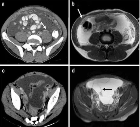
Figure 4: Ileocecal tuberculosis a Cor contrast-enhanced CT: mildly enhancing circumferential symmetrically thickened cecal wall; (a): Thickened cecal wall showing isointense signal, which shows post
contrast enhancement in same patient as in. (b): Cor T2 fat suppressed: Cecal wall thickening showing heterogeneous hyperintense signal; (c, d): Cor Pre-contrast T1 weighted and Pre-contrast-enhanced T1 fat
suppressed: The enhancement on MRI has much better visibility compared to CT.44
Figure 5: Lymph node pathology on MRI a Cor T2 fat suppressed: conglomerate mesenteric lymph node mass showing heterogeneous hyperintense T2 signal
with hypointense rim and high signal area (arrowhead) (b): Cor contrast-enhanced T1 fat suppressed: Conglomerate lymph node mass same as
in (a), Showing peripheral and central enhancement. (c): Ax T2 fat suppressed: Enlarged
lymph node showing homogeneous hyperintense signal; (d): Ax contrast-enhanced T1 fat suppressed:
Same lymph node as in (a) showing homogeneous enhancement; (e): Ax T2 fat suppressed: Enlarged lymph nodes showing heterogeneous hyperintense signal; (f): Ax contrast-enhanced T1 fat suppressed: Heterogeneous enhancement of the lymph nodes seen
in (e).44
Tubercular strictures are usually short, smooth, and concentric; however, discrimination from other lesions, including fungal infections and malignant lesions of the peritoneal and intestine, requires histological evidence. A recent paper that compared the use of magnetic resonance enterography with small bowel follow-through suggested that magnetic resonance enterography diagnosed a higher number of strictures except when used in the evaluation of extraintestinal lesions.24
barium enema, ultrasound, and CT have been well described. However, very few previous reports have focused on the MR imaging findings of tuberculosis of the gastrointestinal tract with lymph nodes involved in 90 % patients and peripheral and heterogeneously enhancing lymph node groups showing equal incidence. On MRI lymph nodes showed predominantly heterogeneous hyperintense signal with some of them showing perinodal hyperintnesity suggestive of capsular disruption on T2W sequences (Figure 5). On T1 W sequence, hypointense lymph nodes showed peripheral enhancement and isointense lymph nodes showed homogeneous enhancement.44
Figure 6: Sonographic image of (a): enlarged lymph nodes in the periportal area; (b): focal lesions in the spleen of approximately 3 mm in diameter; (c): focal
lesions in the spleen of approximately 10 mm in diameter.45
Ultrasonography may give the characteristic features of early abdominal tuberculosis which include mesenteric thickness of 15 mm or more and an increase in the mesenteric echogenicity (due to fat deposition), combined with mesenteric lymphadenopathy. Presence of dilated small bowel loops and ascites further substantiate the diagnosis.45
Figure 7: Peritoneal tuberculosis. (a): Ax contrast-enhanced CT; (b): Ax T2 weighted; Smoothly
thickened enhancing peritoneum. Thickened peritoneum appears hyperintense on T2 weighted
image. (c): Ax contrast-enhanced CT: Loculated ascites in pelvis with enhancing peritoneum. (d): Ax
T2 fat suppressed: Loculated ascites with multiple thin septae within ascites.44
Diagnostic evaluation for peritoneal tuberculosis
cytological evaluations for malignant cells. Peritoneoscopy may need to be used in some cases and can show tubercles, thickened peritoneum, and adhesions.29
Diagnostic evaluation for intestinal tuberculosis
Colonoscopy is the most important tool for evaluation of ITB as it helps in the characterisation of lesions, as well as in obtaining samples for microbiological and histological analysis. The colonoscopic findings in ITB include intestinal ulcers (usually transverse), pseudopolyps, strictures (usually short), and involvement of the ileocaecal valve. However, none of these are pathognomonic of TB and may be found in other conditions, including Crohn’s Disease (CD). The histological or cytological diagnosis is often based on fine-needle aspiration or biopsy obtained from radiology-guided sampling of the abdominal lymph nodes, peritoneal or omental thickening, or on endoscopic biopsies from the involved intestinal segments. The features suggestive of TB include the presence of granulomas, giant cells, caseating necrosis, and demonstration of acid-fast bacilli. The presence of granulomas is not unique to TB and may occur in other lesions, especially CD, fungal infections, and sarcoidosis; the presence of caseating necrosis is deemed to provide some degree of specificity to the diagnosis. Discrimination from CD is difficult, but the presence of multiple, large, confluent granulomas may be discriminative for TB. However, granulomas are only detected in a minority of cases (20-50%).30
Figure 8: (a): Deep, fissuring ulcer in Crohn's disease - representative picture; (b): patulous ileocecal valve
with ulceration in tuberculosis.46
Microbiological tools for the diagnosis include smear and culture for acid-fast Bacilli and PCR-based tests; the low yield from the peritoneal fluid and intestinal biopsy samples is the Achilles’ heel of microbiological tests. The use of PCR-based tests has been reported in multiple studies; a report on the use of multiplex PCR (using three probes: 16S rRNA, IS6110, and devR) provided excellent sensitivity for the diagnosis of both peritoneal and ITB but the findings await validation.31 Although the Xpert® MTB/Rif has emerged as an important tool for the diagnosis of pulmonary TB and some forms of EPTB (lymphadenitis), the reports on the use for ATB have indicated a limited benefit.32 In a report on the use of Xpert in peritoneal TB, the test was positive in only 4 out of 21 patients who were diagnosed with peritoneal TB.33 Similarly, in a report on ITB, only 3 out of 37 patients had a positive Xpert MTB/Rif test, suggesting that the sensitivity of the test for ATB would be low.32 The yield of an in-house PCR (using three genes: hsp-66, esat-6, and ITS MAC) was also reported to be low.34 The bulk of evidence therefore suggests that PCR-based tests also provide a low sensitivity for the diagnosis of ATB.
A therapeutic trial of ATT can also be used in the diagnosis and discrimination of ATB from other conditions. A recent study from India has shown that endoscopic healing of ulcers in patients started on empirical ATT may allow differentiation of ITB from CD.35 While the global symptomatic response with ATT was 38% and 37% in patients with CD at 3 and 6 months, respectively, 94% and 99% of patients with ITB showed a response at 3 and 6 months, respectively. When endoscopic response was observed at the end of ATT, all ITB patients had mucosal healing, while only 5% of CD patients showed mucosal healing; similar findings were also documented in the validation cohort. Therefore, persistent symptoms after 3 months of ATT may indicate a diagnosis of CD; however, the presence of a clinical response to ATT does not exclude the possibility of CD and mucosal healing should be sought.35 Another concern in the management of ATB is drug resistance and, therefore, the knock-on effect in patients with HIV, those with a previous history of ATT, and those not improving on treatment should be considered and cultures for drug sensitivity must be done.
TREATMENT OF ABDOMINAL TUBERCULOSIS
had received ATT previously as well as those with other forms of ATB (e.g., hepatic and pancreatic) and therefore the results may not be applicable to these patients.
The appropriate method of follow-up of patients with ATB is a challenging issue. Even though the disease may heal, persistence of symptoms may be related to sequelae like peritoneal adhesions or intestinal strictures. Continued symptoms may result in an unwarranted prolongation of treatment. Therefore, the follow-up should include both objective and subjective parameters for assessment of response. For ITB, demonstration of endoscopic healing (especially the ulcers) appears to be an excellent method to document response and may be performed at 2–3 months or later. A recent paper demonstrated the use of this approach to discriminate ITB from CD in patients where a therapeutic trial of ATT was administered in indeterminate lesions.35 In cases of non-healing ulcers, the possibility of drug-resistant TB or an alternative diagnosis must be considered; the frequency of drug-resistant TB varies from one geographic location to another but culture and drug sensitivity should be performed in patients with non-healing mucosal lesions or at the initial evaluation in those with previous history of ATT therapy or patients with HIV.37 For the follow-up of patients with peritoneal TB, abdominal ultra-sonography to look for resolution of ascites could be an appropriate strategy. Other parameters that are often used to assess response include improvement in appetite and general wellbeing, defervesce of fever, and weight gain. A recent paper also suggests that patients with the special form of peritoneal TB, abdominal cocoon, may also benefit from a conservative approach with ATT and the majority of patients can therefore avoid surgery.13 In a recent multicentre Indian study, the treatment completion rates for ATB were lower than most other forms of EPTB (like pleural, lymph-nodal, genitourinary) although the reasons for this are not clear.38 Surgery may play an important role in the diagnosis and treatment of abdominal form of tuberculosis. Often surgical intervention is the only therapeutic option for the patients presenting with complications of abdominal tuberculosis. The choice of surgical procedure depends on site and extent of disease, status of the remaining gut, general condition of the patient, surgeon’s expertise and individual preference. In a considerable number of cases complicated with faecal peritonitis and intraabdominal sepsis, a two stage procedure with creation of a stoma (ileostomy) was preferred to primary anastomosis. Creation of a stoma followed by reversal of stoma in a well prepared gut after 10-12 weeks of ATD therapy reduces the risks of anastomotic leaks and septic complications.39
INTESTINAL TUBERCULOSIS VS CROHN’S DISEASE: HOW TO DIFFERENTIATE?
In countries where ITB is more prevalent, CD is being increasingly recognized.40 Both these chronic
granulomatous disorders have similar clinical, endoscopic, radiologic, and histologic pictures; however, the natural history of both these disorders is strikingly different with serious implications regarding management. Misdiagnosis of one disease as another may be associated with multiple complications, including unnecessary immune suppression, drug toxicity, and delay in appropriate treatment. In one study, the presence of longitudinal or aphthous ulcers, anorectal lesions, and cobblestoning favoured CD, while transverse ulcers, patulous ileocaecal valve, <4 segments involved, and scars or pseudopolyps favoured ITB.41 Addition of CT enterography to colonoscopy increases the diagnostic accuracy and ability to differentiate CD from ITB from 66.7% to 95.2%. Features favouring CD were reported to be male sex, blood in stools, perianal lesions, bowel obstruction, extraintestinal manifestations, longitudinal ulcers on colonoscopy, cobblestone pattern, stricture, mucosal bridging, and rectal involvement. Histology suggesting focally enhanced colitis and CT findings of asymmetrical mural thickening, mural stratification, comb sign, and proliferation of the fibrofatty tissue also indicate the presence of CD. The findings that favoured the diagnosis of ITB were pyrexia, night sweats, pulmonary involvement, ascites, transverse ulcers, patulous ileocaecal valve, and caecal involvement on colonoscopy. Histological findings of submucosal granulomas or confluent granulomas, lymphocyte cuffing, and histiocyte lined ulcers, CT findings of short segmental involvement, and a positive IGRA also favour ITB diagnosis.42
Figure 9: Endoscopic features in a patient with Crohn's disease; (a): erythema and loss of vascular pattern in a patient with Crohn's disease; (b): a few aphthous ulcers in the colon in a patient with Crohn's
disease; (c): discrete ulcers in the colon in a patient with Crohn's disease; (d): multiple deep longitudinal ulcers in colon in a patient with Crohn's disease; (e): longitudinal ulcers with cobblestone appearance in the
Figure 10: Endoscopic lesions in a patient with intestinal tuberculosis; (a): ulcers over the ileocecal
valve in a patient with intestinal tuberculosis; (b) multiple nodules and deep ulcers (with neoplasm-like
appearance) in cecum with patulous ileocecal valve; (c) fixed patulous ileocecal valve with nodules in cecum and ascending colon in a patient with intestinal
tuberculosis; (d) circumferential ulcerative lesion in a patient with tuberculosis.47
CONCLUSION
To conclude, ATB remains a diagnostic challenge, having diverse and nonspecific symptomatology, and there being no specific laboratory tests. Early diagnosis is the decisive factor in avoiding systemic and local complications of ITB. The discrimination of ITB from CD is challenging and often necessitates a trial of ATT. ATT therapy for 6 months is usually adequate for mucosal healing and resolution of ascites, but sequelae like intestinal strictures may result in persistent symptoms needing endoscopic dilatation or surgical intervention. For those patients presenting in emergency, prompt surgical treatment is necessary to avoid further life threatening complications. A definitive procedure in the form of resection of diseased segment and primary anastomosis is safe and has largely been adopted in place of simple bypass of obstructive lesion with administration of ATT to treat the patients successfully with a complete cure.
Funding: No funding sources Conflict of interest: None declared Ethical approval: Not required
REFERENCES
1. Butt T, Karamat KA, Ahmad RN, Mahmood A. Advances in diagnosis of tuberculosis. Pak J Pathol. 2001;12:1-3.
2. Kapoor VK. Modern Management of Abdominal Tuberculosis. In: Taylor I, Johnson CD, (Eds) Recent Advances of Surgery. 2013: 156–169.
3. World Health Orgainzation. Global Tuberculosis report 2016. Available at: http://apps.who.int/iris/
bitstream/10665/250441/1/9789241565394-eng.pdf?ua=1. Accessed on 3 May 2018.
4. Arora VK, Rajnish G. Trends of extra pulmonary tuberculosis under revised national tuberculosis control programme: A study from south Delhi. Indian J Tuberc. 2006;53(2):77-83.
5. Choi EH, Coyle WJ. Gastrointestinal tuberculosis. Microbiol Spectr. 2016;4(6):1.
6. Sharma MP, Bhatia V. Abdominal tuberculosis. Indian J Med Res. 2004;120(4):305-15.
7. Sharma SK, Mohan A. Extrapulmonary tuberculosis. Indian J Med Res. 2004;120(4):316-53.
8. Donoghue HD, Holton J. Intestinal tuberculosis. Curr Opin Infect Dis. 2009;22(5):490-6.
9. Skopin MS, Batyrov FA, Kornilova Z. The prevalence of abdominal tuberculosis and the specific features of its detection. Probl Tuberk Bolezn Legk. 2007;1:22-6.
10. Khan IA, Khattak IU, Asif S, Nasir M, Ziaur R. Abdominal tuberculosis an experience at Ayub Teaching Hospital Abbottabad. J Ayub Med Coll Abbottabad. 2008;20:115-8.
11. Guirat A, Koubaa M, Mzali R, Abid B, Ellouz S, Affes N, et al. Peritoneal tuberculosis. Clini Res Hepatol Gastroenterol. 2011; 5(1):60-9.
12. Vaid U, Kane GC. Tuberculous Peritonitis. Microbiol Spectr. 2017;5(1).
13. Sharma V, Singh H, Mandavdhare HS. Tubercular abdominal cocoon: Systematic review of an uncommon form of tuberculosis. Surg Infect (Larchmt). 2017;18(6):736-41.
14. Mohammed A. Clinical Profile and Surgical Outcome of Abdominal Tuberculosis-A Retrospective Analysis. Int J Med Health Sci. 2013;2:402-6.
15. Patel N, Amarapurkar D, Agal S, Baijal R, Kulshrestha P, Pramanik S, et al. Gastrointestinal luminal tuberculosis: Establishing the diagnosis. J Gastroenterol Hepatol. 2004;19(11):1240-6.
16. Al-Bahrani ZR, Al-Saleem T. Intestinal tuberculosis in Iraq: A study of 50 cases. Int Surg. 1982;67(4 Suppl):483-5.
17. Paustian FF. Tuberculosis of the intestine. Bockus HL (eds.), Gastroenterology, 2nd edition, Philadelphia: WB Saunders Co. 1964.
18. Logan VS. Anorectal tuberculosis. Proc R Soc Med. 1969;62(12):1227-30.
19. Nayak S, Acharjya B. Mantoux test and its interpretation. Indian Dermat Online J. 2012;3(1):2-6.
20. Runyon BA, Hoefs JC, Morgan TR. Ascitic fluid analysis in malignancy-related ascites. Hepatology. 1988;8(5):1104-9.
22. Sharma R, Madhusudhan K, Ahuja V. Intestinal tuberculosis versus Crohn’s disease: Clinical and radiological recommendations. Indian J Radiol Imaging. 2016;26(2):161-72.
23. Kedia S, Sharma R, Bopanna S, Yadav D, Goyal S, Jain S, et al. Accuracy of computed tomographic features in differentiating intestinal tuberculosis from Crohn’s disease: A systematic review with meta-analysis. Intest Res. 2017;15(2):149-59. 24. Krishna S, Kalra N, Singh P, Kochhar R, Gupta R,
Singh R, et al. Small-bowel tuberculosis: A comparative study of MR enterography and small-bowel follow-through. AJR Am J Roentgenol. 2016;207(3):571-7.
25. Tao L, Ning HJ, Nie HM, Guo XY, Qin SY, Jiang HX. Diagnostic value of adenosine deaminase in ascites for tuberculosis ascites: A meta-analysis. Diagn Microbiol Infect Dis. 2014;79(1):102-7. 26. Liao YJ, Wu CY, Lee SW, Lee CL, Yang SS,
Chang CS, et al. Adenosine deaminase activity in tuberculous peritonitis among patients with underlying liver cirrhosis. World J Gastroenterol. 2012;18(37):5260-5.
27. Sanai FM, Bzeizi KI. Systematic review: Tuberculous peritonitis--Presenting features, diagnostic strategies and treatment. Aliment Pharmacol Ther. 2005;22(8):685-700.
28. Chau TN, Leung VK, Wong S, Law ST, Chan WH, Luk IS, et al. Diagnostic challenges of tuberculosis peritonitis in patients with and without end-stage renal failure. Clin Infect Dis. 2007;45(12):e141-6. 29. Bhargava DK, Shriniwas, Chopra P, Nijhawan S,
Dasarathy S, Kushwaha AK. Peritoneal tuberculosis: Laparoscopic patterns and its diagnostic accuracy. Am J Gastroenterol. 1992;87(1):109-12.
30. Du J, Ma Yan-Yan, Xiang Ha, Li You-Ming. Confluent granulomas and ulcers lined by epithelioid histiocytes: New ideal method for differentiation of ITB and CD? A meta analysis. PloS One. 2014;9(10):e103303.
31. Hallur V, Sharma M, Sethi S, Sharma K, Mewara A, Dhatwalia S, et al. Development and evaluation of multiplex PCR in rapid diagnosis of abdominal tuberculosis. Diagn Microbiol Infect Dis. 2013;76(1):51-5.
32. Kumar S, Bopanna S, Kedia S. Evaluation of Xpert MTB/RIF assay performance in the diagnosis of abdominal tuberculosis. Intest Res. 2017;15(2):187-94.
33. Bera C, Michael JS, Burad D, Shirly SB, Gibikote S, Ramakrishna B, et al. Tissue Xpert™ MTB/Rif
assay is of limited use in diagnosing peritoneal tuberculosis in patients with exudative ascites. Indian J Gastroenterol. 2015;34(5):395-8.
34. Rufai SB, Singh S, Singh A, Kumar P, Singh J, Vishal A. Performance of Xpert MTB/RIF on ascitic fluid samples for detection of abdominal tuberculosis. J Lab Physicians. 2017;9(1):47-52. 35. Pratap Mouli V, Munot K, Ananthakrishnan A,
Kedia S, Addagalla S, Garg SK, et al. Endoscopic and clinical responses to anti-tubercular therapy can differentiate intestinal tuberculosis from Crohn’s disease. Aliment Pharmacol Ther. 2017;45(1):27-36.
36. Jullien S, Jain S, Ryan H, Ahuja V. Six-month therapy for abdominal tuberculosis. Cochrane Database Syst Rev. 2016;11:CD012163.
37. Samant H, Desai D, Abraham P, Joshi A, Gupta T, Rodrigues C, et al. Acid-fast bacilli culture positivity and drug resistance in abdominal tuberculosis in Mumbai, India. Indian J Gastroenterol. 2014;33(5):414-9.
38. Cherian JJ, Lobo I, Sukhlecha A, Chawan U, Kshirsagar NA, Nair BL, et al. Treatment outcome of extrapulmonary tuberculosis under Revised National Tuberculosis Control Programme. Indian J Tuberc. 2017;64(2):104-8.
39. Jamal S, Khan ZM, Ahmed I, Shabbir S. Presentation and Outcome of Abdominal Tuberculosis in a Tertiary Care Unit. Ann. Pak Inst Med Sci. 2011;7(1):33–6.
40. Nayagam JS, Mullender C, Cosgrove C, Poullis A. Abdominal tuberculosis: Diagnosis and demographics, a 10-year retrospective review from a single centre. World J Clin Cases. 2016;4(8):207-12.
41. Lee YJ, Yang SK, Byeon JS, Myung SJ, Chang HS, Hong SS, et al. Analysis of colonoscopic findings in the differential diagnosis between intestinal tuberculosis and Crohn’s disease. Endoscopy. 2006;38(6):592-7.
42. Limsrivilai J, Shreiner AB, Pongpaibul A, Laohapand C, Boonanuwat R, Pausawasdi N, et al. Meta-analytic Bayesian model for differentiating intestinal tuberculosis from Crohn’s disease. Am J Gastroenterol. 2017;112(3):415-27.