CARDIAC PACEMAKER FUNCTION IN TH E AVIATION ENVIRONM ENT
A thesis submitted for the degree of Doctor of Medicine
in the University of London
W ILLIAM DANIEL TO FF
ProQuest Number: 10106671
All rights reserved
INFORMATION TO ALL USERS
The quality of this reproduction is dependent upon the quality of the copy submitted.
In the unlikely event that the author did not send a complete manuscript and there are missing pages, these will be noted. Also, if material had to be removed,
a note will indicate the deletion.
uest.
ProQuest 10106671
Published by ProQuest LLC(2016). Copyright of the Dissertation is held by the Author.
All rights reserved.
This work is protected against unauthorized copying under Title 17, United States Code. Microform Edition © ProQuest LLC.
ProQuest LLC
789 East Eisenhower Parkway P.O. Box 1346
ABSTRACT
Experience suggests that patients with implanted cardiac pacemakers may travel safely by air but the exacting medicW standards for pilots invite special consideration, as pacemaker failure may result in subtle or sudden incapacitation that might jeopardise flight safety. Potential hazards in the aviation environment were assessed, with particular regard to the risk o f electromagnetic interference (EMI).
Occasional inhibition was noted in explanted unipolar pacemakers during flight tests and a systematic study was therefore undertaken. Electromagnetic field strengths were measured in 10 aircraft under operational conditions and explanted and implanted pacemakers were exposed to comparable fields. The effect o f aircraft vibration on activity-sensing, rate-adaptive pacemakers was also assessed by monitoring the rate- response during flights.
Aircraft field strengths ranged from 0.20-1.12Vm'\ 0.20-35.OOVm’^ and 1.60- IS.OOVm'^ in the HP, VHP and microwave bands respectively. Explanted unipolar devices were all affected in comparable fields, with thresholds (m ean±sd) for interference and failure (cessation o f pacing ^30s) respectively o f 3.2±1.9V m ’^ and 7.7±1.5V m ‘^ in HP and 5.7± 3.0V m ‘^ and 11.2±6.6Vm‘^ in VHP fields. Modulation with pulsing increased HP susceptibility (0.49±0.2Vm*^ and 1.5 ±0.93Vm'^). Only one device showed sustained interference in microwave fields (l-2GHz at 16Vm’^) but susceptibility was increased by pulsing. Bipolar devices were less susceptible although one device showed interference and failure in modulated HP fields at 18.5Vm'^ and 30Vm'^ respectively and another showed interference in VHP fields at 27V m '\ Implanted unipolar devices were unaffected. Activity-sensing devices showed increased pacing rates during flight. In fixed-wing aircraft, these were modest and confined to specific periods (take-off, turbulence and landing) but in helicopters and hovercraft, they were marked and sustained.
ACKNOWLEDGEMENTS
The work presented in this thesis was performed during my appointment as Clinical Research Fellow in the Medical Department of the UK Civil Aviation Authority (C AA) and in the Department of Cardiological Sciences at St George's Hospital Medical School.
I am deeply indebted to Professor A. John Camm for his enthusiastic support and guidance throughout. My thanks are also due to Dr Geoffrey Bennett and Dr Michael Joy for their support and encouragement and to Mr Alastair Deller, Principal Physicist in the Department of Medical Electronics, St Bartholomew's Hospital, who constructed the test apparatus for evaluating pacemaker susceptibility to electromagnetic interference and provided valuable technical advice.
My thanks are also due to the Procurement Executive of the Ministry o f Defence and in particular, to Dr Nigel Carter, Mr R.A. Hobbs and their colleagues in the Electromagnetic Hazards and Compatibility Section at the Royal Aircraft Establishment (RAE), Farnborough, for assistance with the measurement of aircraft electromagnetic field strengths and the provision of test facilities. I am also grateful to Mr Colin Leeks of the Noise and Vibration Group at RAE who made the measurements of aircraft vibration and operated the vibration test rig at Farnborough.
The flight tests would not have been possible without the generous help o f Alan Mann Helicopters, British Airways, British Airways Helicopters, British Caledonian Airways, British Hover speed. Civil Aviation Authority Flying Unit, Dr Michael Joy, Racal Avionics and Trent Air Services. Pacemakers and equipment were provided by Cordis (UK) Ltd, Intermedics Inc, Medtronic Pacing Products Ltd, Siemens-Pacesetter, Sorin Biomedica (UK) Ltd and Telectronics Ltd to whom I am grateful.
TABLE OF CONTENTS
A bstract
Acknowledgements
Table o f Contents
List of Tables & Figures
Page
2
C hapter 1
C hapter 2
C hapter 3
C hapter 4
C hapter 5
C hapter 6
Introduction and Literature Review
Methods, Materials and Patients
Empirical Studies of Pacemaker Function in Aircraft
Measurement of Aircraft Electromagnetic Field Strengths
65
93
105
The Electromagnetic Interference Susceptibility of Unimplanted 137 Pacemakers
The Electromagnetic Interference Susceptibility of Implanted 212 Unipolar Pacemakers
C hapter 7 Activity-Sensing Pacemaker Function During Air Travel
C hapter 8 Concluding Remarks and Recommendations
References
Presentations & Publications
A bbreviations
234
263
269
290
LIST O F TABLES & FIGURES
TABLES:
Page
1.1: NASPE/BPEG generic (NBG) pacemaker code 51
1.2: Age profile of patients paced in the UK in 1991 52
1.3: Pre-pacing EGG in patients paced in the UK in 1991 53
1.4: BPEG recommended pacing modes 54
1.5: Cumulative survival figures for single and dual chamber pacemakers 55
1.6: The radio-frequency spectrum 56
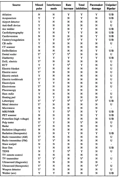
1.7: NRPB reference levels for exposure to electromagnetic fields 57 1.8: Commonly encountered sources of EMI and pacemaker responses 58 1.9: Civil and military aircraft radio-frequency transmitters 59
2.1: Fixed-wing aircraft 68
2.2: Rotary-wing aircraft and air-cushioned vehicle 69
2.3: Cardiac pacemakers 70
4.1: Aircraft equipment specification and test environment 116 4.2: Maximum recorded electric field strengths in aircraft 117 4.3: Measured audio frequency field strengths in Concorde 118 4.4: Radio-frequency emitters contributing to the total (severe) environment 119 4.5: Airworthiness terms and their meaning in relation to the probability and
consequence of an adverse event 120
5.1: Pacemakers studied 151
5.2: Responses o f explanted pacemakers to radio-frequency fields 152 5.3: Minimum field strengths for interference and failure in explanted
pacemakers in radio-frequency fields 153
5.4: Reactance and attenuation of test apparatus aerial and a standard pacing
electrode at different frequencies 154
5.5: Pacing characteristics before and after test exposures 155
7.1: Aircraft studied 248
FIGURES:
Page
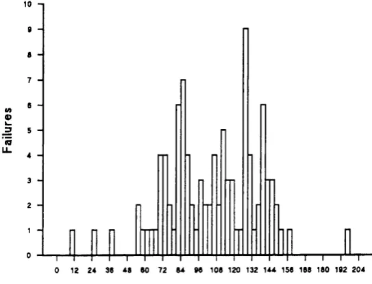
1.1: Pacemaker implantation rates in Europe & the USA during 1989 60 1.2: Trends in the use of bipolar pacing in the UK 1981-1991 (BPEG data) 61 1.3: Single & dual chamber pacemaker survival curves (BPEG data) 62 1.4: Single & dual chamber pacemaker survival curves (Bilitch data) 63 1.5: Histograms showing implant duration at time of failure for single & dual
chamber pacemakers (Bilitch data) 64
2.1: Fixed-wing aircraft: Slingsby Firefly; Gulfstream Grumman Cougar;
Beechcraft Baron 71
2.2: Fixed-wing aircraft: Piper Turbo Navajo; British Aerospace Jet Stream;
Hawker Siddely HS125 72
2.3: Fixed-wing aircraft: British Aerospace BAG 1-11; Boeing 737 73
2.4: Fixed-wing aircraft: Boeing 757; Lockheed Tristar 74
2.5: Fixed-wing aircraft: McDonnell Douglas DCIO; Boeing 747 75 2.6: Fixed-wing aircraft: British Aerospace/Aerospatiale Concorde 76
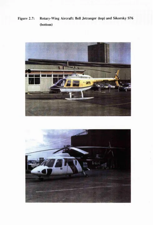
2.7: Rotary-wing aircraft: Bell Jetranger; Sikorsky S76 77
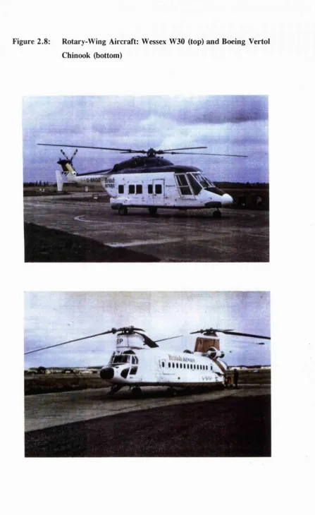
2.8: Rotary-wing aircraft: Wessex W30; Boeing Vertol Chinook 78 2.9: Hovercraft: British Hoverspeed SRN4 Mk3 Mountbatten Class 79
3.1: Circuit diagram of test apparatus 1(X)
3.2: Test apparatus 101
3.3: Electrical equipment on Boeing 747: Electric warming ovens; cart-lift to
upper deck; concealed electrical equipment bay 102
3.4: View from starboard wing root of Boeing 747 showing HF aerial at wing tip 103 3.5: Ground radar installation at London Heathrow airport 104 4.1 : Three Eaton receivers during in-flight recording on Boeing 747 121 4.2: Recording aerials: Triaxial dipole (HF electric field component); triaxial
shielded loop (HF magnetic field component) 122
4.3: Recording aerials: Monoconical (VHF electric field component); Eaton log
periodic (microwave electric field component) 123
FIGURES (...cont'd/):
Page
5.1: Detail of test apparatus 156
5.2: Circuit diagram of test apparatus (1) 157
5.3: Circuit diagram of test apparatus (2) 158
5.4: Circuit diagram of test apparatus (3) 159
5.5: Complete test apparatus in unipolar configuration 160
5.6: Complete test apparatus in bipolar configuration 161
5.7: Exterior of test facility showing screened chamber and instrumentation annex 162 5.8: Interior of screened chamber and instrumentation annex with test equipment 163 5.9: Summary of pacemaker responses to HF CW field exposure 164 5.10: Summary of pacemaker responses to HF modulated field exposure 165 5.11: Summary of pacemaker responses to VHF CW field exposure 166 6.1: Interior of screened chamber and instrumentation annex 221 6.2: PA and lateral chest X-ray of patient 1 (SC) showing pacing lead in situ 222 6.3: PA and lateral chest X-ray of patient 2 (KB) showing pacing lead in situ 223 6.4: PA and lateral chest X-ray of patient 3 (JW) showing pacing lead in situ 224 7.1: Processing of the signal from the activity-sensor in the Medtronic Activitrax 250 7.2: In-flight monitoring of the rate-response of the Medtronic Activitrax 251 7.3: Vibration simulation equipment at the Royal Aircraft Establishment 252
7.4: Vibration rig control and instrumentation annex 253
7.5: Activitrax pacing rates during flight in four fixed-wing aircraft: Grumman
Cougar, Beechcraft Baron, Piper Navajo and British Aerospace Jetstream 254 7.6: Activitrax pacing rates during flight in four fixed-wing aircraft: Hawker
Siddely HS125, McDonnell Douglas DCIO, Boeing 747 and Concorde 255 7.7: Activitrax pacing rates during flight in four rotary-wing aircraft:
Bell Jetranger, Sikorsky S76, Wessex W30 and Boeing Vertol Chinook 256 7.8: Activitrax pacing rates during flight in hovercraft:
British Hoverspeed SRN4 Mk 3 Mountbatten Class 257
7.9 Box and whisker' plot of pacing rates during flight in eight fixed-wing
FIGURES (...cont'd/):
Page
7.10 'Box and whisker' plot of vibration levels during flight in eight fixed-wing
aircraft, four rotary-wing aircraft and a hovercraft 259 7.11: Comparison of implanted and epicutaneous Activitrax pacing rates during
treadmill exercise 260
7.12: Effects of reprogramming on the Activitrax rate response to the simulated
environment of a Chinook helicopter 261
7.13: Pacing rate responses of Sensolog 703 (P48), Sensolog P49 and Activitrax
CHAPTER I:
BACKGROUND TO THIS THESIS
In recent years air travel has become more commonplace and increasingly accessible to large numbers of people. Increasing numbers of passengers are flying on scheduled airlines and private flying for business and pleasure is becoming more popular. Concurrent developments in health care technology have included improvements in the design and performance of implantable cardiac pacemakers such that their recipients can now expect to lead relatively normal lives. A likely corollary of these parallel developments is that increasing numbers of patients with pacemakers will wish to enjoy the benefits of air travel as passengers. A smaller number may wish to fly themselves in light aircraft and inevitably the occasional professional pilot who requires a pacemaker may seek to continue his career.
The environment of the modem aircraft is dramatically safer than in the past but it still contains special hazards, some of which may pose a greater threat to the patient with a pacemaker than to others. Patients who wish to travel by air and their physicians need to know the level of risk engendered by such hazards and those responsible for air
safety are occasionally called upon to provide advice.
The studies presented in this thesis were performed to determine the level o f risk to pacemaker users in the aviation environment. The principal aim was to enable soundly based advice to be offered to those responsible for assessing the fitness to fly of prospective aircrew but the findings are also of relevance to those advising passengers with pacemakers. The results of these studies have led to a series of recommendations to the United Kingdom Civil Aviation Authority regarding the medical certification of pilots with pacemakers (Toff & Camm, 1988; Toff et al, 1992). They have also led to the identification of deficiencies in pacemaker safety standards. In addition, they have identified inherent flaws in the current generation o f activity-sensing rate-adaptive pacemakers and indicated potential design improvements, some of which have been implemented.
and previous data regarding the possibility of an adverse interaction will be reviewed. Finally, the requirements for medical certification of aircrew will be discussed. In chapters 2 to 6 a series of experiments designed to evaluate the risks related to the principal hazard, electromagnetic interference (EMI), will be described and discussed. In chapter 7 an evaluation of the unique risk posed by aircraft vibration to one particular type of rate-responsive pacemaker will be presented. In chapter 8, conclusions from the studies will be drawn and recommendations made in respect of the medical certification of prospective aircrew with pacemakers.
THE EVOLUTION OF CARDIAC PACING
Electrical stimulation of the heart was attempted as early as the late eighteenth century (Aldini, 1804). In a series of macabre experiments carried out on decapitated criminals it was demonstrated that electrical stimulation could initiate cardiac contraction. It was not until the 19th century, however, that it was recognised that bradycardia might be caused by degeneration of the heart and that this could cause cardiac syncope. Various experimenters at the time recognised and developed the possibility that electrical energy might usefully be employed to stimulate the heart therapeutically. Notable among them was John McWilliam who, in 1889, suggested that repeated electrical stimulation between saline-soaked pads placed on the anterior and posterior chest might be used to maintain the heart rhythm in patients with bradycardia and certain forms o f cardiac arrest (McWilliam, 1889). Successful application of the method in resuscitation of a patient with apparent cardiac arrest was subsequently reported in 1908 (Robinovitch,
1909).
Restoration of normal cardiac rhythm by electrical stimulation following ventricular fibrillation during cardiac surgery was suggested in 1940 (Wegria & Wiggers, 1940) and successfully implemented in 1947 (Beck et al, 1947). In the same year, two patients were resuscitated by stimulation of the sinus node using a pulse generator connected to mains electricity (Sweet, 1947). A battery-operated pacemaker for use in animal experiments was subsequently described by Callaghan and Bigelow in 1950 (Callaghan & Bigelow, 1950).
The first application of cardiac pacing to the treatment of bradycardia and Stokes Adams' attacks in man is attributed to Zoll who, in 1952, paced between oesophageal and precordial electrodes using an external pacemaker (Zoll, 1952). Hie treatment could only be used for limited periods as the electrical impulses o f 1(X)-200V that were required caused painful muscle jerks and skin burns. In 1957 Lillehei and colleagues demonstrated that by applying a stainless steel suture to the ventricular wall of a dog, pacing could be accomplished with an externalized lead and external pulse generator using impulses of about 2.3V. The method was soon extended to temporary use in patients following open heart surgery (Weirich et al, 1957 & 1958).
In 1958 Furman (Furman & Robinson, 1958) introduced a bipolar catheter transvenously to the right ventricle and used this with an external pulse generator to stimulate the patient's heart. In the same year the first permanent cardiac pacemaker, a transistor-operated device powered by a rechargeable nickel-cadmium battery, was implanted using two myocardial electrodes sutured to the left ventricle (Elmqvist & Senning, 1959; Lagergren, 1978). Contemporaneous work by Greatbach and Chardack led to the development of an implantable pacemaker used successfully in animal studies in 1958 (Greatbatch & Chardack, 1959) and in patients two years later (Chardack et al, 1960). Implanted pacemakers using transvenous leads soon followed (Furman et al 1961; Lagergren & Johansson, 1963; Lagergren et al 1965) marking the start o f the current era of cardiac pacing.
towards better emulation of normal cardiac physiology with the evolution of different pacing modes. Initially, all pacemakers delivered impulses to the ventricle at a fixed rate pre-determined by the manufacturer (VOO pacing [vide infra]). Recognition of the importance of the normal sequential depolarisation of atrium and ventricle in maximizing cardiac output (Kahn et al, 1960) led to the development of an implantable atrio-ventricular (AV) synchronous pacemaker in which sensing of atrial depolarisation was used to trigger the delivery of a stimulus to the ventricle by a separate electrode (VAT pacing) (Nathan et al, 1963(a) & (b)). This had the further advantage of matching the ventricular rate to the physiologically determined atrial rate. If atrial activation failed, a standby self-pacer oscillator initiated fixed-rate ventricular stimulation. This mode of pacing introduced both the concept of a sensing function and rate variability. Although successful, the device had the disadvantage that, at that time (1962), thoracotomy was required for its insertion. There was also a risk o f competition between a spontaneous ventricular contraction and a pacemaker stimulus. Recognition that pacing during the vulnerable period of ventricular repolarization could provoke ventricular tachycardia and fibrillation (Lemberg et al, 1965; Bilitch et al, 1967) led to the development and implementation of ventricular inhibited (W I) pacing in which a ventricular stimulus was delivered only in the absence of spontaneous ventricular depolarisation (Castellanos et al, 1964; Lemberg et al, 1965). The term 'demand pacing' has become associated with modes in which pacing occurs only when spontaneous cardiac activation fails.
Recognition that the atrium cannot always be used to determine the appropriate ventricular rate led to the development of devices using alternative biosensors to preserve the cardiac rate response to changing metabolic needs. The possibility of sensing respiratory changes during exercise to determine the appropriate pacing rate was proposed in 1975 (Funke, 1975) and the first implanted rate-adaptive devices using a blood pH sensor were reported shortly afterwards (Camilli, 1977 & 1978). A wide variety of sensor driven pacemakers have since been developed (Alt et al, 1986(a); Anderson & Moore, 1986; Rossi, 1987). Pivotal to the development of single chamber rate-adaptive pacing was recognition that the heart rate response during exercise is a more important determinant of cardiac output than AV synchrony (Karlof, 1975).
A further development was the introduction of pacemaker programmability (Hehrlein et al, 1974). This describes the facility whereby an external programming device is used to make reversible adjustments in one or more pacing parameters by non-invasive means. Initially, coded magnetic pulses were used but more recently these have been replaced by radio-frequency signals. Pacemakers incorporating a wide and increasing variety of programmable parameters are now available (Hayes, 1989(a)). More recently, a variety of anti-tachycardia devices (Camm & Ward, 1983) and a fully implantable automatic defibrillator (Mirowski et al, 1970) have been developed.
MODERN PACEMAKER DESIGN AND FUNCTION
TH E PACEMAKER:
The modern implantable pacemaker is a battery powered pulse generator capable of delivering an electrical stimulus o f fixed characteristics at regular intervals. The generator is housed in an hermetically sealed can, usually of titanium. Typical dimensions are 4-6cm height and width and 1cm thickness but the current trend is towards smaller devices. Weight is typically 30-50g. The integral power source is usually a lithium battery which offers good longevity before depletion. The unit is implanted in a subcutaneous pocket in the anterior or lateral chest wall or abdomen and it is connected by a transvenous lead to the endocardium. Occasionally, the lead is attached to the epicardial surface of the heart following open chest surgery or limited thoracotomy. Either ventricular or atrial chambers may be paced or both. For safety and convenience the right side of the heart is paced. Ventricular leads are typically 58- 60cm in length; atrial leads are approximately 10cm shorter. The rate of pulse delivery is either preset by the manufacturer to around 70ppm to emulate the normal heart rate or is adjustable along with other programmable characteristics of the pacing pulse and sensing circuitry.
ELECTRODE SYSTEMS:
1982). Bipolar leads also offer an advantage in pacing modes dependent on atrial sensing in that the ventricular signal amplitude on the atrial electrogram using a bipolar lead is less than one tenth of that using a unipolar lead (Griffin, 1983). The risk of double-sensing and pacemaker recycling is thus considerably lower with a bipolar lead. There are wide regional variations in the type of lead system most commonly used. With improved lead technology enabling the size o f bipolar leads to be reduced and increased recognition of their theoretical advantages, their use should become more widespread (Furman, 1987).
MODE O F OPERATION:
Almost all modern pacemakers incorporate a sensing function to detect endogenous cardiac electrical activity and most pacemakers are designed to pace the heart only when endogenous activity is inadequate (demand pacing). The detection of an endocardial signal inhibits the output of the pacemaker and resets the timing circuit. Some devices are programmed to pace in response to the sensed cardiac signal (triggered pacing) and either deliver a pulse during the refractory period following a spontaneous cardiac depolarisation or, in the absence of spontaneous activity, at the end of their timing cycle. The high power consumption and potentially deleterious effects of pacing during spontaneous cardiac electrical activity militate against the use of this mode of pacing except in dual chamber pacemakers in patients with complete heart block but normal sinus node function, in whom triggering of ventricular pacing by atrial activity may restore physiological function.
PHYSIOLOGICAL PACING:
The aim of physiological cardiac pacing is to emulate the behaviour of the normal sinus node and conduction system and the selection of pacemaker and mode of pacing for each patient should reflect this (Furman, 1987). The emulation should include rate variability according to the patient's metabolic requirements and a mechanism for the preservation of AV synchrony which may contribute up to 20% of cardiac output at rest (Samet et al, 1966). Physiological pacing may offer a patient greater exercise capacity and a better quality of life than conventional ventricular pacing (Kruse et al, 1982; Perrins et al, 1983) and may also be associated with reduced mortality (Perrins et al, 1983; Alpert et al, 1986; Alpert et al, 1987).
The most physiological pacemaker is the normal sinus node and if its function is preserved, as in isolated conducting system disease, it should be harnessed by atrial sensing and used to drive the ventricle by means o f a dual chamber pacemaker. If the sinus node is diseased or in the presence of chronic or frequent atrial fibrillation or flutter, rate-modulation using atrial sensing is clearly inappropriate. When the sinus node is diseased but conduction is intact, it is logical to pace the atrium. When neither sinus node nor conduction are intact, AV sequential pacing most closely mimics normal physiology.
SENSOR-DRIVEN RATE-ADAPTIVE PACING:
1984), minute ventilation (Lan et ai, 1988(c)), ventricular depolarisation gradient (Callaghan et al, 1987) and first-derivative of right ventricular pressure (Sutton et al, 1987). Each of the sensors has deficiencies or limitations and attention has recently turned to the possibility of combining multiple sensors to optimise the advantages and circumvent the shortcomings of the individual sensors (Lau, 1992(a) & 1993). A further development has been the introduction of dual chamber rate-adaptive pacemakers (DDDR) (Kappenberger & Herpers, 1986). These are indicated in patients with AV block and chronotropic incompetence, to combine the benefits of rate responsiveness with restoration of AV synchrony.
The most widely used alternative (non-atrial) sensor in current clinical practice is the activity sensor. The reasons for its popularity include efficacy, reliability, ease o f implantation (no extra procedure is required) and simplicity of programming. The majority o f the devices presently in clinical use incorporate a piezoelectric crystal bonded to the inner surface o f the pacemaker can, the output from which is used to determine the appropriate pacing rate. These devices are susceptible to extraneous vibration, such as that associated with various forms of transport, and may develop inappropriately high pacing rates in response. Rises of 10 to 30 pulses per minute (ppm) have been demonstrated during travel by car, bus, tram, subway and railway train (Humen et al, 1985; Rankin and Lindemans, 1985; Lindemans et al, 1986; Heuer et al, 1986; Stangl et al, 1986). One study (Heuer et al, 1986) refers to a rise of 5ppm during travel in an unspecified type of aircraft. Similar observations have been made by Lau (1987) who described pacing rate responses in a patient with an Activitrax pacemaker during a flight in a BAG 1-11 Jet aircraft. Rises of up to 10 ppm were observed during flight and 28 ppm during taxying. Rate rises have also been reported during horse-riding (Lamas & Keefe, 1990) and dental treatment (osteotomy and preparation of cavities) (Rahn et al, 1988).
limitations. Inappropriate sensor-mediated tachycardias have also been described in other devices but these are usually resolved by reprogramming and rarely give rise to clinically significant problems (Lau, 1991; Lau, 1993).
THE EPIDEMIOLOGY OF PACEMAKER USE
DEM OGRAPHIC DATA:
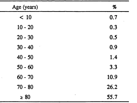
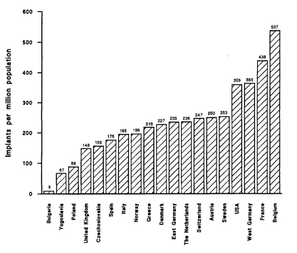
There are approximately one million patients with implanted cardiac pacemakers worldwide and approximately 200,000 new patients receive implants annually (Feruglio et al, 1987). Europe and North America account for 50.12% and 47.04% o f the world's implants respectively. European statistics for 1989 have recently been presented, relating to 18 countries with a total population o f 440 million inhabitants (Edhag, 1991). A total of 125,000 pacemaker systems were implanted, of \\bich 18% were replacements. There was considerable variation in implant rates between different countries, with the number of first implants ranging from 9 to 537 per million population. Only Belgium, France and West Germany matched or exceeded the corresponding rate in the USA, which was 359 per million (Bernstein & Parsonnet, 1992) (figure 1.1). Possible reasons for the wide variation include differences in the age distribution o f the populations, the proportion of patients being paced for conditions other than AV block and socio-economic factors. Variations in medical attitudes and competence may also be relevant. In the United'Kingdom there are thought to be some 40,000 patients with pacemakers, which approximates to 1 per thousand of the adult population and the annual rate of new implants is 148 per million people. This is less than half the number of patients whom it has been estimated would benefit from a pacemaker and it is likely that improved education of physicians will result in increased implantation rates in the future. The majority of patients are elderly, the mean age at first implantation being 72.8 years. The age distribution of patients at implantation is shown in table 1.2 (Rickards, 1992).
INDICATIONS FOR PACING:
American College of Cardiology (ACC) and the American Heart Association (AHA) in the USA (Dreifus et al, 1991) and by a working party o f the British Pacing and Electrophysiology Group (BPEG) in the UK (Clarke et al, 1991). Although some areas remain controversial, such as asymptomatic type I (Wenckebach) second degree AV block, there is broad agreement on the indications for pacing.
The majority of pacemakers are implanted for the treatment of symptomatic bradyarrhythmia, the most common symptoms being syncope (38%) and dizzy spells (27%) (Rickards, 1992). In a typical population o f patients paced for bradyarrhythmia (Clarke et al, 1991), 42% had AV block, 25% sinus node dysfunction, 10% AV block and sinus node dysfunction, 13% atrial fibrillation and AV block and 10% carotid sinus or vasovagal syndromes. Data from the BPEG database concerning the pre-pacing ECG of 7254 patients receiving implants in 1991 (Rickards, 1992) are presented in table 1.3.
The relative prevalence of the different indications for pacing varies with age, as does the aetiology of the underlying disease. Indirect evidence for the latter is offered by the observation that the mortality ratio in middle aged patients paced for complete heart block is higher (4.5:1) than that of similar elderly subjects, who can expect to live as long as their peers. It is most likely that this reflects a greater prevalence of underlying coronary artery disease in the former group and of idiopathic fibrosis in the latter (Ginks et al, 1979). The pilot population is generally younger than typical paced populations and the importance of excluding co-existent coronary artery disease in prospective aircrew with pacemakers has been stressed (Toff & Camm, 1988).
CH OICE OF PACING MODE:
rate-adaptation to changing metabolic requirements, if required (Furman, 1987). The guiding principles are: i) The ventricle should be paced if there is actual or threatened AV block; ii) the atrium should be paced/sensed unless contra-indicated; iii) rate response is not essential if the patient has a normal chronotropic response; iv) rate hysteresis may be valuable if bradycardia is intermittent (Clarke et al, 1991; Nathan & Davies, 1992). The BPEG recommendations regarding 'optimal', 'alternative' and 'inappropriate' pacing modes in different conditions are presented in table 1.4.
The selection of inappropriate pacing modes may not only deprive the patient of optimal benefit but may actually increase morbidity and mortality. In sinus node disease, for example, there is increasing evidence that ventricular pacing is associated with a greater risk of atrial fibrillation, thromboembolism and congestive heart failure than atrial pacing (Camm & Katritsis, 1990). In patients capable o f retrograde ventriculo-atrial conduction, ventricular pacing may allow the development of the pacemaker syndrome with hypotension and symptoms such as weakness, dizziness, pre-syncope or syncope (Mitsui et al, 1971; M candri et al, 1978; Johnson et al, 1978; Ausubel & Furman, 1985). When a dual chamber pacemaker is implanted, atrial sensing o f retrograde ventriculo-atrial conduction may result in an 'endless loop' pacemaker mediated tachycardia which may be rapid (often at the programmed upper rate) and sustained (Furman, 1989(a)). Once recognised, the problem can usually be dealt with by appropriate reprogramming. Dual chamber pacemakers may also be hazardous when atrial tachyarrhythmias occur, resulting in ventricular tracking at rapid and potentially dangerous rates. Sensor-driven dual chamber devices may avert this problem by allowing sensor-mediated regulation of the ventricular rate under such circumstances. In all systems, optimal pacemaker programming is essential to ensure maximum benefit at minimum risk.
that may cause inhibition or triggering of the pacemaker, according to its programmed mode. Exogenous electromagnetic signals may also be inductively coupled to the unipolar pacing lead, which effectively acts as an aerial. Bipolar systems, in which both electrodes are remote from the skeletal muscles, are practically immune to myopotential interference unless insulation failure occurs (Hauser et al, 1985). The small electrode separation also confers a high degree of immunity to exogenous electromagnetic interference (Imich, 1984). The advantages of bipolar leads are reflected in implantation practice in the USA, where up to 75 % of implants are bipolar (Bernstein & Parsonnet, 1992) and data from the BPEG database indicate a trend to increasing use of the bipolar configuration in the UK (figure 1.2). With the increased use of dual chamber pacemakers with high atrial sensitivity, myopotential interference has assumed greater significance. Over half of all unipolar systems may be affected and in about 10% of these, symptoms may be sufficient to warrant major intervention (Furman, 1986).
RELIABILITY AND SAFETY OF PACING SYSTEMS
PACEM AKER DEPENDENCE:
In considering the risk of a pacing system failure, it is important to recognise that the consequences of failure will depend on the extent to which the patient is reliant upon the system. The term 'pacemaker dependence' is used to indicate a high degree of reliance although it has no universally agreed definition. For practical purposes, it may be defined as the occurrence of symptoms or failure to develop an adequate escape rhythm in response to abrupt cessation of pacing. Dependence may be graded as follows (Furman, 1989(b)): Class I - No escape rhythm (pacemaker dependent); Class II - escape rhythm consists of complete AV block with atrial fibrillation or sinus rhythm (substantially pacemaker dependent); Class III - escape rhythm consists o f a lesser degree of AV block, an arrhythmia or a sinus bradycardia of 30 beats per minute or less (moderately dependent); Class IV - escape consists of regular sinus rhythm of normal rate (50 beats per minute or more) (not dependent).
minutes each month and a number, in whom implantation has been solely for prognostic benefit, rather than to ameliorate symptoms, may never pace. In such patients the risks o f an adverse event resulting from pacemaker failure are likely to be negligible. In contrast, as many as 40% of patients paced for symptomatic bradycardia may exhibit a paced rhythm at follow-up, precluding assessment o f the underlying cardiac rhythm (Edhag, 1979). In such patients, the consequences of abrupt pacemaker failure may be observed if the pacemaker is inhibited by chest wall stimulation using an external pacemaker or stimulator connected to chest electrodes (Barold et al, 1970; Samet et al, 1970; Grendahl et al, 1978; Staessen et al, 1982). Typically, 20V stimuli are delivered at about 20 beats per minute above the pacing rate. Faster rates may cause reversion to fixed rate pacing in the 'interference mode' (Castellanos et al, 1979). An alternative protocol has been described, in which the pacing rate is progressively reduced by serial décrémentai rate inhibition (Crick et al, 1985). This tends to evoke a more adequate and consistent escape rhythm than abrupt inhibition. Although this may better reflect the degree o f the underlying disturbance of rhythm or conduction, it may be less appropriate for the assessment of 'worst case' responses to sudden pacemaker malfunction.
Between 5% and 30% o f patients have symptoms or fail to develop a satisfactory escape rhythm in response to chest wall stimulation (Rokas et al, 1981; Staessen et al, 1982; Edhag et al, 1983; Crick et al, 1985; Furman, 1989(b)). The response may vary at different times and the value of a single assessment is üierefore limited. It has been suggested that dependency status should be defined by the worst ever response (Furman, 1989(b)). A number of characteristics prior to pacing have been shown to be more prevalent in subjects with a higher degree o f pacemaker dependence, including syncope, AV block and slower ventricular rates but none is sufficiently specific to be o f clinical use (Staessen et al, 1982; Crick et al, 1985).
MODES O F PACING SYSTEM FAILURE:
considerably Influenced by the mode of failure and the clinical context in which it occurs. Abrupt or unpredictable failure is clearly o f greatest significance.
The pacemaker:
Pacemaker batteries have a finite life span and depletion will inevitably occur. Although pacemaker longevity of 10-15 years has been achieved, a reasonable expectation of modem devices would be for 95% cumulative survival at about 7 years. At present, many sophisticated dual chamber devices and sensor-driven rate-adaptive devices, particularly those using active rather than passive sensors, fall well short of this (Furman, 1989(c)). Normal battery depletion poses little risk as it is usually predictable in broad terms from the expected device longevity and more particularly, by end of life indicators incorporated in the pacemaker circuitry. Even premature battery depletion may pose little risk, provided that it is detected early during routine follow-up.
O f greater concern, are abrupt or unpredictable component failures. These may be idiosyncratic and a chance occurrence in a particular device but more commonly, they result from a manufacturing fault and may affect a complete batch o f pacemakers. In such circumstances, a small number of failures may be sufficient to draw attention to the problem. The remainder of the batch can then be withdrawn and recipients o f affected implants or their physicians can be advised, thereby reducing the impact on safety o f a batch failure. It is, however, important to recognise that until the problem is identified, all patients with affected devices are at risk. It is noteworthy that a substantial proportion of pacemaker failures (40% o f those occurring in the first 3 years) occur within 3 months of implantation (Toff & Camm, 1988).
The pacing electrode:
around the electrode tip and tends to occur late. Damage to the wire or insulation of the pacing lead may be caused by traumatization during implantation or the use of excessively tight ligatures to secure the lead in place. Late conduction failure may arise from sub-optimal placement of the lead, allowing it to be crushed between the clavicle and the first rib. Insulation failure may similarly arise from wear or tear. It may result in current leakage causing increased battery drain, skeletal muscle stimulation, myopotential inhibition and failure of sensing or capture.
Silicone rubber was the principal material used for lead insulation until polyurethane was introduced in 1977. The latter rapidly gained favour due to the leads being of smaller calibre and having superior handling properties. A number of specific polyurethane lead models were subsequently shown to be particularly susceptible to insulation failure due to environmental stress cracking and metal ion oxidatioiL Hie fault was largely confined to specific models from one manufacturer (Medtronic ventricular leads 6972 and 4(X)2 and atrial leads 6990U and 6991U) and is thought to reflect deficiencies specific to the particular polyurethane type and manufacturing process rather than being a generic problem (Phillips et al, 1986; Stokes & Church, 1986). All but one of the affected lead models were bipolar. In only one case was the unipolar lead of the same model affected and that was to a lesser extent than its bipolar counterpart. It has been suggested that poor performance of the bipolar leads may have been due to insulation failure between the two metal conductors (Furman & Benedek, 1990).
INHERENT RELIABILITY OF PACING SYSTEMS:
Modern pacemakers are generally highly reliable. Very few deaths are known to have occurred as a result of pacemaker failure. Data derived from the Department of Health (DOH)/BPEG database in 1985 were presented to the Second UK Workshop in Aviation Cardiology in 1987 (Toff & Camm, 1988). These indicated mean major failure rates o f 1.7% per annum for pacemakers and 0.7% per annum for leads. The assumptions made and possible sources of error in deriving these figures were indicated. Subsequent progress in pacing technology and practice invites review o f the data. Of particular interest are the relative reliability of newer and more complex devices compared to their predecessors and the relative performance of single versus dual chamber systems. It is also relevant to assess variation in reliability between different manufacturers and models.
Methodological considerations:
There are a number o f sources of information regarding the reliability of implanted devices and the strengths and limitations of each must be recognised in interpreting the data. Hie preferred mode of data presentation is the actuarial life table method (Cutler & Ederer, 1958) which allows for different lengths o f follow-up. The method can be . extended to allow inclusion of devices for which there is a delay between implantation and commencement of follow-up, as often occurs in studies from commercial pacemaker monitoring services. Failure to make appropriate adjustments in such studies may result in an overestimate of the survival probability (Kim & Sugalski, 1990; Broste & Kim, 1987).
M anufacturers' data:
Manufacturers have details of the number of devices sold and are likely to be told of equipment failure although they may not have details of implantation dates and follow- up for all of their products. Implant information may be returned in the form o f warranty registration and in Europe, by return of the manufacturer's page o f the European Pacemaker Registration Card. An assumption that all devices sold have been implanted at or soon after the date of sale may overestimate the size o f the population at risk and inflate estimates of reliability. Similarly, allowance must be made for a h i ^ expected mortality in the predominantly elderly paced population and an unknown number o f unreported explants. Manufacturers' data are usually limited to hardware performance and they may be unaware o f the prevalence of clinical problems such as myopotential inhibition, erosion and infection.
Variation in the methods o f data collection, analysis and presentation may confound attempts to compare differences in reliability between manufacturers. In the USA, proposals have been put forward to implement legislation requiring manufacturers to adopt effective systems for tracking their products through the distribution chain to the patient and to enable 6 monthly audit of system performance (Anon, 1992). These may ultimately provide a valuable source of information regarding device reliability, provided that the data are reported in an appropriate and standardised form.
were derived where possible and ranged from 0.024% to 0.324% for single chamber devices and from 0.144% to 0.6% for dual chamber devices. The format in which the data were provided generally precluded detailed assessment of trends in reliability with time. One manufacturer, however, drew attention to a relatively high early failure rate with early dual chamber pacemakers (1.84% per annum) which only became evident as the number of implants rose. The failure rate subsequently fell shaiply and remained low, which was felt to reflect the end of the Teaming curve' for these devices.
Lead reliability is considerably more difficult for manufacturers to assess. Damage may occur during implantation and it may be difficult to determine whether the lead or the implanter is at fault. Not all leads that fail or are presumed to have failed are explanted and of those that are, not all are returned. Furthermore, many leads sustain damage due to traction and handling during the explantation procedure, which inevitably confounds the analysis. Of the 7 manufacturers providing information on lead reliability, 2 had no data but believed lead failure to be too rare to permit statistical analysis and unlikely to influence the overall system reliability. One manufacturer was aware of only 3 failures in over 91,(XX) leads produced during the past 6 years and another reported only 4 known failures in 52,000 leads implanted in a similar period. Three manufacturers provided detailed survival data. One reported failures o f 0.0055% and 0.0097% per armum for each of two lead types, both of which use silicone rubber insulation (CPI,
impression is supported by data from a multi-centre lead registry published elsewhere (Furman & Benedek, 1990).
Im planting centres:
Implanting centres may keep details locally which inevitably involve relatively small numbers of patients and reflect local preferences for particular devices. Data collected locally have the advantage that they are more likely to be accurate, complete and accessible for review or clarification than in large multi-centre studies. Specific questions may be addressed. Thus Mueller et al (1990) analyzed 337 consecutive procedures over a 3 year period and demonstrated similar complication rates after the implantation of single and dual chamber devices. Local databases are also useful for audit, including the comparison of complication rates with those from larger series. Patient numbers are, however, rarely sufficient to contribute to the overall assessment o f device reliability, unless the data are pooled with those o f other centres. A number o f multi-centre studies are ongoing and many centres register implants, explants and adverse events with national or supra-national databases such as the Department of Health (DOH) sponsored BPEG database in the UK and the European Registry. In the USA, there is no comparable database but cumulative survival data are published at intervals by a multi-centre study group (the 'Bilitch reports') (Song, 1992).
The complication rate of atrial leads has been reported to be considerably higher than that for ventricular leads although it can be reduced to some extent by the use of active fixation (Brownlee & Hirst, 1986). Recent data have shown a 92% survival for pacing and an 88% survival for sensing (or pacing and sensing) at 5 years using polyurethane screw-in atrial leads (Parsonnet et al, 1991). Over half of the reported lead failures occurred within 2 months of implantation, typical problems being electrode displacement and loss of sensing. The survival figures quoted are better than those previously reported by others, yet they suggest that atrial leads may be insufficiently reliable for use in aircrew dependent on atrial sensing or pacing.
The European pacing registry:
A number of National Registration Centres have been established in Europe, in collaboration with the European Working Group on Cardiac Pacing (EWGCP) and the International Association o f Pacemaker Manufacturers (LAPM). All implants and explants in participating countries are intended to be registered with the National Registry and the manufacturer and a multi-part Registration Card is supplied for that purpose with all pacemakers. In the UK, data are collated in the DOH/BPEG computer database. Data have recently been analyzed relating to the reliability o f the devices implanted in the past 5 years firom each of the 3 manufacturers with the greatest number of registered implants (Toff et al, 1992). This represents a total experience of 17,823 implants. Cumulative survival curves, with respect to freedom from major failure, are presented in figures 1.3 (a) and (b) for single and dual chamber devices respectively. Mean major failure rates per annum were between 0.010% and 0.096% for single chamber devices and between 0.039% and 0.254% for dual chamber devices.
The Bilitch reports:
failure. The data thus reflect longevity or overall performance rather than reliability in the sense used earlier, in respect of the data from the DOH/BPEG database and most o f the manufacturers', to indicate freedom from major failure. Devices withdrawn for a variety of other reasons, including recalls, are excluded from the analysis thereby decreasing the size of the database but not the actuarial survival. This may result in an overestimate of reliability. Cumulative survival curves plotted from the data for the models with the largest number of implants for each o f the manufacturers represented are presented in figures 1.4 (a) and (b) for single and dual chamber devices respectively. Significant attrition typically commences at about 60 months for single chamber devices and at about 48 months for dual chamber devices. The Bilitch reports also indicate the implant duration (or time to failure) for devices failing in the preceding year. The data for 1991 are displayed graphically in figure 1.5 (a) and (b) for single and dual chamber devices respectively. It can be seen that failures were uncommon in devices that had been implanted for less than 4 years. The data presented are intended to be illustrative and should not be taken to represent the overall performance o f a particular manufacturer's products as the models selected may not be typical. Any inference made from the data should also take account o f the standard errors. These are omitted from the g r^h s for clarity but range from 0% to 1.4% at 1 year, 0% to 9.8% at 5 years and 5.2% to 40.3% at 10 years. At the leading edge o f the data, the numbers of devices surviving are often small and the data become unreliable. The authors rightly state that it would be inappropriate to conclude from the data that any presently implanted device would have a particular functional reliability. Comparison of the data from the Bilitch report with that provided by the manufacturers often reveals striking discrepancies. These may partly be explained by differences in the methods of collection and analysis of the data but they nonetheless serve to highlight its limitations.
Commercial monitoring services:
Dreifus et al (1989) presented data on trans-telephonic monitoring of 25,919 pacemakers by Cardiac Datacorp Inc. (Connecticut, USA). They characterised a pattern of failure with a high incidence of problems immediately and up to three months post implant, followed by a relatively trouble free period until the 36th month for single chamber devices and the 28th month for dual chamber devices, when capture, sensing and battery problems began to occur at a steady rate.
The CardioCare study database (Medtronic, 1991; Kim & Sugalski, 1990) uses trans- telephonic monitoring to identify sensing or capture problems with ventricular leads from one manufacturer (Medtronic) occurring more than one month after implantation. Atrial leads are not reported due to the limited accuracy with which atrial activity can be monitored by telephone. Excluding the specific polyurethane leads known to be prone to premature failure (vide supra), actuarial survival probabilities range from 98.5% to 100% at 1 year and from 98.3% to 99.4% at 5 years. Analysis of data on all o f the 41,370 leads in the database (Kim & Sugalski, 1990) indicated that the performance of polyurethane unipolar leads is similar to that o f silicone unipolar leads but that polyurethane bipolar leads are more susceptible to insulation failure than their silicone counterparts, reflecting the manufacturing problems discussed above.
The Chronic Lead Study (Medtronic, 1991) is a collaborative study between a manufacturer (Medtronic) and 11 implanting centres in the USA. The total number of devices involved (19,446) is less than in the CardioCare study but closer follow-iç) and review o f the clinical records enhance the quality o f the data. All lead problems occurring more than one month after implantation are noted, including lead displacement, perforation, exit block or other sensing problems, as well as failure of lead integrity. Atrial and ventricular leads are included. Excluding those leads known to be at increased risk, actuarial survival probabilities range from 99.0 to 100% at 1 year and 95.8% to 98.6% at 5 years for ventricular leads and from 99.4% to 100% at 1 year and 96.3% to 98.6% at 5 years for atrial leads.
Advisory and warning notices:
France (Godin et ai, 1992). There have been 317 such reports in the past 5 years, of which over half were considered to be dangerous. The size of the population at risk from which these cases are drawn, however, is not known. Thus, although the data may serve to draw attention to emerging patterns of failure, they are of limited value in the numerical assessment of risk. From time to time manufacturers, distributors and other responsible parties also issue Medical Device Safety Alerts voluntarily or Medical Device Notifications at the request of the Food and Drug Administration (FDA) in the USA. Formal reporting systems also exist in a number o f European countries, including the issue of Hazard Notices in the UK. Tyers (1989) has reviewed FDA device recalls during a 13 year period from 1974 to 1987. Almost 250,000 devices were recalled during the period with considerable variation between different years and for different manufacturers. There was no evidence of improvement with time, with 40% o f the recalls during the 13 year period occurring in the last 30 months o f the study.
EXTRANEOUS CAUSES O F PACING SYSTEM MALFUNCTION:
Apart from the propensity to failure due to component malfunction, inherent in any electronic device, there may be special risks of failure due to exposure to physical elements or hostile environments. For the most part, these are unlikely to affect a pacemaker in situations when they would not, in any event, threaten the user. Thus, extremes of heat and humidity, for example, pose little threat. Direct mechanical trauma may damage the pacemaker or cause lead displacement or fracture and pacemaker users are generally advised to refrain" from contact sports or activities associated with a risk of direct impact or excessive movement at the site o f lead entry to the venous system (Hayes, 1989(c)). Discomfort from direct pressure has been described in association with the wearing o f seat belts in motor cars (Wallis et al,
is posed by electromagnetic interference which may arise either from endogenous sources, in the form of skeletal muscle myopotentials or from external electromagnetic radiation. The latter has the capacity to disturb the function of the pacing system at levels o f intensity below those that would otherwise affect the user and is therefore of particular importance.
ENDOGENOUS ELECTROM AGNETIC INTERFERENCE:
All demand pacemakers with a sensing function are potentially susceptible to interference from the effects of electrical impulses other than those intended to be sensed. In attempting to detect the R wave in the intracardiac electrogram, the sensing mechanism may be erroneously activated by the P wave, the T wave or by skeletal muscle myopotentials. The T wave spectral content is predominantly below 3Hz and it can be adequately distinguished from the P and R waves by appropriate filters (Kleinert et al, 1979) but myopotentials have spectral densities and amplitudes that overlap with those of the P and R waves and band-pass filters can only partially resolve the problem. The unipolar intracardiac electrogram has frequency components in the range 20-100Hz and myopotential signal frequency components have been reported in the range 30- 200Hz (Watson, 1985).
Inhibition of demand pacemakers by skeletal muscle myopotentials was first described by Wirtzfeld (1972) and remains a problem in unipolar systems today (Gross et al,
EXOGENOUS ELECTROM AGNETIC INTERFERENCE:
ELECTROM AGNETIC COUPLING:
There are a variety of ways in which electrical or electromagnetic energy from external sources may be coupled to an implanted pacing system (Irnich et al, 1978; Irnich, 1984). Some understanding of the mechanisms involved, facilitates recognition o f the circumstances in which problems may arise.
Galvanic (conductive) coupling:
Direct contact with a live electric circuit will induce current flow within the body, with an associated electric potential gradient. If the potential difference between the two pacing electrodes is sufficient (ImV at 50Hz), the pacemaker may be affected, particularly if the applied current is pulsed or amplitude modulated. This threshold at the pacemaker input may be achieved in unipolar pacing systems when the patient is exposed to an applied current ^20pA or a voltage ^0.2V, either o f which may be below the threshold for cutaneous perception. This form of 'closed current path' effect may be caused by touch-activated switches such as those used in certain domestic ^ lia n c e s , lifts and digital electronic equipment and by current leaks in faulty electrical appliances (Imich, 1984). It may also occur with certain medical applications such as electrocautery or coagulation (Leraer, 1973; Krull et al, 1975; Batra & Bali, 1978; Domino & Smith, 1983), electro-acupuncture (Fujiwara et al, 1980), transcutaneous nerve stimulators (Erikson et al, 1978) and dental pulp testers (Wooly et al, 1974).
Magnetic coupling:
responsible for the pacemaker inhibition reported with the use of electric toothbrushes (Escher et al, 1976) and razors (Furman, 1972).
Electrical coupling:
Close proximity to the strong electric fields associated with high voltage sources may induce a charge on the surface of the human body. In alternating electric fields, current flow may be induced within the body associated with the development of electric potential gradients. If the gradient across the pacemaker leads is sufficient, the device will be affected. The majority of pacemaker users rarely encounter sources of sufficiently high voltage (> 1.5kV at 50Hz) to be affected but high-intensity, power- frequency fields o f the type found in electricity substations may be problematic for workers or visitors with implanted pacing systems (Butrous, 1983(a) & (b)). EMI has been observed in proximity to certain electric motors and petrol engine ignition systems (Imich, 1984).
Electromagnetic coupling:
principal theme of the experimental work presented in this thesis and is discussed further in subsequent chapters.
Magnetostatic coupling:
Modern pacemakers incorporate magnetically-activated reed switches which are used for testing and in some cases, to activate the circuitry for receiving and transmitting programming information by telemetry. Closure of the reed switches usually also results in asynchronous (fixed-rate) pacing, although inhibition may occur if the switch is repeatedly activated by moving a magnet to and fro (Irnich, 1984). The field strength required to activate the switch is in the order of 2mT and the magnetic fields associated with some electrical equipment, such as hand-held drills, may be sufficient to cause EMI (Irnich, 1984). The risk is greater in proximity to the more powerful static and time-varying magnetic fields of the type used in magnetic resonance imaging (MRI) (Pavlicek et al, 1983; Fetter et al, 1984; Zimmerman & Paul, 1984; Erlebacher et al, 1986; Hayes et al, 1987). In addition to the closure of reed switches, there may be a risk of magnetization and damage of pacemaker components, with possible displacement by magnetic torque. Similarly, there may be attraction o f the metal pacing electrode, with a consequent risk o f displacement. For these reasons, MRI is best avoided in pacemaker users (Wamowicz-Papp, 1983; Hayes, 1989(c)).
INTERFERENCE PROTECTION METHODS:
Pacemaker casing:
The early pacemakers were embedded in bio-compatible epoxy materials cast into appropriate anatomical shapes. These were permeable to tissue fluid which adversely affected longevity. Hermetic sealing was advocated (Tyers et al, 1976; Tyers & Brownlee, 1976) and made feasible by the advent of the lithium iodine battery, which was free from the build-up of gas associated with previous power sources (Lillehei et al, 1974). Carleton et al (1964) provided experimental evidence that encapsulation with metals of high magnetic permeability, such as nickel alloys, could reduce susceptibility to EMI. This eliminated most of the direct effects of radio-frequency electromagnetic radiation on the pacemaker, although it remained vulnerable to diathermy. With demand for improved interference protection and the possibility of hermetic sealing, metal encapsulated devices, similar to those in current use, were introduced (Milroy, 1971; Michaelson & Moss, 1971; Blaser et al, 1972; Neu et al, 1980). Although encapsulation attenuates some forms o f EMI that enter the pacemaker directly, it has no effect on EMI mediated by the pacing lead and electrical methods o f interference protection in the pacemaker input stage are also required (Grand et al, 1977).
Signal filters:
Ih e use of band pass filters at the iiput of the pacemaker's sensing amplifier has been usefully employed to enhance interference rejection at frequencies above lOOkHz. In the low frequency range, however, filtering is o f little if any value, due to the broad spectral range of the intracardiac electrogram and its overlap with the spectral range of myopotentials and low frequency exogenous EMI. Filters with a narrow band pass and sharp cut-off, of the type that would be required, would diminish the amplitude o f the signal excessively and result in the phenomenon known as 'ringing' at the cut-off frequencies (a form of electrical echo due to oscillation in circuits with a high ratio of reactance to resistance). The result would potentially be to decrease the signal to noise ratio, rather than increase it as required (Irnich & Barold, 1985). Filtering is also ineffective in attenuating the effects o f modulated or pulsed signals.
Programmable sensitivity:
events if the latter of high amplitude. There is inevitably a risk, however, that a reduction in sensitivity may result in the intermittent or sustained loss of intracardiac sensing (Watson, 1985). One manufacturer (Medtronic Inc) has developed a discriminating amplifier, that automatically changes the sensitivity detection threshold in response to the average amplitude of continuous signal passing through the input filter. Thus, in the presence of continuous interference, the threshold is raised slightly above the interference level, avoiding oversensing and enhancing detection o f the relatively infrequent intracardiac signal (Watson, 1983).
Noise recognition:
When electrical noise prevents reliable detection of the intracardiac signal, most demand pacemakers are designed to revert to asynchronous (fixed-rate) pacing at or above the programmed rate if interference is recognised. This avoids the risk of prolonged inhibition due to oversensing. Similarly, in triggered devices, excessively high rates may be avoided. A potential hazard, however, is the risk that competitive pacing may provoke ventricular tachycardia or fibrillation, due to pacing in the vulnerable period of ventricular repolarization (Tavel & Fisch, 1964; Bilitch et al, 1967). Although generally small, the risk may be significantly increased in patients with myocardial ischaemia or electrolyte imbalance and in those on certain drugs, such as class III anti-arrhythmic agents. The risk will also be influenced by the firequency and duration of competitive pacing. In addition, it has been suggested that hypotension may result from the reduced diastolic filling and stroke volume during competitive pacing. Appropriate reversion to the interference mode clearly requires effective recognition of interference signals. A variety o f electronic methods have been devised including application of the principles of the relative refractory period, peak detection and time analysis (Imich, 1985; Imich & Barold, 1985). Each has advantages and disadvantages which vary with different types of interference but none is wholly effective. Their variety may partly explain the heterogeneity of pacemaker responses to interference.
Lead type:
et al, 1985; Furman, 1986). For conductively and electrically coupled interference, the improvement in interference rejection between bipolar and unipolar leads is proportional to the ratio of the respective distances between electrodes. The same applies for electromagnetically coupled interference, provided that the wavelength in the body is long, compared to the distance between the pacemaker and the distal electrode. As the wavelength approaches that distance, as occurs at about lOOMHz, the protective effect o f a bipolar system is lost. For magnetically coupled interference, the interference rejection of bipolar leads is even better as long as the lead segment between the electrodes is not curved although when it is, the rejection ratio is determined by the ratio of the square of the distance between electrodes. Overall, the rejection ratio is in the order of 10:1 (Imich & Barold, 1985).
Environmental control:
to a possible source of EMI, they are generally advised to move away. Turning away, changing posture or shielding with the hands or by standing behind another person might also be effective.
External barriers:
The use o f protective clothing with electrically conductive properties has been shown to reduce susceptibility to EMI in high intensity electric fields and may facilitate the return to work of pacemaker recipients whose work involves exposure to such fields as prevail at electricity generating sub-stations (Butrous et al, 1983b). Similarly, Djordjevic et al (1986) have demonstrated the ability o f a metallized fabric ('Baymetex', Bayer AG, Germany) to prevent EMI by microwave radiation at 2.45GHz at field strengths o f 10-60mWcm*^. A commercially available range of garments made o f cotton interwoven with 8pm diameter steel fibre is now marketed for pacemaker users, health care workers and military personnel (Promicro, Handels-GmbH, Munich, Germany). The fabric is claimed to reduce electromagnetic radiation by 20-40dB at frequencies between lOOMHz and lOGHz. Although o f little evident benefit for the majority of pacemaker users, these products may be o f particular value for employees in electromagnetically hostile working environments.