R E S E A R C H
Open Access
Targeted massive parallel sequencing: the effective
detection of novel causative mutations associated
with hearing loss in small families
Jeong-In Baek
1, Se-Kyung Oh
1, Dong-Bin Kim
1, Soo-Young Choi
1, Un-Kyung Kim
1*, Kyu-Yup Lee
2*and Sang-Heun Lee
2Abstract
Background:Hereditary hearing loss is one of the most common heterogeneous disorders, and genetic variants that can cause hearing loss have been identified in over sixty genes. Most of these hearing loss genes have been detected using classical genetic methods, typically starting with linkage analysis in large families with hereditary hearing loss. However, these classical strategies are not well suited for mutation analysis in smaller families who have insufficient genetic information.
Methods:Eighty known hearing loss genes were selected and simultaneously sequenced by targeted next-generation sequencing (NGS) in 8 Korean families with autosomal dominant non-syndromic sensorineural hearing loss.
Results:Five mutations in known hearing loss genes, including 1 nonsense and 4 missense mutations, were identified in 5 different genes (ACTG1, MYO1F, DIAPH1, POU4F3andEYA4), and the genotypes for these mutations were consistent with the autosomal dominant inheritance pattern of hearing loss in each family. No mutational hot-spots were revealed in these Korean families.
Conclusion:Targeted NGS allowed for the detection of pathogenic mutations in affected individuals who were not candidates for classical genetic studies. This report is the first documenting the effective use of an NGS technique to detect pathogenic mutations that underlie hearing loss in an East Asian population. Using this NGS technique to establish a database of common mutations in Korean patients with hearing loss and further data accumulation will contribute to the early diagnosis and fundamental therapies for hereditary hearing loss.
Keywords:Hearing loss, Heterogeneous, Next-generation sequencing, Mutation, Gene
Background
A number of hereditary disorders that follow a Mendelian inheritance pattern are genetically heterogeneous. Heredi-tary hearing loss is one such heterogeneous disorder, and it may be caused by a multitude of genes. Currently, muta-tions in 63 genes have been found to be associated with hearing loss. However, there are 54 candidate chromosomal loci at which causative genes have not yet been identified, although classical genetic studies such as linkage analysis have predicted that these loci contain novel hearing
loss-associated genes (Hereditary Hearing loss Homepage, http://hereditaryhearingloss.org). Due to the limitation of the Sanger sequencing method, which is highly expensive and time-consuming, it has been difficult to sequence the hundreds of genes in these candidate chromosomal loci. Therefore, it has been nearly impossible to identify the pre-cise pathogenic mutations in affected individuals from small families who cannot be examined either through linkage analysis or standard capillary sequencing analysis. Next-generation sequencing (NGS) can overcome these limitations through its ability to perform parallel sequen-cing of billions of nucleotides at a low cost and high speed [1-4]. The capacity to simultaneously screen thousands of target genes makes this technique an especially powerful tool for detecting pathogenic mutations that * Correspondence:[email protected];[email protected]
1Department of Biology, College of Natural Sciences, Kyungpook National
University, Daegu 702-701, South Korea
2Department of Otolaryngology, College of Medicine, Kyungpook National
University, Daegu 700-721, South Korea
cause heterogeneous disorders such as hereditary hearing loss. In 2010, Walsh et al. performed whole-exome sequen-cing in a Palestinian family with hereditary hearing loss and identified a novel mutation in the gene,GPSM2, which had been hidden in the autosomal recessive hearing loss locus (DFNB32) first identified by Masmoudi et al. in 2003 [5]. However, there have been no reports of the detection of pathogenic mutations by screening candidate hearing loss genes in small families without using linkage analysis.
Therefore, based on the hypothesis that causative mutations of hearing loss are more likely to exist in known hearing loss associated-genes than in novel genes, we performed exon capture and resequencing using Illumina library generation and Solexa sequencing methods.
Subjects and methods
Families and clinical evaluation
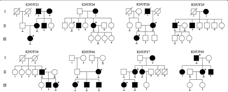
Eight small Korean families with hereditary hearing loss were recruited from the Department of Otorhinolaryngology-Head and Neck Surgery at the Kyungpook National University Hospital in Daegu, South Korea (Figure 1). A total of 31 individuals, including 18 affected and 13 unaffected members, participated in this study. A clinical questionnaire excluded any history of other diseases and environmental factors, including infec-tion, ototoxicity and noise. Physical examinations ruled out the probability of syndromic hearing loss. The hearing level in all of the participants was evalu-ated by audiological tests, including pure-tone audi-ometry (PTA) and auditory brainstem response (ABR). PTA was calculated as an average of the
threshold measured at 0.5, 1.0, 2.0 and 4.0 KHz [6], and air-conduction threshold measurements were performed at 125-8000 Hz. The level of hearing loss is described as follows depending on PTA: normal hearing, below 20 dB; mild hearing impairment, 21 to 40 dB; moderate hearing impairment, 41 to 70 dB; severe hearing impairment, 71 to 95 dB; and pro-found hearing impairment, above 95 dB. In addition, bone conduction thresholds were measured at 250-4000 Hz to check conductive hearing loss in affected individuals. One hundred unrelated Koreans with normal hearing were recruited from Kyungpook Na-tional University Hospital as normal controls. All of the participants provided written informed consent before the study according to the protocol approved by the Ethics Committee of Kyungpook National University Hospital. Genomic DNA from 31 indivi-duals, including 18 patients from 8 families and 100 normal controls, was extracted from peripheral blood or buccal cells using either the FlexiGene DNA extraction kit (QIAGEN, Hilden, Germany) or the Puregene Buccal Cell Core kit, respectively (QIAGEN, Hilden, Germany). All of the subjects were examined for theGJB2and theSLC26A4 gene by Sanger sequencing and were negative for both of the genes.
Targeted resequencing and variation analysis
Using previously published literature, we selected 80 genes that are associated with sensorineural hearing loss as the target genes for sequencing (Additional file 1: Table S1). Five microgram of genomic DNA was frag-mented to approximately 250 base pairs, which was
followed by end-repair, adenylation and adapter ligation for library generation using an Illumina sample prepar-ation kit (Illumina, Inc., San Diego, CA, USA). All protein-coding regions and exon-intron boundaries of the target genes were captured by hybridization with designed nucleotide probes. The captured target DNA fragments were sequenced using the Illumina HiSeq 2000 paired-end read sequencing system for 90 cycles per fragment.
All bioinformatics processing and data analysis, includ-ing genome alignment, variation detection, filterinclud-ing and visualization, were performed on the DNAnexus plat-form (DNAnexus, Inc., Mountain View, USA, http:// www.dnanexus.com). The millions of reads that were derived from the targeted sequencing were aligned to the human reference genome hg19 (University of California, Santa Cruz). dbSNP Build 134 was used as a reference for recorded SNPs. The 1000 genome database (http:// www.1000genomes.org) and the Washington University exome database (http://evs.gs.wathington.edu) were used as references to investigate the novelty and probable pathogenicity of the allelic variations detected in our se-quencing approach. The reliability of variations was esti-mated by the allele frequency and Ref score, which represents the PHRED-encoded probability (quality score) [7,8]. For a probability p, the PHRED-encoded quality score Q is given by the formula Q= -10log10p.
Variations with a quality score of less than 20 were regarded as sequencing errors and discarded, and only alleles that appeared in more than 20% of heterozygotes from 8 DNFA families were recognized as potentially real allelic variations. To analyze the possible functional pathogenic effects of the variants, 2 types of prediction programs, SIFT (http://sift.jcvi.org) and Mutation taster (http://www.mutationtaster.org), were used.
Sanger sequencing
Specific exons containing candidate variations were amplified by polymerase chain reaction (PCR) using primer sets designed via Primer3 software (http:// www-genome.wi.mit.edu/cgi-bin/primer/primer3_www. cgi). Each variation was sequenced by the Sanger se-quencing method using an ABI PRISM Big Dye Ter-minator Cycle Sequencing Kit v3.1 and an ABI 3130xl DNA sequencer (Applied Biosystems Corps., Foster City, CA, USA). Finally, the accurate genotype of the variations was confirmed by sequence analysis with the Sequencing Analysis software v5.2 (Applied Biosys-tems Corps., Foster City, CA, USA) in both the family members and 100 unrelated normal controls. To investi-gate the significance of the variations, the conservation level of amino acids between other species was esti-mated using a PhyloP score obtained from the UCSC genome browser (http://genome.ucsc.deu).
Results
Clinical features of the 8 Korean families
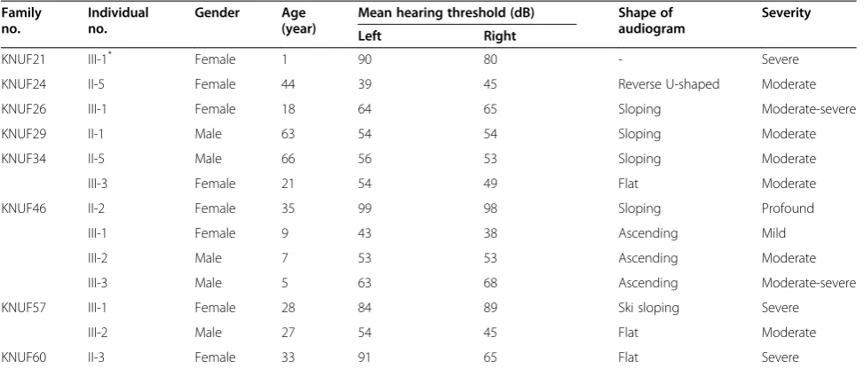
All 8 families exhibited a typical autosomal dominant in-heritance pattern of hearing loss (Figure 1). Audiological assessments of the affected individuals revealed symmet-rical, bilateral, progressive, sensorineural hearing loss that affected both genders (Additional file 2: Figure S1). Patient II-3 of KNUF60 showed asymmetric features (Table 1). According to PTA tests, the severity of hearing loss varied among affected individuals, ranging from mild to profound. The audiogram patterns from all of the patients were distinct, even between members of the same family. None of the affected individuals displayed symptoms of tinnitus, vestibular dysfunction or other clinical abnormalities indicating syndromic hearing loss.
Targeted resequencing and variation analysis
One (KNUF21) or two members per family were selected for targeted sequencing (Figure 1). For these 15 individuals, including 13 patients and 2 normal individuals, we screened 80 candidate deafness genes, including 46 reported hearing loss genes (Additional file 1: Table S1). Target enrichment and massively parallel sequencing using the Illumina HiSeq 2000 paired-end sequencing system produced hundreds of thousands of target mapped reads (tens of millions of bases) with an average length of 90 bases. This se-quencing covered more than 90% of the target regions with a mean depth ranging from 43× to 337×. More than 78% of the target regions were cov-ered by 10 or more reads, demonstrating the high quality of the sequencing (Table 2).
gov) and HAPMAP project (http://www.hapmap.org). This approach yielded an average of 12 novel non-synonymous variations located in coding regions in each individual (Additional file 3: Table S2). Most of these novel non-synonymous variations were mis-sense or nonmis-sense mutations caused by single nu-cleotide substitutions, and a few variations were frame-shift insertions or deletions of a single nucleotide. These variations were checked for co-segregation with hearing loss in the two family members who had been subjected to targeted sequencing, and 10 variations satis-fied this criterion.
Identification of candidate mutations of hearing loss in each family
Variant filtering was performed to identify the most probable causative variants, and 10 non-synonymous variants were found to be significant candidates in 7 families, with the exception of the KNUF26 family. A second co-segregation analysis was performed using the Sanger sequencing method in 14 additional individuals (underlined in Figure 1) from the 7 families, and 3 varia-tions that were not co-inherited with hearing loss were subsequently ruled out from the candidate mutations. Ultimately, 7 variations were confirmed for their
co-Table 2 Run statistics and target coverage of NGS in each individual
Family no. Individual no. On-target sequenced bases (bp)
Covered target region (bp)
Target coverage (%)
Mean depth (×)
Depth≥10×(%)
KNUF21 III -1 11,555,406 193,337 90.05 54 82.22
KNUF24 II -2 54,927,039 201,347 93.78 256 92.21
II-5 16,525,672 196,024 91.30 77 85.82
KNUF26 II-3 67,166,957 202,456 94.30 313 92.41
III-1 9,251,859 192,854 89.83 43 78.26
KNUF29 II-1 12,384,809 195,104 90.87 58 83.71
III-1 44,392,335 201,109 93.67 207 92.14
KNUF34 II-5 57,045,380 201,906 94.04 263 92.39
III-3 10,551,804 194,536 90.61 50 82.51
KNUF46 II-2 72,336,591 201,693 93.94 337 92.55
III-1 14,635,372 195,693 91.15 68 83.51
KNUF57 II-2 51,755,311 200,756 93.51 241 92.20
III-1 12,911,952 194,467 90.58 60 84.60
KNUF60 I-1 59,181,391 202,284 94.22 276 92.47
II-3 10,591,967 194,615 90.65 49 82.61
Table 1 Clinical features of patients in 8 Korean families carrying autosomal dominant hearing loss
Family no.
Individual no.
Gender Age (year)
Mean hearing threshold (dB) Shape of audiogram
Severity
Left Right
KNUF21 III-1* Female 1 90 80 - Severe
KNUF24 II-5 Female 44 39 45 Reverse U-shaped Moderate
KNUF26 III-1 Female 18 64 65 Sloping Moderate-severe
KNUF29 II-1 Male 63 54 54 Sloping Moderate
KNUF34 II-5 Male 66 56 53 Sloping Moderate
III-3 Female 21 54 49 Flat Moderate
KNUF46 II-2 Female 35 99 98 Sloping Profound
III-1 Female 9 43 38 Ascending Mild
III-2 Male 7 53 53 Ascending Moderate
III-3 Male 5 63 68 Ascending Moderate-severe
KNUF57 III-1 Female 28 84 89 Ski sloping Severe
III-2 Male 27 54 45 Flat Moderate
KNUF60 II-3 Female 33 91 65 Flat Severe
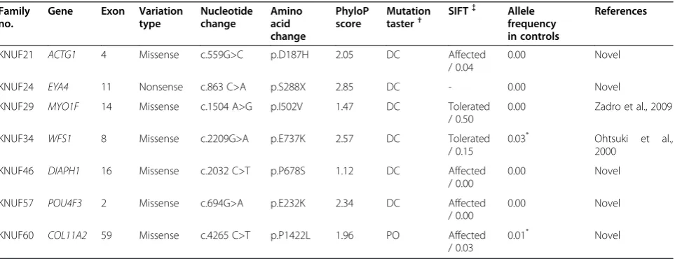
segregation with hearing loss in these families (Table 3). Although all of these variations were found in genes pre-viously known to be associated with hearing loss, 5 of the 7 variations were novel missense (p.D187H in ACTG1, p.P678S inDIAPH1, p.E232K inPOU4F3and p. P1422L in COL11A2) or nonsense mutations (p.S288X inEYA4) that had not been recorded in any public data-bases [12-18]. Sanger sequencing confirmed that all 7 variations in the 7 DFNA families were heterozygous in the patients with hearing loss, which was consistent with the inheritance pattern of the disorder.
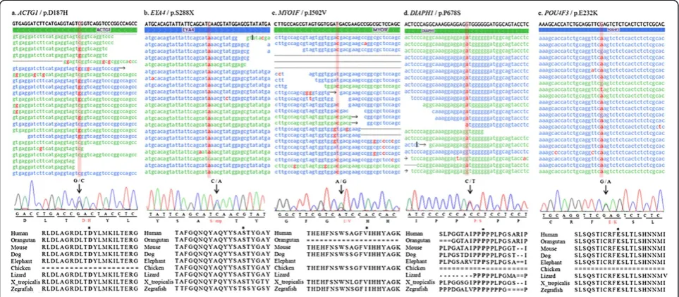
To estimate the probable pathogenicity of the variants, evolutionary conservation and the predicted damaging effects of the amino acid substitution were analyzed. In most of the variants, the amino acid where the substitu-tion occurred was highly conserved in a number of ver-tebrates showing positive PhyloP scores (Figure 2). In addition, two prediction programs, Mutation Taster and SIFT, strongly predicted that the variants would inter-rupt normal protein function (Table 3). However, one variant, proline at position 1422 in the COL11A2 gene, was not conserved between other species and was pre-dicted to be a probable polymorphism using Mutation Taster. Control sequencing of 100 unrelated normal individuals practically supported these predictions. How-ever, we excluded two variants, the p.E737K variant in WFS1 and the p.P1422L variant in COL11A2 from the candidate mutations, because these two variants were detected in 5 and 2 normal controls, respectively (Table 3). In addition, p.E737K in WFS1 was also detected in normal European American population (rs147834269) in the Washington University exome database (http://evs.gs.wathington.edu). Furthermore, it has been reported to be a non-pathogenic polymorphism
in several studies that provided strong evidence that this variant was not the genetic cause of hearing loss in the KNUF34 family [19,20]. In contrast, the other 5 variations (p.D187H inACTG1, p.S288X inEYA4, p.I502V inMYO1F, p.P678S inDIAPH1 and p.E232K inPOU4F3) were ab-sent in all 100 normal controls in this study. In addition, none of the healthy controls in the 1000 genomes project and the Washington University exome database carried these variants, suggesting that they could be pathogenic mutations.
The p.D187H variation in ACTG1was detected in pa-tient III-1 of the KNUF21 family and followed an auto-somal dominant inheritance pattern (Figure 2a). This variation was observed in 17 of 39 reads (44%) and had a quality score of 35, indicating heterozygosity. Genotype confirmation using Sanger sequencing revealed that two patients, II-2 and III-1, were heterozygous for this muta-tion. A normal individual, II-1, was homozygous for the wild type, consistent with the autosomal dominant in-heritance pattern of the disorder.
The p.S288X mutation in EYA4 was identified in the KNUF24 family (Figure 2b). In the targeted sequencing, the p.S288X mutation appeared in 45% of reads (21 of 47) and had a variation score of 35, indicating heterozygosity. Sanger sequencing revealed that this mutation is co-segregated with hearing loss in one patient (II-5) and was absent from two unaffected members (II-2 and III-2).
The KNUF29 family had dominant hearing loss that was confirmed to be caused by the p.I502V mutation in MYO1F (Figure 2c). The heterozygosity and reliability of this mutation was supported by a 35% variation frequency (17 of 46) and a variation score of 35. This result was con-firmed by Sanger sequencing, and only patient II-1 had the mutation, confirming its co-segregation with the disorder.
Table 3 Overview of 7 variants showing co-segregation with hearing loss in the families
Family no.
Gene Exon Variation type
Nucleotide change
Amino acid change
PhyloP score
Mutation taster†
SIFT{{ Allele frequency in controls
References
KNUF21 ACTG1 4 Missense c.559G>C p.D187H 2.05 DC Affected
/ 0.04
0.00 Novel
KNUF24 EYA4 11 Nonsense c.863 C>A p.S288X 2.85 DC - 0.00 Novel
KNUF29 MYO1F 14 Missense c.1504 A>G p.I502V 1.47 DC Tolerated
/ 0.50
0.00 Zadro et al., 2009
KNUF34 WFS1 8 Missense c.2209G>A p.E737K 2.57 DC Tolerated
/ 0.15
0.03* Ohtsuki et al.,
2000
KNUF46 DIAPH1 16 Missense c.2032 C>T p.P678S 1.12 DC Affected
/ 0.00
0.00 Novel
KNUF57 POU4F3 2 Missense c.694G>A p.E232K 2.34 DC Affected
/ 0.00
0.00 Novel
KNUF60 COL11A2 59 Missense c.4265 C>T p.P1422L 1.96 PO Affected
/ 0.03
0.01* Novel
* p.E737K inWFS1and p.P1422L inCOL11A2found in normal controls were eliminated from the final candidate mutations. The p.S288X variant inEYA4was not predicted by SIFT because it is a nonsense variant.
The KNUF46 family had low-to-mid-frequency non-syndromic hearing loss (Additional file 2: Figure S1). This type of hearing loss is uncommon and has been associated with only the DIAPH1, MYO7A and WFS1 genes [21]. Interestingly, we identified the missense mu-tation, p.P678S of DIAPH1 in this family (Figure 2d), and it showed coincident audiographical configuration with known DFNA1 (DIAPH1) hearing loss. This mis-sense mutation was observed in 53 of 121 reads, with a variation score of 40. Site-directed sequencing confirmed that only the hearing loss patients in this family (of 4 patients and 2 controls sequenced) were heterozygous for the mutation.
A novel missense mutation, p.E232K in POU4F3, was identified in the KNUF57 family (Figure 2e). Thirty-six percent of all reads had this mutation, with a variation score of 35 in the targeted sequencing. Five family members, including 4 patients, were sequenced for the mutation by Sanger sequencing, which verified co-segregation.
In summary, the targeted sequencing of 15 individuals from 8 DFNA families yielded 10 candidate non-synonymous mutations as the primary causes of hearing loss. Sanger sequencing was carried out in 31 indivi-duals, including 16 additional family members, to con-firm whether these 10 variations are co-segregated with hearing loss. This sequencing allowed us to confirm the
7 co-inherited variations. Finally, variation screening in 100 normal controls using Sanger sequencing confirmed 5 variations as probable pathogenic mutations in 5 of the families.
Discussion
The purpose of this study was to identify the genetic basis of hereditary hearing loss using next-generation se-quencing technology in small families who could not be analyzed using the current genetic approaches. Eight small families with autosomal dominant, non-syndromic, sensorineural hearing loss were selected, and 80 target genes associated with hearing loss were screened using target capture and massively parallel sequencing meth-ods. In current study, 5 non-synonymous mutations were confirmed in 5 of the 8 families. The causative genes underlying the hearing loss in the 3 other families are still awaiting discovery. There are 2 possible explana-tions for the hearing loss in these families: (1) patho-genic mutations exist in 1 of the 80 candidate genes studied but in an exon that was not covered by our se-quencing (approximately 6-10%) or within intronic regu-latory sequences, (2) their causative mutation is in an as-yet-unidentified hearing loss gene. However, 5 distinct causative mutations were identified in the other 5 fam-ilies, including 4 missense mutations and 1 nonsense mutation. None of these mutations, except the p.I502V Figure 2The identification of 5 non-synonymous variations showing co-segregation with hearing loss in affected families. Top:
mutation in MYO1F, has been identified in previous studies of hereditary hearing loss.
ACTG1 (NM_001199954), which is responsible for
DFNA20/26 hearing loss, encodes γ-actin, one of the non-muscle cytoskeletal proteins. This protein is pre-dominantly expressed in cochlear hair cells and contri-butes to the structural maintenance of stereocilia, cuticular plates and adherens junctions [22].γ-actin con-sists of 4 sub-domains (sub-domains 1-4), and the novel missense mutation p.D187H that is caused by a c.559 G > C transversion results in the substitution of a basic amino acid (histidine) for an acidic amino acid (aspartic acid) in sub-domain 4. Otterbein et al. found that even minor changes in this domain may lead to major effects on the structural stability of the actin poly-mer [23]. Using 2D gel electrophoresis, Verrills et al. identified that p.D187H in ACTG1 leads to the expres-sion of more basic gamma-actin in leukemia cells, and they reported that Asp187 is a surface residue in close proximity to the ATP-binding cleft of the protein. The substitution of the histidine for aspartic acid inACTG1 changes the charge from negative to positive, which results in reduced hydrophobicity and electrostatic inter-actions in this region. According to their research, mu-tant gamma-actin expressing leukemia cells display resistance to anti-microtubule drugs. The mutant gamma-actin expressing cells intactly retained their morphology, whereas the cells expressing wild-type gamma-actin were contracted and destroyed. These data suggest that the Asp187 residue in gamma-actin contri-butes to the interaction with microtubules, and the p.D187H variant inhibits depolymerization of tubulin in leukemia cells. This result suggests that the p.D187H vari-ant could collapse the polymerization-depolymerization balance of microtubules, which leads to the destruction of cellular homeostasis in normal hair cells [24]. These clusions from previous biochemical studies provide con-vincing evidence that p.D187H is a novel mutation that has pathogenic effects on the normal functions of γ-actin in the hair cells.
Mutation p.S288X in EYA4 (NM_004100.4) was the only nonsense mutation identified in this study. The protein encoded byEYA4(DFNA10) is a member of the vertebrate Eya family of transcriptional activators, and it consists of two functional domains: the C-terminal EYA homolog domain and the N-terminal transactivation do-main [13]. The EYA homolog dodo-main and SIX family transcription factors interact to form transcriptional complexes that regulate the expression of target genes that are required for the development and maturation of the organ of Corti [13]. Most of the reported EYA4 mutations produce truncated proteins missing a part of the EYA homolog domain [25]. The novel nonsense mutation p.S288X changed Ser288 to a stop
codon, which produced a truncated protein lacking the entire EYA homolog domain. It suggests that this nonsense mutation may inhibit normal development and maintenance of the organ of Corti and cause sen-sorineural hearing loss.
MYO1F(NM_012335.3) has been frequently proposed
as a candidate hearing loss gene, because several myosin genes have been demonstrated as causative genes of non-syndromic hearing loss andMYO1Fis expressed in cochlea. Recently, Zadro et al. reported that MYO1F mutations were identified in hearing loss patients, and one of the reported mutations, p.I502V, was detected in this study [14]. According to their study, the mutated residue, Ile502, is located near the actin-binding site in the motor domain of myosin-1 f. Through homology modeling, it was predicted that the Ile502 residue contri-butes to the structural stabilization of the protein by forming hydrophobic interactions with the Val444, Leu447 and Ile448 residues. Additionally, there is an ATP-binding site near Ile502. Therefore, the authors concluded that the substitution of isoleucine for valine may interrupt the hydrophobic interaction with other residues, resulting in structural instability of the protein and disturbance of ATP binding. Based on these predic-tions, it can be hypothesized that the p.I502V mutation likely has a pathogenic effect on the cellular function of myosin-1 F [14].
Human diaphanous 1 protein, encoded by DIAPH1 (NM_005219.4), belongs to the formin protein family, which regulates various cellular mechanisms such as cytoskeleton remodeling and the maintenance of cell po-larity in hair cells [16,26-29]. The formins have several functional domains, including formin homology-1 and 2 domains (FH1, FH2), which play a key role in the polymerization of unbranched actin filaments by inter-acting with profilin [26,30,31]. The FH1 domain is char-acterized by consecutive proline residues. Profilin-actin complexes bind to the poly-L-proline stretch of the FH1 domain and are assembled into unbranched actin fila-ments allowing barbed end elongation [32]. The novel mutation p.P678S is located in the poly-L-proline stretch of the FH1 domain. This mutation changes a hydropho-bic non-polar residue (proline) to a hydrophilic polar residue (serine) in the poly-L-proline stretch, and it will likely adversely affect actin polymerization at the barbed end. Therefore, it can be hypothesized that this mutation may disturb the interactions with partner proteins, which interrupts actin polymerization and collapses cel-lular polarity in hair cells of cochlea.
Transcription factors bind directly to DNA and regu-late expression of target genes. Brn-3.1, encoded by the
POU4F3 gene (NM_002700.2), is a well-known
is composed of two functional DNA-binding domains: a POU-specific domain and a POU-homeodomain. In the DNA binding domains, the molecular features of amino acids such as electric property and acidity significantly influence the maintenance of the structural conform-ation of the protein [34,35]. The p.E232K mutconform-ation detected in this study is caused by a single nucleotide substitution of adenine for guanine at nucleotide pos-ition 694, resulting in the replacement of a negatively charged glutamic acid with a positively charged lysine in an α-helix (α3) in the POU-specific domain. According to 3D modeling by Collin et al., among the 4 α-helix structures in the POU-specific domain, the α3 helix where the variant p.E232K occurs has the most direct interaction with target DNA [34]. Thus, altered electric properties of the Glu232 residue would directly reduce the structural stability of the domain or the strength of the interaction with the target DNA. It suggests that this missense mutation might have pathogenic effects on the maturation and survival of hair cells by failing to regu-late the expression of downstream genes.
Although the pedigrees have insufficient genetic infor-mation, various probable pathogenic mutations were successfully detected by NGS technique and molecular genetic analysis in current study. Moreover, it will com-pletely overcome the weakness of this study ensuring pathogenicity of the mutations when segregation study for the detected mutations can be performed with all of the members in the family.
According to a number of previous genetic studies, her-editary hearing loss shows distinct spectrums and preva-lence of mutations in different ethnic groups [36-40]. For example, the GJB2 gene accounts for approximately 20-40% of genetic hearing loss in Caucasian popula-tions but only 10% of genetic hearing loss in Korean populations [37,38,41-43]. In addition, the most fre-quent GJB2 mutation also varies among populations: c.35delG, c.235delC and c.167delT are the most com-mon variants in Caucasian, Asian and Jewish popula-tions, respectively [38,44-46]. However, this feature may not be applicable for autosomal dominant hear-ing loss because major hearhear-ing loss genes such as
GJB2 and SLC26A4 predominantly cause autosomal
recessive hearing loss. To date, no major genes have been described that cause a significant proportion of dominant hearing loss in any ethnic population. In this study, 5 mutations were detected in 5 different genes in 5 Korean families with autosomal dominant, non-syndromic, sensorineural hearing loss. Addition-ally, we provide the first evidence of pathogenic muta-tions in theACTG1, EYA4, DIAPH1andMYO1Fgenes in a Korean population. These results, as well as previous genetic studies performed by our group, suggest that there are no mutational hot spots for dominant hearing loss in
the Korean population [33,47-50]. This conclusion is con-sistent with the idea that the genetic causes of autosomal dominant hearing loss are more heterogeneous than those of autosomal recessive hearing loss in most ethnic groups.
Because of these characteristics, it is difficult to diag-nose and establish the exact causes of hereditary hearing loss, although it accounts for approximately 50% of all hearing loss [51]. Currently, several simple DNA tests are performed in medical institutions in many countries to diagnose hereditary hearing loss. However, the test evaluates only few major genes, such asGJB2, SLC26A4 and mitochondrial genes, and is unable to detect other genetic causes of hereditary hearing loss. Considering that the ultimate goal of genetic disease research is to establish basic information and genetic databases for clinical diagnosis and treatment, the use of population genetic studies to accurately understand the genetic background of diseases is essential. Although current genetic technologies, such as linkage analysis and Sanger sequencing, are very reliable methods for identifying genomic variations associated with genetic disorders, they are not well suited for the analysis of heterogeneous diseases. Next-generation sequencing (NGS) is acceler-ating the qualitative improvement of mutational studies for numerous heterogeneous disorders due to its ability to perform simultaneous and massively parallel se-quencing. Although the current NGS technique is too expensive to be widely used, the cost of NGS is grad-ually decreasing, which will lead to the increased ap-plicability of this technology. Two recent population genetic studies on hereditary hearing loss have been performed in American and Jewish populations [52,53]. In these studies, various genetic mutations and a founder mutation were detected using next-generation sequencing. Our study is the first report of dominant hearing loss causative gene mutations identified by the targeted-sequencing of affected individuals in an East Asian population. Our successful identification of several pathogenic mutations using target-capture and massively parallel sequencing demonstrates that gene targeted se-quencing is a highly effective and powerful tool for clin-ical and population genetic studies of heterogeneous disorders.
Additional files
Additional file 1: Table S1.Eighty genes targeted for the next-generation sequencing.
gene which causes low frequency hearing loss. It provides a strong possibility that p.P678S is the pathogenic mutation causing hereditary hearing loss in family KNUF46.
Additional file 3: Table S2.Summary of genetic variations detected in targeted sequencing.
Competing interests
The authors declare that they have no competing interests.
Authors’contributions
JI participated in the selection of target genes, performed bioinformatic interpretation of the NGS data, confirmed the NGS data through Sanger sequencing and has drafted the manuscript. SK interpreted the NGS data and confirmed the NGS data by Sanger sequencing. DB extracted genomic DNA of all participants from their whole blood and performed bioinformatic interpretation of the NGS data. SY participated in the selection of target genes and predicted probable pathogenic effects of the mutations by several software programs. UK has made general study design and wrote the manuscript. KY has directed all clinical parts including clinical evaluation and hearing test, has made study design. SH clinically investigated the patients and collected genetic information of all families participated in this study. All authors read and approved the final manuscript.
Acknowledgements
We are grateful to the families for their collaboration in this study. This research was supported by the Basic Science Research Program through the National Research Foundation of Korea (NRF) funded by the Ministry of Education, Science and Technology (2011-0009674).
Received: 30 March 2012 Accepted: 21 August 2012 Published: 3 September 2012
References
1. Mardis ER:The impact of next-generation sequencing technology on genetics.Trends Genet: TIG2008,24(3):133–141.
2. Metzker ML:Sequencing technologies - the next generation.Nat Rev Genet2010,11(1):31–46.
3. Shendure J, Ji H:Next-generation DNA sequencing.Nat Biotechnol2008, 26(10):1135–1145.
4. Shendure J, Mitra RD, Varma C, Church GM:Advanced sequencing technologies: methods and goals.Nat Rev Genet2004,5(5):335–344. 5. Walsh T, Shahin H, Elkan-Miller T, Lee MK, Thornton AM, Roeb W, Abu Rayyan A,
Loulus S, Avraham KB, King MC,et al:Whole exome sequencing and homozygosity mapping identify mutation in the cell polarity protein GPSM2 as the cause of nonsyndromic hearing loss DFNB82.Am J Hum Genet2010,87 (1):90–94.
6. Liu X, Xu L:Nonsyndromic hearing loss: an analysis of audiograms.Ann Otol Rhinol Laryngol1994,103(6):428–433.
7. Ewing B, Green P:Base-calling of automated sequencer traces using phred. II. Error probabilities.Genome Res1998,8(3):186–194. 8. Ewing B, Hillier L, Wendl MC, Green P:Base-calling of automated
sequencer traces using phred. I. Accuracy assessment.Genome Res1998, 8(3):175–185.
9. Doi H, Yoshida K, Yasuda T, Fukuda M, Fukuda Y, Morita H, Ikeda S, Kato R, Tsurusaki Y, Miyake N,et al:Exome Sequencing Reveals a Homozygous SYT14 Mutation in Adult-Onset, Autosomal-Recessive Spinocerebellar Ataxia with Psychomotor Retardation.Am J Hum Genet2011,89(2):320–327.
10. Gilissen C, Arts HH, Hoischen A, Spruijt L, Mans DA, Arts P, van Lier B, Steehouwer M, van Reeuwijk J, Kant SG,et al:Exome sequencing identifies WDR35 variants involved in Sensenbrenner syndrome.Am J Hum Genet
2010,87(3):418–423.
11. Otto EA, Hurd TW, Airik R, Chaki M, Zhou W, Stoetzel C, Patil SB, Levy S, Ghosh AK, Murga-Zamalloa CA,et al:Candidate exome capture identifies mutation of SDCCAG8 as the cause of a retinal-renal ciliopathy.Nat Genet2010,42(10):840–850.
12. Morell RJ, Friderici KH, Wei S, Elfenbein JL, Friedman TB, Fisher RA:A new locus for late-onset, progressive, hereditary hearing loss DFNA20 maps to 17q25.Genomics2000,63(1):1–6.
13. Wayne S, Robertson NG, DeClau F, Chen N, Verhoeven K, Prasad S, Tranebjarg L, Morton CC, Ryan AF, Van Camp G,et al:Mutations in the transcriptional activator EYA4 cause late-onset deafness at the DFNA10 locus.Hum Mol Genet2001,10(3):195–200.
14. Zadro C, Alemanno MS, Bellacchio E, Ficarella R, Donaudy F, Melchionda S, Zelante L, Rabionet R, Hilgert N, Estivill X,et al:Are MYO1C and MYO1F associated with hearing loss?Biochim Biophys Acta2009,1792(1):27–32. 15. Young TL, Ives E, Lynch E, Person R, Snook S, MacLaren L, Cater T, Griffin A,
Fernandez B, Lee MK,et al:Non-syndromic progressive hearing loss DFNA38 is caused by heterozygous missense mutation in the Wolfram syndrome gene WFS1.Hum Mol Genet2001,10(22):2509–2514.
16. Lynch ED, Lee MK, Morrow JE, Welcsh PL, Leon PE, King MC:Nonsyndromic deafness DFNA1 associated with mutation of a human homolog of the Drosophila gene diaphanous.Science1997,278(5341):1315–1318. 17. Vahava O, Morell R, Lynch ED, Weiss S, Kagan ME, Ahituv N, Morrow JE, Lee MK,
Skvorak AB, Morton CC,et al:Mutation in transcription factor POU4F3 associated with inherited progressive hearing loss in humans.Science1998, 279(5358):1950–1954.
18. McGuirt WT, Prasad SD, Griffith AJ, Kunst HP, Green GE, Shpargel KB, Runge C, Huybrechts C, Mueller RF, Lynch E,et al:Mutations in COL11A2 cause non-syndromic hearing loss (DFNA13).Nat Genet1999,23(4):413–419. 19. Gomez-Zaera M, Strom TM, Rodriguez B, Estivill X, Meitinger T, Nunes V:
Presence of a major WFS1 mutation in Spanish Wolfram syndrome pedigrees.Mol Genet Metab2001,72(1):72–81.
20. Ohtsuki T, Ishiguro H, Yoshikawa T, Arinami T:WFS1 gene mutation search in depressive patients: detection of five missense polymorphisms but no association with depression or bipolar affective disorder.J Affect Disord
2000,58(1):11–17.
21. Bramhall NF, Kallman JC, Verrall AM, Street VA:A novel WFS1 mutation in a family with dominant low frequency sensorineural hearing loss with normal VEMP and EcochG findings.BMC Med Genet2008,9:48.
22. Zhu M, Yang T, Wei S, DeWan AT, Morell RJ, Elfenbein JL, Fisher RA, Leal SM, Smith RJ, Friderici KH:Mutations in the gamma-actin gene (ACTG1) are associated with dominant progressive deafness (DFNA20/26).Am J Hum Genet2003,73(5):1082–1091.
23. Otterbein LR, Graceffa P, Dominguez R:The crystal structure of uncomplexed actin in the ADP state.Science2001,293(5530):708–711. 24. Kavallaris M, Verrills NM, Po'uha ST, Liu MLM, Liaw TYE, Larsen MR, Ivery MT,
Marshall GM, Gunning PW:Alterations in gamma-actin and tubulin-targeted drug resistance in childhood leukemia.J Natl Cancer I2006,98 (19):1363–1374.
25. Makishima T, Madeo AC, Brewer CC, Zalewski CK, Butman JA, Sachdev V, Arai AE, Holbrook BM, Rosing DR, Griffith AJ:Nonsyndromic hearing loss DFNA10 and a novel mutation of EYA4: evidence for correlation of normal cardiac phenotype with truncating mutations of the Eya domain. Am J Med Genet Part A2007,143A(14):1592–1598.
26. Wallar BJ, Alberts AS:The formins: active scaffolds that remodel the cytoskeleton.Trends Cell Biol2003,13(8):435–446.
27. Krebs A, Rothkegel M, Klar M, Jockusch BM:Characterization of functional domains of mDia1, a link between the small GTPase Rho and the actin cytoskeleton.J Cell Sci2001,114(Pt 20):3663–3672.
28. Chesarone MA, DuPage AG, Goode BL:Unleashing formins to remodel the actin and microtubule cytoskeletons.Nat Rev Mol Cell Biol2010,11(1):62–74.
29. Lalwani AK, Jackler RK, Sweetow RW, Lynch ED, Raventos H, Morrow J, King MC, Leon PE:Further characterization of the DFNA1 audiovestibular phenotype. Archives of otolaryngology–head.Neck Surg1998,124(6):699–702. 30. Mattila PK, Lappalainen P:Filopodia: molecular architecture and cellular
functions.Nat Rev2008,9(6):446–454.
31. Paul AS, Pollard TD:The role of the FH1 domain and profilin in formin-mediated actin-filament elongation and nucleation.Curr Biol2008,18 (1):9–19.
32. Kovar DR:Molecular details of formin-mediated actin assembly.Curr Opin Cell Biol2006,18(1):11–17.
33. Lee HK, Park HJ, Lee KY, Park R, Kim UK:A novel frameshift mutation of POU4F3 gene associated with autosomal dominant non-syndromic hearing loss.Biochem Biophys Res Commun2010,396(3):626–630. 34. Collin RW, Chellappa R, Pauw RJ, Vriend G, Oostrik J, van Drunen W, Huygen PL,
35. Sturm RA, Herr W:The POU domain is a bipartite DNA-binding structure. Nature1988,336:601–604.
36. Yuan Y, You Y, Huang D, Cui J, Wang Y, Wang Q, Yu F, Kang D, Yuan H, Han D,
et al:Comprehensive molecular etiology analysis of nonsyndromic hearing impairment from typical areas in China.J Transl Med2009,7:79.
37. Ouyang XM, Yan D, Yuan HJ, Pu D, Du LL, Han DY, Liu XZ:The genetic bases for non-syndromic hearing loss among Chinese.J Hum Genet2009, 54(3):131–140.
38. Ito T, Noguchi Y, Yashima T, Ohno K, Kitamura K:Hereditary hearing loss and deafness genes in Japan.J Med Dent Sci2010,57(1):1–10. 39. Brownstein Z, Avraham KB:Deafness genes in Israel: implications for
diagnostics in the clinic.Pediatr Res2009,66(2):128–134.
40. Mahdieh N, Rabbani B, Wiley S, Akbari MT, Zeinali S:Genetic causes of nonsyndromic hearing loss in Iran in comparison with other populations.J Hum Genet2010,55(10):639–648.
41. Green GE, Scott DA, McDonald JM, Woodworth GG, Sheffield VC, Smith RJ: Carrier rates in the midwestern United States for GJB2 mutations causing inherited deafness.JAMA: J Am Med Assoc1999,281(23):2211–2216. 42. Khidiiatova IM, Dzhemileva LU, Khabibulin RM, Khusnutdinova EK:
Frequency of the 35delG mutation of the connexin 26 gene (GJB2) in patients with non-syndromic autosome-recessive deafness from Bashkortostan and in ethnic groups of the Volga-Ural region. Molekuliarnaia biologiia2002,36(3):438–441.
43. Lee KY, Choi SY, Bae JW, Kim S, Chung KW, Drayna D, Kim UK, Lee SH: Molecular analysis of the GJB2, GJB6 and SLC26A4 genes in Korean deafness patients.Int J Pediatr Otorhinolaryngol2008,72(9):1301–1309. 44. Choi SY, Lee KY, Kim HJ, Kim HK, Chang Q, Park HJ, Jeon CJ, Lin X, Bok J,
Kim UK:Functional evaluation of GJB2 variants in nonsyndromic hearing loss.Mol Med2011,17(5–6):550–556.
45. Morell RJ, Kim HJ, Hood LJ, Goforth L, Friderici K, Fisher R, Van Camp G, Berlin CI, Oddoux C, Ostrer H,et al:Mutations in the connexin 26 gene (GJB2) among Ashkenazi Jews with nonsyndromic recessive deafness. N Engl J Med1998,339(21):1500–1505.
46. Sobe T, Vreugde S, Shahin H, Berlin M, Davis N, Kanaan M, Yaron Y, Orr-Urtreger A, Frydman M, Shohat M,et al:The prevalence and expression of inherited connexin 26 mutations associated with nonsyndromic hearing loss in the Israeli population.Hum Genet2000,106(1):50–57.
47. Sagong B, Park R, Kim YH, Lee KY, Baek JI, Cho HJ, Cho IJ, Kim UK, Lee SH: Two novel missense mutations in the TECTA gene in Korean families with autosomal dominant nonsyndromic hearing loss.Ann Clin Lab Sci
2010,40(4):380–385.
48. Baek JI, Park HJ, Park K, Choi SJ, Lee KY, Yi JH, Friedman TB, Drayna D, Shin KS, Kim UK:Pathogenic effects of a novel mutation (c.664_681del) in KCNQ4 channels associated with auditory pathology.Biochim Biophys Acta2011,1812(4):536–543.
49. Baek JI, Cho HJ, Choi SJ, Kim LS, Zhao C, Sagong BR, Kim UK, Jeong SW:The Trp117Arg mutation of the COCH gene causes deafness in Koreans. Clin Genet2010,77(4):399–403.
50. Park HJ, Cho HJ, Baek JI, Ben-Yosef T, Kwon TJ, Griffith AJ, Kim UK:Evidence for a founder mutation causing DFNA5 hearing loss in East Asians. J Hum Genet2010,55(1):59–62.
51. Denoyelle F, Weil D, Maw MA, Wilcox SA, Lench NJ, Allen-Powell DR, Osborn AH, Dahl HH, Middleton A, Houseman MJ,et al:Prelingual deafness: high prevalence of a 30delG mutation in the connexin 26 gene.Hum Mol Genet1997,6(12):2173–2177.
52. Brownstein Z, Friedman LM, Shahin H, Oron-Karni V, Kol N, Rayyan AA, Parzefall T, Lev D, Shalev S, Frydman M,et al:Targeted genomic capture and massively parallel sequencing to identify genes for hereditary hearing loss in Middle Eastern families.Genome Biol2011,12(9):R89. 53. Shearer AE, DeLuca AP, Hildebrand MS, Taylor KR, Gurrola J 2nd, Scherer S,
Scheetz TE, Smith RJ:Comprehensive genetic testing for hereditary hearing loss using massively parallel sequencing.Proc Natl Acad Sci U S A
2010,107(49):21104–21109.
doi:10.1186/1750-1172-7-60
Cite this article as:Baeket al.:Targeted massive parallel sequencing: the effective detection of novel causative mutations associated with hearing
loss in small families.Orphanet Journal of Rare Diseases20127:60.
Submit your next manuscript to BioMed Central and take full advantage of:
• Convenient online submission
• Thorough peer review
• No space constraints or color figure charges
• Immediate publication on acceptance
• Inclusion in PubMed, CAS, Scopus and Google Scholar
• Research which is freely available for redistribution