R E S E A R C H
Open Access
Updated Oxford classification and the
international study of kidney disease in
children classification: application in
predicting outcome of Henoch-Schönlein
purpura nephritis
Xiaohan Huang
1,2,3,4, Lili Ma
1,5, Pingping Ren
1,2,3,4, Hongya Wang
1,2,3,4, Liangliang Chen
1,2,3,4, Haidongqin Han
1,2,3,4,
Jianghua Chen
1,2,3,4and Fei Han
1,2,3,4*Abstract
Background:Henoch-Schönlein purpura nephritis (HSPN) shares many similarities with IgA nephropathy. We aimed to analyze the predictive value of the International Study of Kidney Disease in Children (ISKDC) classification and the updated Oxford classification for IgA nephropathy in HSPN patients.
Methods:Data of 275 HSPN patients (aged≥14 years) were retrieved, and all of them underwent a renal biopsy. We re-classified the biopsies according to the ISKDC classification and the updated Oxford classification to analyze their correlations with clinical features and renal outcomes. The renal endpoints were defined as≥30% reduction in baseline estimated glomerular filtration rate (eGFR) in 2 years, doubling of serum creatinine (Scr) or end stage renal disease.
Results:During follow-up period of 56(30,86) months, 30(10.9%) patients reached renal endpoints. Segmental sclerosis was the only pathological feature independently associated with renal endpoints (HR 4.086, 95%CI 1.111– 15.026,P= 0.034). Tubular atrophy/ interstitial fibrosis was associated with eGFR and Scr levels, and its correlation with renal endpoints was found by univariate analysis. Endocapillary hypercellularity was associated with 24 h urine protein and is of prognostic value in univariate analysis. Mesangial hypercellularity was not associated with clinical features or renal endpoints. Crescents were associated with 24 h urine protein, Scr and eGFR levels, but both ISKDC and updated Oxford classifications of crescents were not associated with renal endpoints by multivariate analysis. Conclusions:The updated Oxford classification can help in disease management and renal outcome prediction of HSPN.
Keywords:IgA vasculitis, Nephritis, Renal biopsy, Crescent, Outcomes
© The Author(s). 2019Open AccessThis article is distributed under the terms of the Creative Commons Attribution 4.0 International License (http://creativecommons.org/licenses/by/4.0/), which permits unrestricted use, distribution, and reproduction in any medium, provided you give appropriate credit to the original author(s) and the source, provide a link to the Creative Commons license, and indicate if changes were made. The Creative Commons Public Domain Dedication waiver (http://creativecommons.org/publicdomain/zero/1.0/) applies to the data made available in this article, unless otherwise stated.
* Correspondence:[email protected]
1Kidney Disease Center, The First Affiliated Hospital, College of Medicine,
Zhejiang University, 79 Qingchun Road, Hangzhou, Zhejiang Province 310003, People’s Republic of China
2Institute of Nephrology, Zhejiang University, Hangzhou, Zhejiang Province,
China
Background
Henoch-Schönlein purpura (HSP), which also known as IgA vasculitis (IgAV), is an immunoglobulin A (IgA)-me-diated disease characterized by a generalized vasculitis mainly involving the skin, joints, gastrointestinal tract
and kidneys [1]. IgAV nephritis (IgAVN) or
Henoch-Schönlein purpura nephritis (HSPN) is the kid-ney damage caused by IgAV. It has been reported that 7–23% of children and 10–27% of adults with HSPN would progress to end stage renal disease (ESRD) [2–5]. For HSP patients, a major factor affecting the long-term outcome is the severity of renal involvement [6], and it has been suggested that some pathologic features may have values in predicting the outcomes of HSPN [7].
Pathologic features of HSPN is usually graded by the International Study of Kidney Disease in Children (ISKDC) classification, mainly according to the existence and the number of crescents [8]. However, the value of crescents in predicting long-term outcome of HSPN is still lack of consensus. IgA nephropathy shares many similarities with HSPN in clinical, immunological and histological features [9]. In some studies, IgA nephropa-thy and HSPN were regarded as different manifestations of a single disease [10,11]. Therefore, the Oxford classi-fication of IgA nephropathy may also be used to group pathologic features of HSPN. The updated Oxford classi-fication uses mesangial hypercellularity (M), endocapil-lary hypercellularity (E), segmental sclerosis (S), tubular atrophy/ interstitial fibrosis (T) and crescents (C) to evaluate renal biopsies [12]. A study published in 2014 suggested that the original Oxford classification was of prognostic value in HSPN [13],while another study in 2018 suggested that updated oxford classification can be used to assess renal outcomes of HSPN in children [14]. Here, we evaluated the utility of both the ISKDC classifi-cation and the updated Oxford classificlassifi-cation as a pre-dictor of renal outcome by retrospectively reviewing a larger cohort (275 patients, aged≥14 years)followed up in our center.
Methods
Patients
Data of 292 HSPN patients diagnosed by the Kidney Disease Center of the First Affiliated Hospital of Zhe-jiang University between August 2004 and November 2015 were retrieved. Criteria for a renal biopsy was HSPN patients who had active urinary sediments (pro-teinuria or hematuria) with or without impairment of glomerular filtration rate. Inclusive criteria included: aged ≥14 years; a history of palpable purpuric eruption, with or without involvement of gastrointestinal tract or joints; biopsy-proven HSPN. Exclusion criteria was as follows: inadequate number of glomeruli (< 8) in biopsy, concomitant with cirrhotic liver disease, IgA-dominant
infection associated glomerulonephritis, human
im-munodeficiency virus associated IgA nephropathy,
anti-neutrophil cytoplasmic antibody associated vascu-litis, hepatitis B virus associated nephritis, idiopathic thrombocytopenic purpura, systemic lupus erythemato-sus and other autoimmune diseases. Levels of antinu-clear antibody (ANA), anti-neutrophil cytoplasmic antibody (ANCA), anti-glomerular basement antibody together with complements were collected to exclude autoimmune disease.
Of these 292 patients, 11 patients were lost to follow-up, and another 6 patients were excluded because of biopsy inadequacy (< 8 glomeruli), the remaining 275 patients were the basis of our study. The study protocols conformed to the provisions of the Declaration of Helsinki. The Research Ethics Committee of the First Affiliated Hospital, College of Medicine, Zhejiang Uni-versity approved the protocols (Reference number: 2017678).
Clinical data collection
Medical records of the patients were reviewed to obtain clinical data at the time of biopsy and during the course of follow-up. The data included age, gender, blood pres-sure, urine protein excretion based on 24-h urine collec-tion, serum creatinine (Scr) and estimated glomerular filtration rate (eGFR). Also, the use of immunosuppres-sant drugs was assessed. The mean arterial pressure (MAP) was calculated by the equation MAP = (2 × dia-stolic pressure + sydia-stolic pressure)/3, while the eGFR was calculated using the Chronic Kidney Disease Epidemi-ology Collaboration (CKD-EPI) formula.
Pathologic data collection
(C2). Group T1 and T2 were merged because there were only 2 patients scored T2. One experienced renal path-ologist who was blinded to the case histories evaluated all renal pathology.
Definitions
The primary renal endpoint was defined as≥30% reduc-tion of eGFR from baseline in 2 years, doubling of Scr or ESRD. ESRD was defined by reaching an eGFR of <15 ml/min/1.72m2 or requiring maintaining renal replace-ment therapy for more than 3 months.
Statistical analysis
Statistical analysis was performed by IBM SPSS 22.0 (IBM, Chicago, IL, USA). Normal distributed data were expressed as mean ± standard deviation, and compari-sons were tested by Student’s t test. Non-normal distrib-uted data were expressed as median(interquartile range), and compared by the Mann-Whitney U-test. Chi-square
χ2
test, Fisher’s exact test and non-parametric test were used to compare categorical variables. We used one-way ANOVA and R x C contingency table for comparison of three groups. Student-Newman-Keuls test was used to further compare between two groups. To evaluate the association between the classifications and renal end-point, we used univariate analysis and multivariate Cox regression model, defining the day of renal biopsy as starting point, and the result was presented with hazard ratios (HR).P< 0.05 was considered significant.
Results
Demographic and clinical features
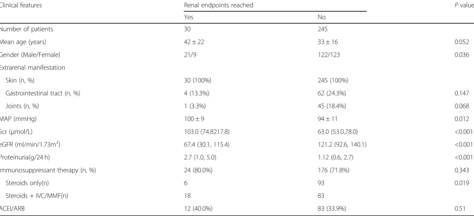
The study sample comprised 275 HSPN patients. At the time of biopsy, their mean age was 33 ± 17 years, and 52.0% were male. All of the patients were the Han popula-tion. During a median follow-up of 56(30,86) months, 30(10.9%) patients reached renal endpoints including more than 30% decrease in baseline eGFR in 2 years (11 patients), doubling in Scr (7 patients) and ESRD (12 pa-tients). Comparations between patients with or without renal endpoints were shown in Table1. There were no sig-nificant differences in age and extrarenal manifestations, but patients reaching endpoints had a higher male propor-tion, higher average MAP (100 ± 9 mmHg vs 94 ± 11 mmHg, p= 0.012), higher Scr level [103.0 (74.8217.8)
μmol/L vs 63.0 (53.0,78.0) μmol/L, p< 0.001] and lower eGFR level [67.4 (30.1, 115.4) ml/min/1.73m2 vs 121.2 (92.6, 140.1)ml/min/1.73m2,p< 0.001], and more protein-uria [2.7 (1.0, 5.0) g/24 h vs 1.12 (0.6, 2.7) g/24 h, p< 0.001]. Angiotensin converting enzyme inhibitor (ACEI) / angiotensin II receptor blocker (ARB) and immunosup-pressants were prescribed in our patients. A percentage of 72.7% of patients received immunosuppressive therapy; the percentage was same in patients with or without renal endpoints. However, patients with endpoints received a higher proportion of steroids combining with intravenous cyclophosphamide or mycophenolate mofetil (18/24 vs 83/176,p= 0.019), probably correlating with a higher se-verity of the disease.
The detailed pathologic features (including the severity of mesangial proliferation, the proportion of glomeruli
Table 1Clinical features for patients with or without renal endpoints
Clinical features Renal endpoints reached Pvalue
Yes No
Number of patients 30 245
Mean age (years) 42 ± 22 33 ± 16 0.052
Gender (Male/Female) 21/9 122/123 0.036
Extrarenal manifestation
Skin (n, %) 30 (100%) 245 (100%)
Gastrointestinal tract (n, %) 4 (13.3%) 62 (24.3%) 0.147
Joints (n, %) 1 (3.3%) 45 (18.4%) 0.068
MAP (mmHg) 100 ± 9 94 ± 11 0.012
Scr (μmol/L) 103.0 (74.8217.8) 63.0 (53.0,78.0) <0.001
eGFR (ml/min/1.73m2
) 67.4 (30.1, 115.4) 121.2 (92.6, 140.1) <0.001
Proteinuria(g/24 h) 2.7 (1.0, 5.0) 1.12 (0.6, 2.7) <0.001
Immunosuppressant therapy (n, %) 24 (80.0%) 176 (71.8%) 0.343
Steroids only(n) 6 93 0.019
Steroids + IVC/MMF(n) 18 83
ACEI/ARB 12 (40.0%) 83 (33.9%) 0.51
The data were expressed as mean ± s.d. or as numbers(percentage)
Results of Scr, eGFR, proteinuria were expressed as median (interquartile range)
with each pathologic lesion, and the proportion of intersti-tial inflammation) in patients with or without renal end-points were shown in Table 2. A greater proportion of glomeruli with crescents (p< 0.001), and higher percent-ages of tubular atrophy (p= 0.013) and tubulointerstitial inflammation (P= 0.004) were observed in patients reach-ing renal endpoints. Immunofluorescence deposition fea-ture is provided in Additiobnal file1: Table S1
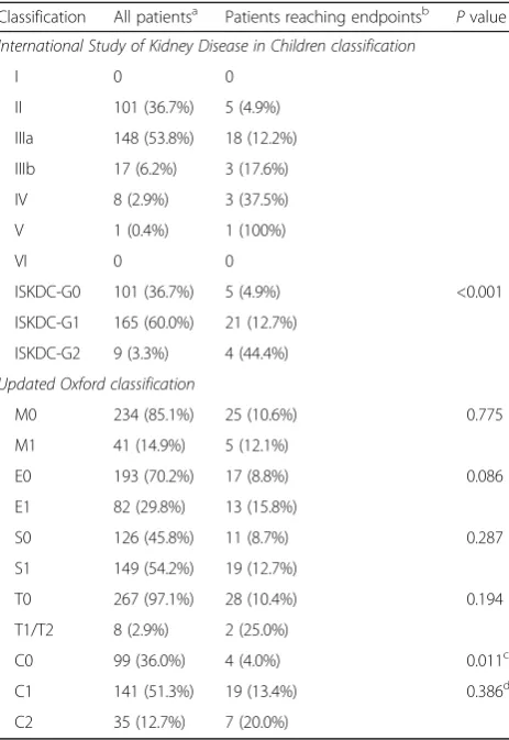
Distribution of the patients categorized by the ISKDC classification and the updated Oxford classification was presented in Table 3. There were 101(36.7%) patients graded ISKDC II, 148(53.8%) patients graded IIIa, 17(6.2%) patients graded IIIb, 8(2.9%) patients graded IV, and only 1(0.4%) patients graded V. No patient was graded I or VI. By the updated Oxford classification, M1, E1, S1, T1/T2 were observed in 41(14.9%), 82(29.8%), 149(54.2%), 8(2.9%) patients respectively. Additionally, there were only 6 pa-tients graded T1, and 2 papa-tients graded T2, so we merged them in a group (T1/T2) for analysis. With regards to cres-cents, 141(51.3%) patients scored C1 and 35(12.7%) patients scored C2. For renal outcomes, the percentage of patients reaching renal endpoints increased with the increasing grade from ISKDC-G0, G1 to G2 (4.9, 12.7, 44.4% respect-ively,p< 0.001), and those in M1, E1, S1, T1/T2, C1 or C2 groups of the updated Oxford classification were 12.1, 15.8, 12.7, 25, 13.4% or 20%, respectively.
Correlations between clinical data and pathologic classifications
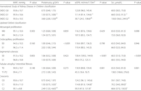
Correlations between clinical data and pathologic classifications were shown in Table 4. According to
the ISKDC classification, with the increase of classifi-cation from ISKDC G0, G1 to G2, patients had more daily urine protein (p< 0.05), higher Scr level (p< 0.05) and lower eGFR level (p< 0.05). According to the updated Oxford classification, no significant dif-ference in MAP, proteinuria, eGFR and Scr levels was found between M0 and M1 group; patients in E1 group had more daily urine protein than E0 group (p < 0.001); patients in S1 group had higher MAP level (p= 0.003), higher Scr level (p< 0.001) and lower eGFR level (p < 0.001) than those in S0 group; pa-tients in T1/T2 group had higher Scr level (p= 0.001) and lower eGFR level (p = 0.001) than those in T0 group; both patients in C1 group (Crescents 0–25%) and C2 group (Crescents ≥25%) had more daily urine protein (p< 0.05), higher Scr level (p< 0.05) and lower eGFR level (p< 0.05) than those in C0 group; further-more, patients in C2 group have more proteinuria than C1 group.
Correlations between outcomes and pathologic classifications
Survival analysis was shown in Table5. By the univariate Cox regression analysis, E1(P= 0.038), S1(p= 0.005), T1/ T2(p < 0.001) and C1/C2(p = 0.005) were risk factors for renal endpoints. In multivariate model, variants includ-ing age, 24-h proteinuria, eGFR, and pathologic features (E1, S1, T1/T2 and C1/C2) were analyzed; and S1 lesion (HR = 4.086 95%CI = 1.111–15.026, p= 0.034) and more proteinuria (HR = 1.191, 95%CI = 1.012–1.401,p= 0.035) were independently risk factors for renal endpoints.
Table 2Detailed pathologic features for patients with or without renal endpoints
Pathologic features Renal endpoints reached Pvalue
Yes No
Mesangial hypercellularitya
, n (%) 0.238
Normal 0 2 (0.4)
Mild 26 (86.7) 207 (84.5)
Moderate 3 (10.0) 35 (14.3)
Severe 1 (3.3) 1 (0.4)
tubulointerstitial inflammationa,b, n (%) 0.004
Absent 4 (13.3) 68 (27.7)
≤25% 12 (40.0) 133 (54.3)
>25% and≤50% 6 (20.0) 12 (4.9)
>50% 8 (26.7) 32 (13.1)
Proportion of glomeruli per biopsy
Segmental sclerosis (%)c 3.4 (0, 16.7) 2.0 (0, 7.5) 0.098
Crescents (%)c
12.9 (3.5, 23.3) 4.0 (0, 13.3) <0.001
Tubular atrophy (%)c 5 (1, 15) 5 (0, 5) 0.013
The data was expressed as numbers (percentages) a
Mesangial hypercellularity was assessed in semi- quantitative way: normal = 3 or less cells per mesangial area; mild = 4 or 5 cells per mesangial area; moderate = 6 or 7 cells per mesangial area; severe = 8 or more cells in mesangial area
b
Tubulointerstitial inflammation applied to inflammation in the biopsy overall (including areas of interstitial fibrosis/tubular atrophy) c
Discussion
We showed that the S lesion was an independent risk factor for poor renal outcome in HSPN. This was in agreement with previous studies in HSPN [3, 14] and IgA nephropathy including the oxford cohort [15–18]. The S lesion is considered to represent chronic and late stage kidney damage; therefore, the presence of S lesion can be valuable in predicting renal outcome of HSPN.
Tubular and interstitial lesions were not mentioned in ISKDC classification but were regarded as independent risk factors of IgA nephropathy according to the Oxford classification [12], validated by following studies [18– 20]. The T lesions were related to higher Scr level and lower eGFR level, but not proven to be an independent risk factor of poor renal outcome in the present study. The degree of tubulointerstitial inflammation was associ-ated with poor outcome, and by univariate Cox analysis, tubular atrophy and interstitial fibrosis were associated
with poor renal prognosis, but Cox multivariate regres-sion analysis failed to demonstrate the correlation. This result agreed with the study of Inagaki et al [21]. How-ever, we must mention that the inadequacy of sample (only 8 patients were categorized in T1/T2 group) may mask the prognostic value of the T lesions. In the study of Kim et al [13] tubular atrophy and interstitial fibrosis were suggested to be an independent risk factor, but the number of cases in their study was also limited (8 pa-tients). This may indicate that T1 or T2 lesion were rela-tively rare in HSPN patients.
In our study, mesangial proliferation was not related with renal outcome. This is consistent with previous studies, including a new classification for predicting out-comes of HSPN raised by Mikael K et al [22]. Thus, al-though the M lesion was confirmed to be a significant predicting factor in IgA nephropathy, its value in HSPN is still lack of evidence.
Patients presented with endocapillary hypercellularity had larger amount of 24 h urine protein excretion. The degree of proteinuria is proved to be a significant prog-nostic factor in our study and studies reported [3,8,23]. And, the E lesion was found of prognostic value in uni-variate cox model in our study. Glomerular endothelial cells are important components of the renal filtration barrier and the E lesion can damage the filtration barrier, letting protein leak into the urine, thus increase urine protein excretion. At the same time, the E lesion is con-sidered to be an acute lesion related with inflammation, which can be reversed by immunosuppressant drugs or steroid therapy, so its chronic effect on GFR may be lim-ited [24], and its correlation with renal outcome is still unclear. Our study, which found the E lesion was not in-dependently associated with outcome, is consistent with the oxford classification study [16] and the VALIGA co-hort [25] of IgA nephropathy. But it is in the contrary to studies with smaller patient population from Korea and Japan [13, 21]. Some studies had suggested that im-munosuppressive therapy might affect the prognostic value of the E lesion in IgA nephropathy [26–28], so the high proportion of immunosuppressive therapy in our study may improve the effect of E lesion on renal outcome.
Crescents were also associated with more daily urine protein excretion, higher Scr and lower eGFR, support-ing that the number of crescents was related with sever-ity of clinical manifestation of HSPN [2, 29, 30]. The utility of crescents as a predictor of renal outcome in HSPN was long-debated. The formation of crescents has been a main grading parameter in the ISKDC classifica-tion since 1977; however, its correlaclassifica-tion with renal out-come was not clear. Crescents were related with poor prognosis in some studies [2,30,31], but it was not con-firmed by others [3, 13, 23]. Similar situation occurs in
Table 3Distribution of patients classified according to the updated Oxford classification or the International Study of Kidney Disease in Children classification
Classification All patientsa Patients reaching endpointsb Pvalue
International Study of Kidney Disease in Children classification
I 0 0
II 101 (36.7%) 5 (4.9%)
IIIa 148 (53.8%) 18 (12.2%)
IIIb 17 (6.2%) 3 (17.6%)
IV 8 (2.9%) 3 (37.5%)
V 1 (0.4%) 1 (100%)
VI 0 0
ISKDC-G0 101 (36.7%) 5 (4.9%) <0.001
ISKDC-G1 165 (60.0%) 21 (12.7%)
ISKDC-G2 9 (3.3%) 4 (44.4%)
Updated Oxford classification
M0 234 (85.1%) 25 (10.6%) 0.775
M1 41 (14.9%) 5 (12.1%)
E0 193 (70.2%) 17 (8.8%) 0.086
E1 82 (29.8%) 13 (15.8%)
S0 126 (45.8%) 11 (8.7%) 0.287
S1 149 (54.2%) 19 (12.7%)
T0 267 (97.1%) 28 (10.4%) 0.194
T1/T2 8 (2.9%) 2 (25.0%)
C0 99 (36.0%) 4 (4.0%) 0.011c
C1 141 (51.3%) 19 (13.4%) 0.386d
C2 35 (12.7%) 7 (20.0%)
Data was expressed as numbers(percentages) a
Number of patients in the group, and the proportion in all patients b
Number of patients reached endpoint and the proportion in this group c
C0 compares to C1 + C2, crescent absent vs crescent formation d
IgA nephropathy [32–34]. Crescents were relatively rare in IgA patients and were not used as a parameter in the Oxford classification until its recent update. The re-searchers also pointed out that only in those who did not receive immunosuppression was the association be-tween crescents and poor prognosis statistically signifi-cant [12]. In our study, crescents graded by both the
ISKDC and the Oxford classification was not independ-ently related with renal outcome, probably correlating with the high proportion of immunosuppressive therapy (72.7%).
Our study shared some similarities with those done by Kim et al [13] and Xu et al [14]. In Kim’s study, E lesion and T lesions were suggested to be of prognostic value,
Table 4Correlations between clinical data and pathologic classifications by the updated Oxford classification or the International Study of Kidney Disease in Children classification
MAP, mmHg P value Proteinuria, g/24 h Pvalue eGFR, ml/min/1.73m2
Pvalue Scr,μmol/L P value
International Study of Kidney Disease in Children classification
ISKDC-G0 93.8 ± 10.7 0.75 (0.40, 1.75) 123.8 (96.0, 145.4) 60.0 (50.5, 75.0)
ISKDC-G1 95.9 ± 10.6 1.50 (0.75, 3.00)a 111.4 (81.4, 134.6)a 68.0 (55.0, 91.5)a
ISKDC-G2 95.0 ± 10.5 3.60 (2.00, 5.50)ab 30.7 (24.5, 138.6)ab 150.0 (56.0, 244.5)ab
Updated Oxford classification
Messangial proliferation
M0 95.1 ± 10.4 0.303 1.23 (0.60, 3.00) 0.859 116.2 (87.6, 139.6) 0.429 65.0 (53.0, 81.3) 0.098
M1 94.3 ± 12.4 1.34 (0.73, 3.01) 107.3 (85.5, 134.7) 72.0 (56.0, 92.0)
Endocapillary proliferation
E0 94.5 ± 10.3 0.160 1.00 (0.46, 2.15) < 0.001 118.6 (89.7, 136.3) 0.798 65.0 (54.0, 84.0) 0.946
E1 96.2 ± 11.4 2.02 (1.00, 3.44) 110.4 (80.5, 145.3) 66.0 (52.0, 84.3)
Segmental sclerosis
S0 91.5 ± 10.2 0.003 1.06 (0.52, 3.00) 0.423 130.4 (109.3, 144.9) < 0.001 60.3 (51.0, 75.0) < 0.001
S1 96.8 ± 10.8 1.50 (0.70, 3.00) 99.0 (75.2, 125.1) 68.5 (59.3, 94.0)
Tubular atrophy/ interstitial fibrosis
T0 94.9 ± 10.7 0.148 1.30 (0.64, 3.00) 0.273 118.9 (89.8, 139.4) 0.001 65.0 (54.0, 81.0) 0.001
T1/T2 99.4 ± 7.1 2.72 (1.00, 3.43) 45.3 (18.4, 76.7) 138.5 (106.8, 276.0)
Crescents
C0 93.6 ± 10.7 0.75 (0.42, 1.97) 124.2 (96. 5, 145.6) 59.1 (50.7, 74.0)
C1 95.9 ± 11.0 1.50 (0.75, 3.05)c 114.8 (81.9, 134.8)c 70.2 (54.0, 89.0)c
C2 95.1 ± 8.8 2.49 (1.33, 4.65)cd 95.9 (41.9, 131.9)c 68.6 (57.0, 150.5)c
MAP, mean arterial pressure; Scr, serum creatinine; eGFR, estimated glomerular filtration rate; ISKDC, International Study of Kidney Disease in Children.aP < 0.05 compared with ISKDC-G0,bP< 0.05 compared with ISKDC-G1;cP< 0.05compared with C0,dP< 0.05 compared with C1
Results of MAP were provided in mean ± standard deviation while results of Scr, eGFR, proteinuria were provided in median and (interquartile range)
Table 5Cox model analysis for renal endpoints
Factors univariate Multivariatea
HR 95%CI Pvalue HR 95%CI Pvalue
ISKDC G1/G2 2.359 0.868–6.411 0.092 – – –
M1 0.979 0.326–3.873 0.854 – – –
E1 2.447 1.051–5.697 0.038 1.527 0.571–4.082 0.399
S1 3.597 1.196–10.717 0.023 4.086 1.111–15.026 0.034
T1/T2 9.834 2.749–35.173 <0.001 2.605 0.546–12.434 0.230
C1/C2 2.387 1.296–4.394 0.005 1.757 0.858–3.598 0.123
Age 1.030 1.007–1.054 0.010 1.007 0.981–1.034 0.590
Proteinuria 1.196 1.059–1.351 0.004 1.191 1.012–1.401 0.035
eGFR 0.979 0.969–0.991 <0.001 0.993 0.980–1.006 0.284
a
while in Xu’s study, S and T lesions were of clinical value in outcome prediction. Possible factors which may lead to the difference were mentioned in previous para-graphs. Additionally, the disparity of patient population, follow-up duration and classification may also lead to the difference. Our study included a larger cohort (273 patients vs 92 patients or 104 patients respectively) and had longer median follow-up duration (56.0 months vs 49.3 months or 40 months respectively). Patients in our study were 14 years old or older (characteristics and sur-vival curve of different age groups were provieded in Additional file 2: Table S2 and Additional file 3: Fig-ure S1), while in Kim’s study the inclusive criteria was aged ≥16 years, and Xu’s study focused on pediatric pa-tients. Also, we used updated Oxford classification in-stead of the original one, and evaluated the value of ISKDC classification as well.
There were several limitations in our study. Despite many similarities shared by HSPN and IgA nephropathy, there are still differences existing between them. Dif-fused endocapillary hypercellularity and crescents for ex-ample, are observed more common in HSPN than IgA nephropathy [9]. Besides, as a single-center retrospective study, selection bias was unavoidable. Also, the median follow-up duration of was 56 months, which was rela-tively shorter to observe more endpoint events. And the inadequacy of patient number, especially patients with T1 or T2 lesions, may restrict the accuracy of our study.
Conclusion
The Oxford classification, especially the S score was helpful to evaluate the renal outcome of HSPN and can be used in disease management together with ISKDC classification.
Additional files
Additional file 1:Table S1.Clinical and pathological features of patients in different age group. (DOCX 17 kb)
Additional file 2:Table S2.Immunofluorescence deposition characteristics. (DOCX 15 kb)
Additional file 3:Figure S1.K-M curve of different age groups. (DOCX 31 kb)
Abbreviations
ANA:antinuclear antibody; ANCA: anti-neutrophil cytoplasmic antibody; eGFR: estimated glomerular filtration rate; ESRD: End stage renal disease; HSP: Henoch-Schönlein purpura; HSPN: Henoch-Schönlein purpura nephritis; IgA: Immunoglobin A; IgAV: IgA vasculitis; IgAVN: IgA vasculitis nephritis; ISKDC: International Study of Kidney Disease in Children; MAP: mean arterial pressure; Scr: serum creatinine
Acknowledgments
Not applicable
Funding
This study was supported by the funds from National Natural Science Foundation of China (81570605, 81770674) to Fei Han.
Availability of data and materials
The datasets used and/or analysed during the current study are available from the corresponding author on reasonable request.
Authors’contributions
All of the authors listed have made substantial contributions to the paper. XH and LM designed the research, conducted analysis and interpretation of data; PR, HW, LC, and HH has contributed to acquisition of data and assisted data analysis; FH drafted the research concept and revised the article critically with JC. All authors read and approved the final manuscript.
Ethics approval and consent to participate
The Research Ethics Committee of the First Affiliated Hospital, College of Medicine, Zhejiang University approved the protocols (Reference number: 2017678). Due to the retrospective nature of the study, informed consent was waived.
Consent for publication
Not applicable.
Competing interests
The authors declare that they have no competing interests.
Publisher’s Note
Springer Nature remains neutral with regard to jurisdictional claims in published maps and institutional affiliations.
Author details
1Kidney Disease Center, The First Affiliated Hospital, College of Medicine,
Zhejiang University, 79 Qingchun Road, Hangzhou, Zhejiang Province 310003, People’s Republic of China.2Institute of Nephrology, Zhejiang
University, Hangzhou, Zhejiang Province, China.3Key Laboratory of Kidney Disease Prevention and Control Technology, Hangzhou, Zhejiang Province, China.4The Third Grade Laboratory under the National State Administration of Traditional Chinese Medicine, Hangzhou, China.5Department of
Nephrology, Shaoxing Traditional Chinese Medicine Hospital, Shaoxing, China.
Received: 25 January 2019 Accepted: 25 April 2019
References
1. D'Amico G. Natural history of idiopathic IgA nephropathy and factors predictive of disease outcome. Semin Nephrol. 2004;24:179–96. 2. Shrestha S, Sumingan N, Tan J, Alhous H, McWilliam L, Ballardie F. Henoch
Schonlein purpura with nephritis in adults: adverse prognostic indicators in a UK population. QJM. 2006;99:253–65.
3. Pillebout E, Thervet E, Hill G, Alberti C, Vanhille P, Nochy D. Henoch-Schonlein Purpura in adults: outcome and prognostic factors. J Am Soc Nephrol. 2002;13:1271–8.
4. Coppo R, Mazzucco G, Cagnoli L, Lupo A, Schena FP. Long-term prognosis of Henoch-Schonlein nephritis in adults and children. Italian Group of Renal Immunopathology Collaborative Study on Henoch-Schonlein purpura. Nephrol Dial Transplant. 1997;12:2277–83.
5. Butani L, Morgenstern BZ. Long-term outcome in children after Henoch-Schonlein purpura nephritis. Clin Pediatr (Phila). 2007;46:505–11. 6. Lau KK, Suzuki H, Novak J, Wyatt RJ. Pathogenesis of Henoch-Schonlein
purpura nephritis. Pediatr Nephrol. 2010;25:19–26.
7. Davin JC. Henoch-Schonlein purpura nephritis: pathophysiology, treatment, and future strategy. Clin J Am Soc Nephrol. 2011;6:679–89.
8. Counahan R, Winterborn MH, White RH, Heaton JM, Meadow SR, Bluett NH, et al. Prognosis of Henoch-Schonlein nephritis in children. Br Med J. 1977;2:11–4. 9. Davin JC, ten Berge IJ, Weening JJ. What is the difference between IgA
nephropathy and Henoch-Schonlein purpura nephritis? Kidney Int. 2001;59: 823–34.
10. Oh HJ, Ahn SV, Yoo DE, Kim SJ, Shin DH, Lee MJ, et al. Clinical outcomes, when matched at presentation, do not vary between adult-onset Henoch-Schonlein purpura nephritis and IgA nephropathy. Kidney Int. 2012;82:1304–12. 11. Waldo FB. Is Henoch-Schonlein purpura the systemic form of IgA
12. Trimarchi H, Barratt J, Cattran DC, Cook HT, Coppo R, Haas M, et al. Oxford classification of IgA nephropathy 2016: an update from the IgA nephropathy classification working group. Kidney Int. 2017;91:1014–21. 13. Kim CH, Lim BJ, Bae YS, Kwon YE, Kim YL, Nam KH, et al. Using the Oxford
classification of IgA nephropathy to predict long-term outcomes of Henoch-Schonlein purpura nephritis in adults. Mod Pathol. 2014;27:972–82. 14. Xu K, Zhang L, Ding J, Wang S, Su B, Xiao H, et al. Value of the Oxford
classification of IgA nephropathy in children with Henoch–Schönlein purpura nephritis. Journal of Nephrology. 2018;31:279–86.
15. Shi SF, Wang SX, Jiang L, Lv JC, Liu LJ, Chen YQ, et al. Pathologic predictors of renal outcome and therapeutic efficacy in IgA nephropathy: validation of the oxford classification. Clin J Am Soc Nephrol. 2011;6:2175–84. 16. Working Group of the International Ig ANN, The renal pathology S, Cattran
DC, Coppo R, Cook HT, Feehally J, et al. The Oxford classification of IgA nephropathy: rationale, clinicopathological correlations, and classification. Kidney Int. 2009;76:534–45.
17. Tanaka S, Ninomiya T, Katafuchi R, Masutani K, Tsuchimoto A, Noguchi H, et al. Development and validation of a prediction rule using the Oxford classification in IgA nephropathy. Clin J Am Soc Nephrol. 2013;8:2082–90. 18. Herzenberg AM, Fogo AB, Reich HN, Troyanov S, Bavbek N, Massat AE, et al.
Validation of the Oxford classification of IgA nephropathy. Kidney Int. 2011; 80:310–7.
19. Alamartine E, Sauron C, Laurent B, Sury A, Seffert A, Mariat C. The use of the Oxford classification of IgA nephropathy to predict renal survival. Clin J Am Soc Nephrol. 2011;6:2384–8.
20. Katafuchi R, Ninomiya T, Nagata M, Mitsuiki K, Hirakata H. Validation study of oxford classification of IgA nephropathy: the significance of extracapillary proliferation. Clin J Am Soc Nephrol. 2011;6:2806–13.
21. Inagaki K, Kaihan AB, Hachiya A, Ozeki T, Ando M, Kato S, et al. Clinical impact of endocapillary proliferation according to the Oxford classification among adults with Henoch-Schonlein purpura nephritis: a multicenter retrospective cohort study. BMC Nephrol. 2018;19:208.
22. Koskela M, Ylinen E, Ukonmaanaho EM, Autio-Harmainen H, Heikkila P, Lohi J, et al. The ISKDC classification and a new semiquantitative classification for predicting outcomes of Henoch-Schonlein purpura nephritis. Pediatr Nephrol. 2017;32:1201–9.
23. Coppo R, Andrulli S, Amore A, Gianoglio B, Conti G, Peruzzi L, et al. Predictors of outcome in Henoch-Schonlein nephritis in children and adults. Am J Kidney Dis. 2006;47:993–1003.
24. Hotta O, Furuta T, Chiba S, Tomioka S, Taguma Y. Regression of IgA nephropathy: a repeat biopsy study. Am J Kidney Dis. 2002;39:493–502. 25. Coppo R, Troyanov S, Bellur S, Cattran D, Cook HT, Feehally J, et al.
Validation of the Oxford classification of IgA nephropathy in cohorts with different presentations and treatments. Kidney Int. 2014;86:828–36. 26. El Karoui K, Hill GS, Karras A, Jacquot C, Moulonguet L, Kourilsky O, et al. A
clinicopathologic study of thrombotic microangiopathy in IgA nephropathy. J Am Soc Nephrol. 2012;23:137–48.
27. Chakera A, MacEwen C, Bellur SS, Chompuk LO, Lunn D, Roberts ISD. Prognostic value of endocapillary hypercellularity in IgA nephropathy patients with no immunosuppression. J Nephrol. 2016;29:367–75. 28. Shen XH, Liang SS, Chen HM, Le WB, Jiang S, Zeng CH, et al. Reversal of
active glomerular lesions after immunosuppressive therapy in patients with IgA nephropathy: a repeat-biopsy based observation. J Nephrol. 2015;28: 441–9.
29. Schillinger F, Denis PS, Dion JJ, Montagnac R, Melin JP, Milcent T, et al. Severe Schonlein-Henoch nephritis in adults. A report of twenty cases. Nephrologie. 2000;21:247–52.
30. Szeto CC, Choi PC, To KF, Li PK, Hui J, Chow KM, et al. Grading of acute and chronic renal lesions in Henoch-Schonlein purpura. Mod Pathol. 2001;14: 635–40.
31. Goldstein AR, White RH, Akuse R, Chantler C. Long-term follow-up of childhood Henoch-Schonlein nephritis. Lancet. 1992;339:280–2. 32. Roberts IS. Oxford classification of immunoglobulin a nephropathy: an
update. Curr Opin Nephrol Hypertens. 2013;22:281–6.
33. Lv J, Shi S, Xu D, Zhang H, Troyanov S, Cattran DC, et al. Evaluation of the Oxford classification of IgA nephropathy: a systematic review and meta-analysis. Am J Kidney Dis. 2013;62:891–9.