Pathogen
Histoplasma capsulatum
Reveals a Core Set of Transcripts
That Specify Infectious and Pathogenic States
Diane O. Inglis,a* Mark Voorhies,aDavina R. Hocking Murray,aAnita Sila,b
Department of Microbiology and Immunology, University of California San Francisco, San Francisco, California, USAa
; Howard Hughes Medical Institute, Chevy Chase, Maryland, USAb
Histoplasma capsulatum
is a fungal pathogen that infects both healthy and immunocompromised hosts. In regions where it is
endemic,
H. capsulatum
grows in the soil and causes respiratory and systemic disease when inhaled by humans. An interesting
aspect of
H. capsulatum
biology is that it adopts specialized developmental programs in response to its environment. In the soil,
it grows as filamentous chains of cells (mycelia) that produce asexual spores (conidia). When the soil is disrupted, conidia
aero-solize and are inhaled by mammalian hosts. Inside a host, conidia germinate into yeast-form cells that colonize immune cells
and cause disease. Despite the ability of conidia to initiate infection and disease, they have not been explored on a molecular
level. We developed methods to purify
H. capsulatum
conidia, and we show here that these cells germinate into filaments at
room temperature and into yeast-form cells at 37°C. Conidia internalized by macrophages germinate into the yeast form and
proliferate within macrophages, ultimately lysing the host cells. Similarly, infection of mice with purified conidia is sufficient to
establish infection and yield viable yeast-form cells
in vivo
. To characterize conidia on a molecular level, we performed
whole-genome expression profiling of conidia, yeast, and mycelia from two highly divergent
H. capsulatum
strains. In parallel, we used
homology and protein domain analysis to manually annotate the predicted genes of both strains. Analyses of the resultant data
defined sets of transcripts that reflect the unique molecular states of
H. capsulatum
conidia, yeast, and mycelia.
H
istoplasma capsulatum
is a thermally dimorphic fungus that
causes the disease histoplasmosis, which is a respiratory or
systemic illness that can affect both healthy and
immunocompro-mised individuals. Infections due to
H. capsulatum
are on the rise
(
1
). Although
H. capsulatum
infection can be asymptomatic in
healthy hosts, it is estimated that over 50,000 infections cause
significant morbidity in immunocompetent individuals each year
in the United States alone (
2
).
H. capsulatum
propagates in the soil as an infectious mold that
releases asexual spores, known as conidia, into the environment.
Conidia are considered the natural infectious particle for
Histo-plasma
. The smallest conidia are termed microconidia (ranging
from 2 to 6
m in diameter) and are thought to aerosolize easily
and enter pulmonary alveoli due to their small size (
3
). Once
inside the lungs, conidia germinate and give rise to a pathogenic
yeast form. Both conidia and the resultant yeast cells are thought
to parasitize alveolar macrophages. In contrast, conidia that
re-main in the environment give rise to filamentous cells after
ger-mination. Thus, conidia have the capacity to respond to
environ-mental cues by altering their developenviron-mental program.
Very little is known about the biology of
H. capsulatum
conidia, in part because of the difficulty inherent in producing
them under laboratory conditions. In previous work, we showed
that host macrophages induce different innate immune responses
when infected with conidia or yeast cells (
4
), suggesting that
mac-rophages recognize and respond to an unknown factor(s) that is
unique to conidia. Here we describe the development of robust
conditions for production and purification of
Histoplasma
conidia, with the goal of characterizing these cells and comparing
their transcriptome to those of yeast and mycelia. To identify
genes with conserved patterns of expression, we performed these
experiments with two highly divergent
H. capsulatum
strains,
G217B and G186AR, both of which are commonly studied in the
laboratory and have been shown to have diverged evolutionarily
by phylogenetic analysis (
5
). These two strains differ in virulence,
cell wall composition, colony morphology, and transformation
properties in the laboratory (
6
,
7
). G217B, the more virulent of the
two strains, is designated chemotype I and is part of the North
American class II (NAm II) clade, characterized by the absence of
alpha-1,3 glucan in the cell wall. G186AR is a chemotype II strain
and a member of the Panama clade (PAm), which, in contrast to
the NAm II clade, is characterized by alpha-1,3 glucan in the cell
wall. This carbohydrate polymer contributes to the evasion of
im-mune cell recognition of G186AR (
8
,
9
) but is apparently
dispens-able for virulence of G217B (
10
).
Here we used whole-genome oligonucleotide microarrays
de-signed for analysis of the predicted gene set for either the G217B or
G186AR strain to compare the expression profiles of the conidia,
mycelia, and yeast-form cells of the organism. In parallel, we used
BLASTP (
11
) and protein domain homology results obtained
from the NCBI nonredundant (nr) database to manually annotate
the entire predicted gene sets from both strains. Whereas
large-Received12 March 2013Accepted26 March 2013
Published ahead of print5 April 2013
Address correspondence to Anita Sil, [email protected].
* Present address: Diane O. Inglis, Stanford University School of Medicine, Department of Genetics, Stanford, California, USA.
Supplemental material for this article may be found athttp://dx.doi.org/10.1128 /EC.00069-13.
Copyright © 2013, American Society for Microbiology. All Rights Reserved. doi:10.1128/EC.00069-13
on September 8, 2020 by guest
http://ec.asm.org/
scale analyses of yeast and mycelial enriched transcripts of the
G217B strain have been performed previously in our laboratory
(
12
,
13
), the data we present herein represent the first analysis of
the transcript profile of the infectious conidial form of
H.
capsu-latum
and the first large-scale analysis of the infectious- and
par-asitic-phase enriched genes from the G186AR strain. This work
defines a core set of conidial, yeast, and mycelial enriched
tran-scripts whose expression pattern is conserved between these two
divergent
H. capsulatum
isolates.
MATERIALS AND METHODS
Histoplasmastrains and media.Histoplasmastrains G217B and G186AR in the yeast form were thawed from frozen stocks and routinely cultured onHistoplasmamacrophage medium (HMM) at 37°C with 5% CO2(14). Since we found that prolonged passaging of cultures generally decreased conidial production, cells were passaged no more than 3 times on plates to maintain high levels of conidium production.
Yeast cultures.A fresh plate culture was used to inoculate a 3-day starter culture in Sabouraud dextrose broth (Difco). The starter culture was used to inoculate 50 ml of Sabouraud dextrose broth to a final optical density at 600 nm (OD600) of 0.1. The culture was incubated with shaking at 37°C with 5% CO2, harvested after 3 days of growth (OD600⫽6) by vacuum filtration, and snap-frozen with liquid nitrogen.
Mycelial cultures.Yeast cultures were converted to mycelia by plating a heavy inoculum of yeast (from the yeast starter culture described above) onto Sabouraud dextrose agar (Difco) and incubating the plate at 25°C for ⬃2 weeks. The resulting mycelia were inoculated into Sabouraud dextrose broth and grown for 2 weeks at room temperature with shaking to estab-lish a nonconidiating, vegetative mycelial culture. The shaking mycelial culture was used to inoculate 200 ml of Sabouraud dextrose broth at a 1:50 dilution. One-hundred-milliliter cultures were grown at room tempera-ture with shaking (110 rpm). The mycelia were collected by vacuum fil-tration, divided into eight 5-ml polypropylene tubes with a cell scraper, and snap-frozen with liquid nitrogen.
Conidial culture and purification.Conidia from the G217B and G186AR strains were obtained by plating approximately 3⫻107yeast cells on 15-cm petri plates containing soil agar. Soil agar plates were pre-pared by autoclaving 200 ml soil extract, 1 g yeast extract, 2 g glucose, 15 g Noble agar, and 10 ml 100⫻Pen/Strep (1⫻Pen/Strep is 100 U/ml peni-cillin and 100g/ml streptomycin) with the pH adjusted to 6.8. Soil ex-tract was prepared by autoclaving 770 g African violet potting soil with 2 g Na2CO3in 1 liter of water and then filtering the mixture through cheese-cloth and Whatman paper. The plates were sealed with Parafilm and stored in plastic tubs at room temperature for 4 to 6 weeks in a biosafety level 3 (BSL3) facility. Conidia were harvested by flooding the plates with phosphate-buffered saline (PBS) and dislodging the conidia with a bent glass rod. Mycelial fragments were removed from the conidial suspension by filtration through sterile glass wool or through 6 to 8 layers of sterile Miracloth (EMD Biosciences). Both microconidia (⬎95%⫾standard deviation [SD]) and macroconidia were routinely obtained from the G217B strain by use of the standard purification protocol. An additional low-speed spin to pellet the large macroconidia improved the final yields of microconidia to⬎99%. The G186AR strain produced predominantly microconidia (⬎99%⫾SD) from the standard protocol, without the need for additional purification steps. Conidia were pelleted by centrifu-gation at 2,000⫻gand 4°C for 10 min, washed in PBS, resuspended in PBS or PBS with 1⫻Pen/Strep, and stored at 4°C until use. Conidia were enumerated on a hemacytometer following 1:1 dilution in a solution of 37% formaldehyde with a small amount of lactophenol blue. Subsequent dilutions of the conidia were made with PBS. Conidial viability was con-firmed by plating serial dilutions on brain heart infusion (BHI) agar with 10% sheep blood, 0.05% cysteine-HCl, and 10g/ml gentamicin and incubating them for 10 days or more at 30°C. Twenty to 30% of conidia gave rise to colonies under these conditions. Images of purified spores were obtained with a Zeiss Axiovert 200 inverted microscope using
Axio-vision 4.4 software and a black-and-white camera. For microscopic stud-ies of germination, spores were transferred into Bird medium (15) sup-plemented with 84 mg cystine per liter of medium. Germination was assessed visually by quantifying the appearance of yeast or hypha-like protrusions (at 37°C or 27°C, respectively) 48 h after transfer into Bird medium. Images of live cultures of conidia germinatingin vitroat 27°C and 37°C were taken in a BSL3 facility on a Zeiss Axiovert 200 inverted microscope using Axiovision 4.4 software and a color camera.
Infection of macrophages and mice.Bone marrow-derived macro-phages (BMDM) from 8-week-old female C57BL/6 mice were obtained as previously described (4,16). BMDM were seeded at 2⫻105cells/well in 24-well dishes in bone marrow-derived macrophage medium and then were incubated overnight at 37°C in 5% CO2. After 16 to 20 h of growth, macrophages were infected at a multiplicity of infection (MOI) of 10 with conidia that had been washed and resuspended in PBS. Conidia were centrifuged onto macrophages at 50 rpm for 5 min and then incubated at 37°C in 5% CO2. At the indicated time points, the wells were washed twice in cold PBS and fixed for 5 min in 3.7% formaldehyde diluted in 95% ethanol. The coverslips were then washed two times in double-distilled water (ddH2O) and stored at 4°C in PBS until ready for staining with periodic acid-Schiff (PAS) base (Sigma-Aldrich) and light methyl green counterstain. Color images of conidia-infected macrophages were ob-tained on a Zeiss Axiovert 200 inverted microscope using Axiovision 4.4 software and prepared for presentation with Adobe Photoshop.
Mice of the C57BL/6 background (Charles River Laboratories) were anesthetized with isoflurane and infected intranasally with 2⫻106 conid-ial CFU of the G217B strain in a volume of 25 to 40l sterile PBS. At the indicated time points, mice were euthanized using CO2inhalation fol-lowed by cervical dislocation. Postmortem, the trachea was cannulated and the lungs were inflatedin situwith 0.7 ml of 10% formalin-PBS. The lungs were then removed and postfixed in 10% formalin-PBS before de-hydration in serial alcohols and embedding in paraffin. Five-micrometer parasagittal sections were taken from the right lungs at 100-m intervals. Sections were stained with hematoxylin and eosin (HE) or PAS and he-matoxylin. Sections were analyzed using light microscopy with a Leica DM1000 microscope and were photographed with a Leica DFC290 color camera. All mice were handled according to protocols approved by the UCSF Institutional Animal Care and Use Committee.
RNA preparation.Frozen conidial pellets were homogenized by bead beating with 0.5-mm zirconia-silica beads (Biospec Products, OK) and TRIzol (Invitrogen) in 2-ml screw-cap tubes. RNA was isolated according to the manufacturer’s instructions. On average, 3g conidial RNA was obtained from each 15-cm plate. Yeast and mycelial RNAs were prepared as previously described (12), with the following modification: pellets were homogenized by bead beating with 0.5-mm zirconia-silica beads in gua-nidinium lysis buffer. RNA quality was measured with an Agilent Bioana-lyzer using RNA Nano LabChips.
Microarray analysis.One microgram of total RNA per conidial, my-celial, or yeast sample was amplified according to the manufacturer’s in-structions, using a MessageAmp II kit (Ambion). The average yield was 75 g amplified cRNA (aRNA). Four to five micrograms of aRNA was frag-mented with RNA fragmentation reagent (Ambion) prior to hybridizing the samples to microarrays. Samples were labeled with Cy5 or Cy3 and competitively hybridized to custom glass slide 70-mer oligomer microar-rays in a closed-circuit experimental design (e.g., direct, pairwise compar-isons of yeast and mycelial samples, mycelial and conidial samples, and yeast and conidial samples). Dye-swap hybridizations were performed for these pairwise comparisons. The whole-genome oligonucleotide microar-rays representing the gene models predicted by Washington University, St. Louis, MO, were printed at the UCSF Center for Advanced Technol-ogy. The microarrays were scanned using Gene PixPro 6.0 software on an Axon 4000B scanner (Molecular Devices). The microarrays were gridded with Gene Pix 6.0 (Molecular Devices) and uploaded to the NOMAD database (http://ucsf-nomad.sourceforge.net/) for quality control, nor-malization, and data storage. To eliminate the analysis of microarray
on September 8, 2020 by guest
http://ec.asm.org/
tures with low signal intensities, we excluded features from the analysis for which the sum of the medians for the 635-nm and 532-nm channels was ⱕ500 intensity units. These data were transformed into relative expres-sion levels and analyzed for statistical significance with Bayesian Analysis of Gene Expression Levels (BAGEL) software (17). In the case of G217B, biological replicates were compared to ensure that data were consistent between samples, and representative data were subjected to BAGEL anal-ysis. In the case of G186AR, data from one set of hybridizations were subjected to BAGEL analysis. The data were organized for presentation with Cluster 3 (http://bonsai.hgc.jp/~mdehoon/software/cluster/) and Java Treeview 1.1.4r5 (http://sourceforge.net/projects/jtreeview/).
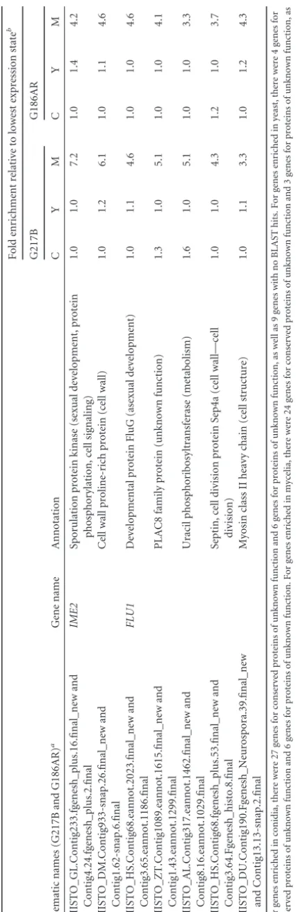
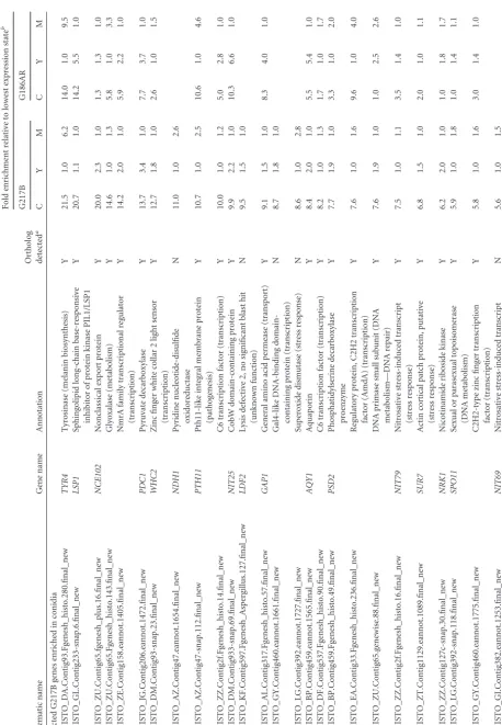
Conserved yeast, mycelial, and conidial enriched gene sets were de-fined as orthologous gene pairs with at least 3-fold enrichment in the phase of interest relative to both other phases in both strains. Genes that passed the analogous criterion in only one of the two strains were consid-ered strain specific in their enrichment. For genes probed with two probes, the gene was considered enriched if either probe passed the enrichment criterion.
Northern blots.For Northern blot hybridization, 1g of DNase I-treated RNA was loaded onto a MOPS (morpholinepropanesulfonic acid) gel, transferred to nitrocellulose membranes, and subjected to hybridiza-tion with a radiolabeled probe as previously described (12). Northern blot probes were generated by PCR with the following primers: CATA-ex3FW3, 5=-TGAAGCCGGAACCTCATAAC-3=; and CATA-ex3RV3, 5=
-ATCGTAACCATCCCCAATCA-3=.
Expression constructs.A sequence containing 1,050 bp of the G217B TYR1promoter was amplified by PCR using the primer pair OAS1040 (5=-agcacggcgccGATATCTTGTTTGCAGGAAGCCG-3=; NarI site shown in lowercase) and OAS1041 (5=-acggcgtcgacGGGTGACGATATG AAGTTGAGG-3=; SalI site shown in lowercase), cloned into pCR2.1, and sequenced. Positive clones were digested with NarI and SalI and cloned into the NarI-SalI sites of a promoterless green fluorescent protein (GFP)-containing entry vector (Invitrogen) containing the hygromycin resistance gene. Entry vectors were recombined in an LR recombination reaction with the H. capsulatum episomal vector pMA35B to generate PTYR1-GFP followed by the terminator of the CATBgene (PTYR1-GFP-CATBt). PCBP1-GFP-CATBtwas generated by cloning GFP into the SalI-NotI sites of a PCBP1(p)-CATBtentry vector, pLH125 (which contains 1,111 bp of the G186ARCBP1promoter), followed by recombination with pMA35B. Expression vectors were digested with PacI and transformed into both G217B and G186AR as previously described (16), using hygromycin resistance as a selectable marker. The resultant yeast-phase transformants were grown in HMM medium at 37°C with 5% CO2as described above. To observe GFP expression in mycelia, yeast cultures were inoculated into HMM, aer-ated at room temperature at 110 rpm, and observed 2 days later. Data are shown only for the G186AR strain (seeFig. 5).
Ortholog prediction and annotation of theH. capsulatumgene sets.
Orthologous genes between the G186AR and G217B strains as well as between the G217B strain andAspergillus nidulans,Aspergillus fumigatus, Candida albicans,Candida glabrata, andSaccharomyces cerevisiaewere determined by InParanoid, version 1.35, software, with no outgroup, to determine the best reciprocal BLAST hits betweenH. capsulatumand these species (18). To manually annotate theHistoplasmapredicted gene sets, BLASTP analysis was performed with the predicted gene models, identified at Washington University, St. Louis, MO, against the NCBI nr database (http://www.ncbi.nlm.nih.gov/GenBank/index.html) between April 2011 and August 2011. These manual gene annotations were further refined using InterProScan protein domain hits (19–21). tRNAs for the G217B strain were predicted by tRNAScan-SE (22). Descriptions for or-thologous genes provided in Tables S1 and S2 in the supplemental mate-rial were obtained from theAspergillusGenome Database (AspGD) (23; http://www.aspergillusgenome.org/download/chromosomal_feature _files/) forA. nidulansandA. fumigatus, and gene descriptions forS. cerevisiaeorthologs were downloaded from theSaccharomycesGenome
Database (24; http://www.yeastgenome.org/download-data/curation). The annotations from this paper will be made available through HistoBase (http://histo.ucsf.edu/), which will also serve as a tracker for revisions and corrections to the annotation set.
Microarray data accession number.Microarray data are available at the NCBI Gene Expression Omnibus (GEO) under accession number GSE45432.
RESULTS AND DISCUSSION
Purified conidia are viable and infectious.
Histoplasma
mycelia
produce two types of spores: microconidia (ranging in size from 2
to 6
m in diameter) and macroconidia (8 to 14
m in diameter).
The microconidia are thought to be the primary infectious
parti-cles due to their small size and potential to enter the small alveoli
in the lungs of a host. We found that the strains used in this study
made both microconidia and macroconidia (
Fig. 1
). The conidia
produced by the G217B strain were brownish, whereas the conidia
produced by the G186AR strain were white or colorless.
Purifica-tion of
H. capsulatum
conidia typically yielded 95% or more
mi-croconidia from the G217B strain, whereas the G186AR strain
produced
⬎
99% microconidia (data not shown). Conidial
viabil-ity was routinely assessed by plating for CFU (see Materials and
Methods).
In the laboratory, temperature is a sufficient signal to trigger
growth in either the filamentous form (at room temperature) or
the yeast form (at 37°C) (
25
), though this temperature-dependent
developmental regulation has largely been observed with
vegeta-tively growing cultures. To determine whether conidial
germina-tion yields filaments or yeast-phase cells in a
temperature-depen-dent manner, we performed a time course of germination at 27°C
and 37°C (
Fig. 2
) by placing purified conidia in fresh medium. We
observed that germination in liquid culture was asynchronous
and that, over the course of the experiment, some conidia failed to
germinate irrespective of temperature. Those that did germinate
displayed initial morphological changes at approximately 24 h
postinoculation at 27°C and 37°C. Visual inspection revealed that
48 h after transfer to germination medium at 27°C, 71% of conidia
(
n
⫽
78) displayed hyphal projections or protuberances.
Simi-larly, 86% of conidia (
n
⫽
237) gave rise to one or more yeast cells
by 48 h after transfer to germination medium at 37°C. Hence, the
majority of conidia that germinated at 27°C generated
filamen-tous cells, whereas the majority of the conidia germinated at 37°C
generated yeast-phase cells, indicating that conidial
differentia-tion was temperature responsive.
To determine whether purified conidia germinated into the
yeast form within macrophages, we infected murine BMDM
with conidia and monitored fungal cellular morphology over
time. At early time points, the conidia readily bound to and
10 µm
A
B
G217B G186AR
FIG 1Purified microconidia and macroconidia were generated fromH. cap-sulatum. Purified macroconidia and microconidia of the G217B (A) and G186AR (B) strains are shown.
on September 8, 2020 by guest
http://ec.asm.org/
were phagocytosed by the macrophages (
Fig. 3A
and
B
).
Ger-mination of conidia into the yeast form within macrophages
was observed by 24 h postinfection (hpi) (
Fig. 3C
). By 72 hpi
(
Fig. 3D
), the resultant yeast cells had proliferated robustly
within macrophages, ultimately causing host cell lysis.
Occa-sionally, conidia also produced mycelial-form cells within
macrophages incubated at 37°C (
Fig. 3E
).
To examine the fate of purified conidia in the context of an
infection, we infected mice intranasally with G217B conidia and
monitored fungal cell morphology and dissemination over time.
Although the only cell type present in the inoculum was conidia,
by 5 days postinfection (dpi) both conidia and yeast were observed
in the lung (
Fig. 3F
), strongly suggesting that yeast cells arose from
germinating conidia. Since multiple yeast cells were observed in
close association with conidia at day 5 (
Fig. 3F
), it is possible that
at least some conidia are capable of continued production of yeast
cells in the lungs. Interestingly, histological analysis showed that
conidia continued to persist in the lung at later time points,
indi-cating that conidial cells are resistant to destruction by the host.
Spleens from conidia-infected mice ultimately became colonized
with yeast cells, as we observed previously (
4
), indicating that
conidia from a purified laboratory strain are capable of causing
disseminated disease in a mouse model of pulmonary
histoplas-mosis. The conidia themselves were never observed in the spleens
of infected mice over a range of time points (data not shown),
suggesting that conidial cells cannot transit from the lung and that
germination into the yeast form is required for dissemination to
the spleen.
Identification of differentially expressed transcripts in two
divergent
H. capsulatum
strains.
Once we established that
puri-fied conidia were viable and infectious, we wanted to determine
which transcripts showed enrichment in conidia versus
yeast-form or mycelial cells. We reasoned that we could identify
phase-specific enriched transcripts that showed conserved patterns of
expression by comparing the transcriptomes of conidia, yeast, and
mycelia from two divergent
H. capsulatum
strains, i.e., G217B and
G186AR. In parallel with these studies, we also manually
anno-tated the gene predictions from each genome, as shown in Table
S1 (G217B) and Table S2 (G186AR) in the supplemental material.
We utilized two approaches to annotate the
Histoplasma
predicted
gene sets. The first was analysis and manual annotation of the
predicted genes based on BLASTP homology to protein matches
at the NCBI nr database. The second was the assignment of Gene
Ontology (GO) annotations for orthologs of characterized
pro-teins of
Saccharomyces cerevisiae
,
Aspergillus nidulans
,
A.
fumiga-tus
,
Candida albicans
, and
C. glabrata
. These orthology-based
computational annotations were examined manually and used to
refine the gene descriptions.
To determine which transcripts were enriched in
H.
capsula-tum
conidia, mycelia, and yeast, we compared the gene expression
profiles of purified conidia with those of yeast-form and mycelial
cells of both the G217B and G186AR strains by using the BAGEL
algorithm (
17
). This algorithm transforms replicate
measure-ments of expression ratios into relative estimates of per-state
ex-pression, where the state of least expression is assigned an
expres-sion value of 1.0. Additionally, BAGEL estimates confidence
12h 24h 48h 72h 96h
37oC
27oC
FIG 2Conidia germinatein vitrointo filaments at 27°C and into yeast-phase cells at 37°C. A time course of germination of G217B conidia was performed at the indicated temperatures.
A
B
C
D
E
F
}
10 µmFIG 3Germination of conidia in macrophages and host tissues. PAS-stained images of conidia-infected bone marrow-derived macrophages are shown at 2 hpi (A), 6 hpi (B), 24 hpi (C), and 72 hpi (D). (E) Occasional mycelial forms observed at 24 hpi. (F) A lung section from a conidia-infected mouse was stained with PAS, hematoxylin, and eosin. Arrows indicate conidia, black arrowheads indicate yeast cells, and the white arrowhead indicates a macroconidium.
on September 8, 2020 by guest
http://ec.asm.org/
intervals and probabilities of differential expression between
states by sampling possible fits of the data in a Bayesian
frame-work. The relative gene expression profiles of orthologous G217B
and G1686AR genes (filtered for data present from the BAGEL
statistical analysis of both strains [see Materials and Methods]) in
the three phases (conidia, yeast, and mycelia) are shown as a heat
map where relative conidial expression is highlighted in blue,
rel-ative mycelial expression is in green, and relrel-ative yeast expression
is in red (
Fig. 4A
). Despite the evolutionary divergence of G217B
and G186AR, we noted a remarkable conservation of
phase-spe-cific enriched expression between orthologs of the two strains. To
visually illustrate the relative expression of a given transcript in the
three phases,
Fig. 4B
and
C
show 3-axis plots of the log
2relative
expression values for each gene in the G217B and G186AR data
sets, respectively. Specific genes of interest, described below or
mentioned in
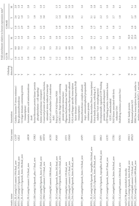
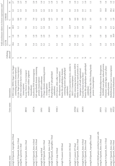
Tables 1
and
2
, are highlighted on the plots.
According to the BAGEL analysis, a total of 3,404 genes were
differentially expressed in one of the three cell types of G217B,
with
P
values of
ⱖ
0.985 (see Table S3 in the supplemental
mate-rial): 1,275 were enriched in conidia, 1,006 were enriched in
my-celia, and 1,123 were enriched in yeast. In the G186AR strain,
3,179 genes were differentially regulated by the same criteria (see
Table S4): 1,183 were enriched in conidia, 925 were enriched in
mycelia, and 1,071 were enriched in yeast. Table S5 reports gene
expression data across the three phases for both strains in a
gene-centric manner, providing ortholog information across both
strains. To define the set of most highly enriched transcripts in
conidia in each strain, the results of the BAGEL analysis were
filtered with a 3-fold cutoff, such that conidial enriched genes were
at least 3-fold enriched relative to those in both yeast and mycelia.
Yeast and mycelial enriched genes were defined analogously.
Us-ing this strUs-ingent 3-fold cutoff, we determined the numbers of
strain-specific and conserved phase-specific enriched genes for
each strain (
Fig. 4D
). By these criteria, we identified 456 and 300
FIG 4Relative expression of G217B and G186AR transcripts in yeast, mycelia, and conidia. (A) Heat map of relative conidial (C), yeast (Y), and mycelial (M) expression levels for pairs of orthologous genes with data present in both strains. Intensities are log2BAGEL-estimated relative expression levels from 0 (black)to 3 (saturated). Genes are ordered by phase specificity, defined as the average angular coordinate from plots B and C. Phase-specific genes are indicated by the bracketed regions to the left of the heat map. Relative enrichment plots are shown for G217B (B) and G186AR (C). Yeast, mycelial, and conidial axes are drawn in red, green, and blue, respectively; axis ticks indicate log2units of enrichment. Genes were plotted by projecting the BAGEL-estimated relative expression values
on the corresponding axes (the condition of lowest expression always has a log2enrichment of zero). Yeast, mycelial, and conidial enriched genes, based on a
3-fold enrichment criterion, are colored red, green, and blue, respectively. Genes of interest mentioned in the text and/or tables are highlighted with black circles and labeled. (D) Venn diagrams showing conserved expression of the 3-fold differentially expressed conidial-, mycelial-, and yeast-specific transcripts of the G217B and G186AR strains.
on September 8, 2020 by guest
http://ec.asm.org/
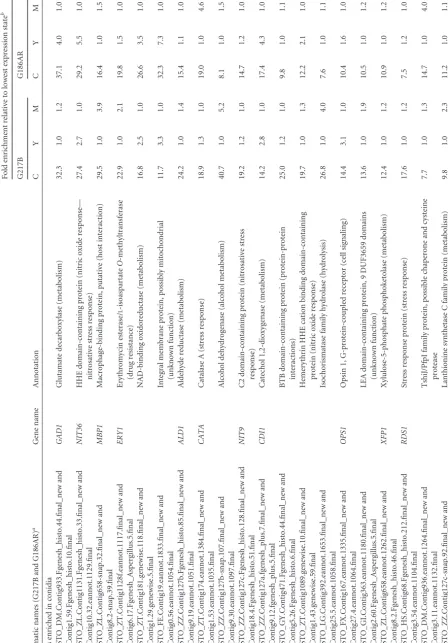
TABLE
1
Conserved
differentially
expressed
transcripts
of
strains
G186AR
and
G217B
Systematic
names
(G217B
and
G186AR)
a
Gene
name
Annotation
Fold
enrichment
relative
to
lowest
expression
state
b
G217B
G186AR
CY
M
C
Y
M
Genes
enriched
in
conidia
HISTO_DM.Contig933.Fgenesh_histo.44.final_new
and
Contig1.59.Fgenesh_histo.10.final
GAD1
Glutamate
decarboxylase
(metabolism)
32.3
1.0
1.2
37.1
4.0
1.0
HISTO_ZL.Contig1131.Fgenesh_histo.33.final_new
and
Contig10.32.eannot.1129.final
NIT36
HHE
domain-containing
protein
(nitric
oxide
response—
nitrosative
stress
response)
27.4
2.7
1.0
29.2
5.5
1.0
HISTO_ZL.Contig658-snap.32.final_new
and
Contig8.2-snap.39.final
MBP1
Macrophage-binding
protein,
putative
(host
interaction)
29.5
1.0
3.9
16.4
1.0
1.5
HISTO_ZT.Contig1128f.eannot.1117.final_new
and
Contig6.17.Fgenesh_Aspergillus.5.final
ERY1
Erythromycin
esterase/
L
-isoaspartate
O
-methyltransferase
(drug
resistance)
22.9
1.0
2.1
19.8
1.5
1.0
HISTO_ZT.Contig181.genewise.118.final_new
and
Contig1.29.genewise.5.final
NAD-binding
oxidoreductase
(metabolism)
16.8
2.5
1.0
26.6
3.5
1.0
HISTO_FE.Contig19.eannot.1833.final_new
and
Contig0.56.eannot.1054.final
Integral
membrane
protein,
possibly
mitochondrial
(unknown
function)
11.7
3.3
1.0
32.3
7.3
1.0
HISTO_ZZ.Contig127b.Fgenesh_histo.85.final_new
and
Contig9.19.eannot.1051.final
ALD1
Aldehyde
reductase
(metabolism)
24.2
1.0
1.4
15.4
1.1
1.0
HISTO_ZT.Contig174.eannot.1384.final_new
and
Contig1.53.eannot.1035.final
CATA
Catalase
A
(stress
response)
18.9
1.3
1.0
19.0
1.0
4.6
HISTO_ZZ.Contig127b-snap.107.final_new
and
Contig9.30.eannot.1097.final
Alcohol
dehydrogenase
(alcohol
metabolism)
40.7
1.0
5.2
8.1
1.0
1.5
HISTO_ZZ.Contig127c.Fgenesh_histo.128.final_new
and
Contig18.4.Fgenesh_histo.51.final
NIT9
C2
domain-containing
protein
(nitrosative
stress
response)
19.2
1.2
1.0
14.7
1.2
1.0
HISTO_ZZ.Contig127a.fgenesh_plus.7.final_new
and
Contig9.12.fgenesh_plus.5.final
CDI1
Catechol
1,2-dioxygenase
(metabolism)
14.2
2.8
1.0
17.4
4.3
1.0
HISTO_GY.Contig471.Fgenesh_histo.44.final_new
and
Contig5.26.Fgenesh_histo.6.final
BTB
domain-containing
protein
(protein-protein
interactions)
25.0
1.2
1.0
9.8
1.0
1.1
HISTO_ZT.Contig1089.genewise.10.final_new
and
Contig1.43.genewise.59.final
Hemerythrin
HHE
cation
binding
domain-containing
protein
(nitric
oxide
response)
19.7
1.0
1.3
12.2
2.1
1.0
HISTO_LG.Contig392.eannot.1653.final_new
and
Contig25.5.eannot.1058.final
Isochorismatase
family
hydrolase
(hydrolysis)
26.8
1.0
4.0
7.6
1.0
1.1
HISTO_FX.Contig167.eannot.1335.final_new
and
Contig27.4.eannot.1064.final
OPS1
Opsin
1,
G-protein-coupled
receptor
(cell
signaling)
14.4
3.1
1.0
10.4
1.6
1.0
HISTO_GI.Contig363.eannot.1180.final_new
and
Contig2.60.Fgenesh_Aspergillus.5.final
LEA
domain-containing
protein,
9
DUF3659
domains
(unknown
function)
13.6
1.0
1.9
10.5
1.0
1.2
HISTO_ZL.Contig658.eannot.1262.final_new
and
Contig8.2.Fgenesh_histo.46.final
XFP1
Xylulose-5-phosphate
phosphoketolase
(metabolism)
12.4
1.0
1.2
10.9
1.0
1.2
HISTO_HS.Contig68.Fgenesh_histo.212.final_new
and
Contig3.54.eannot.1104.final
RDS1
Stress
response
protein
(stress
response)
17.6
1.0
1.2
7.5
1.2
1.0
HISTO_DM.Contig936.eannot.1264.final_new
and
Contig3.11.eannot.1132.final
TshiJ/PfpI
family
protein,
possible
chaperone
and
cysteine
protease
7.7
1.0
1.3
14.7
1.0
4.0
HISTO_ZZ.Contig127c-snap.92.final_new
and
Contig13.16-snap.1.final
Lanthionine
synthetase
C
family
protein
(metabolism)
9.8
1.0
2.3
11.2
1.0
1.1
on September 8, 2020 by guest
http://ec.asm.org/
HISTO_GI.Contig382.Fgenesh_histo.57.final_new
and
Contig2.67.eannot.1129.final
MFS11
MFS
multidrug
transporter
(drug
resistance)
6.9
1.6
1.0
14.9
1.0
1.9
HISTO_ZU.Contig171-snap.5.final_new
and
Contig31.3-snap.18.final
HhH-GPD
domain-containing
protein,
DUF488
domain
12.0
2.4
1.0
8.3
1.5
1.0
HISTO_HS.Contig68-snap.98.final_new
and
Contig3.65.Fgenesh_histo.45.final
Glyoxal
oxidase
(lignin
degradation)
11.1
1.0
1.3
7.8
1.5
1.0
HISTO_DA.Contig93.Fgenesh_histo.10.final_new
and
Contig0.103.Fgenesh_histo.10.final
F-box
domain-containing
protein,
putative
(unknown
function)
12.2
3.6
1.0
6.8
1.2
1.0
HISTO_LG.Contig392.fgenesh_plus.37.final_new
and
Contig25.9.fgenesh_plus.2.final
Glutathione
S
-transferase
family
protein
(metabolism)
5.6
1.0
1.2
12.1
1.0
1.7
HISTO_GI.Contig382.Fgenesh_histo.91.final_new
and
Contig2.67.eannot.1136.final
PSP1
Parasitic-phase-specific
protein
of
Coccidioides
(unknown
function)
6.4
1.0
2.1
10.5
1.0
3.0
HISTO_AL.Contig317.eannot.1531.final_new
and
Contig8.21.Fgenesh_histo.12.final
TPS3
Alpha,alpha-trehalose
phosphate
synthase
subunit
(trehalose
metabolism)
5.2
1.3
1.0
11.4
1.0
1.2
HISTO_ZL.Contig658.eannot.1243.final_new
and
Contig8.2.eannot.1201.final
AKI1
Acetate
kinase
(metabolism)
10.2
1.1
1.0
5.2
1.0
1.0
HISTO_JG.Contig207.genewise.57.final_new
and
Contig6.25.Fgenesh_histo.22.final
ISH1
Stress
response
protein
(stress
response)
8.4
1.2
1.0
6.3
1.0
1.3
HISTO_ZY.Contig518.Fgenesh_histo.22.final_new
and
Contig5.7.eannot.1127.final
Zinc-binding
oxidoreductase,
ToxD-like
(metabolism)
5.9
1.2
1.0
8.5
1.0
2.3
HISTO_EA.Contig33.genewise.94.final_new
and
Contig12.16.Fgenesh_Aspergillus.36.final
Transcription
factor
IIIc-like
protein,
putative
(PolIII
transcription)
7.4
1.0
1.0
6.4
1.1
1.0
HISTO_DU.Contig190.Fgenesh_histo.12.final_new
and
Contig13.7.Fgenesh_Aspergillus.15.final
Carboxypeptidase
(proteolysis)
5.7
1.1
1.0
7.8
2.2
1.0
HISTO_ZY.Contig571f.Fgenesh_histo.26.final_new
and
Contig5.14.Fgenesh_histo.6.final
F-box
domain-containing
protein,
putative
(unknown
function)
4.3
1.0
1.0
10.0
1.3
1.0
HISTO_ZL.Contig1131.eannot.2111.final_new
and
Contig10.33.eannot.1051.final
NmrA
family
transcriptional
regulator
(transcription)
5.8
1.8
1.0
7.3
1.0
2.2
HISTO_ZL.Contig1161b.Fgenesh_histo.87.final_new
and
Contig0.33.Fgenesh_histo.6.final
GAL4-like
Zn2Cys6
transcription
factor
(transcription)
6.6
1.0
1.1
5.7
1.0
1.2
HISTO_GB.Contig114.eannot.1124.final_new
and
Contig26.4.Fgenesh_histo.3.final
Similar
to
YT521-B-like
splicing
factor
(RNA
metabolism)
6.3
1.0
1.2
5.8
1.1
1.0
HISTO_FE.Contig19.Fgenesh_Aspergillus.214.final_new
and
Contig0.57.Fgenesh_Aspergillus.19.final
FBD1
MYB
family
conidiophore
development
protein
FlbD
(asexual
development)
5.7
1.0
1.5
6.1
1.4
1.0
HISTO_LF.Contig359.eannot.2006.final_new
and
Contig2.51.eannot.1195.final
DNL4
NHEJ
DNA
ligase
(DNA
repair)
7.7
1.9
1.0
4.1
1.0
1.0
HISTO_EA.Contig33.fgenesh_plus.53.final_new
and
Contig12.9.genewise.4.final
FET3
Iron
transport
multicopper
oxidase
(iron
transport)
6.2
1.8
1.0
5.0
1.5
1.0
HISTO_HS.Contig68.eannot.2168.final_new
and
Contig3.54.eannot.1111.final
Serine
protein
kinase
(protein
phosphorylation)
4.2
1.1
1.0
5.1
1.7
1.0
HISTO_ZT.Contig181.eannot.1629.final_new
and
Contig1.12.eannot.1068.final
CBS
domain-containing
protein
(unknown
function)
4.1
1.0
1.2
5.0
1.0
1.6
HISTO_GY.Contig471.Fgenesh_Neurospora.27.final_new
and
Contig5.26.eannot.1025.final
FURA
Uracil
permease
(transport)
4.9
1.0
1.4
4.0
1.1
1.0
HISTO_LG.Contig392.genewise.34.final_new
and
Contig25.9.fgenesh_plus.10.final
TGL1
Triglyceride
lipase-cholesterol
esterase
(lipid
metabolism)
4.6
1.0
1.3
3.9
1.0
1.0
HISTO_DA.Contig93-snap.60.final_new
and
Contig0.89-snap.22.final
Alpha/beta
hydrolase
(hydrolysis)
3.9
1.1
1.0
3.9
1.0
1.0
(Continued
on
following
page)
on September 8, 2020 by guest
http://ec.asm.org/
TABLE
1
(Continued)
Systematic
names
(G217B
and
G186AR)
a
Gene
name
Annotation
Fold
enrichment
relative
to
lowest
expression
state
b
G217B
G186AR
CY
M
C
Y
M
Genes
enriched
in
yeast
HISTO_AL.Contig317.eannot.1542.final_new
and
Contig8.3.eannot.1103.final
Highly
yeast-phase-specific
transcript
(unknown
function)
1.4
47.3
1.0
5.0
90.2
1.0
HISTO_GY.Contig460.eannot.1679.final_new
and
Contig37.5.eannot.1055.final
Highly
yeast-phase-specific
transcript
(unknown
function)
1.0
44.8
1.9
1.0
77.3
1.0
HISTO_ZL.Contig1131.eannot.1938.final_new
and
Contig20.3.eannot.1101.final
YPS21
Yeast-phase-specific
protein
(unknown
function)
2.6
41.5
1.0
1.0
14.0
1.6
HISTO_EA.Contig33.eannot.1524.final_new
and
Contig12.3.eannot.1252.final
MET16
3
=
-Phosphoadenosine-5
=
-phosphosulfate
reductase
(metabolism)
5.2
24.8
1.0
2.4
18.1
1.0
HISTO_JG.Contig206.fgenesh_plus.57.final_new
and
Contig24.6.eannot.1053.final
SID4
Acid
coenzyme
A
(coA)
ligase
(siderophore
biosynthesis)
1.0
24.1
2.7
1.0
18.6
1.7
HISTO_ZT.Contig1089.eannot.1639.final_new
and
Contig1.44.Fgenesh_Aspergillus.6.final
GNT1
␣
N
-Acetylglucosamine
transferase
2.0
16.7
1.0
3.1
15.2
1.0
HISTO_KF.Contig597.eannot.1481.final_new
and
Contig11.25.eannot.1100.final
CCP2
Cytochrome
c
peroxidase
(metabolism)
4.6
18.6
1.0
1.0
11.9
1.4
HISTO_JG.Contig206.eannot.1534.final_new
and
Contig24.6.eannot.1061.final
OXR1
Oxidoreductase
(siderophore
biosynthesis)
1.0
11.7
2.5
1.0
17.2
5.0
HISTO_BP.Contig459-snap.79.final_new
and
Contig7.33.eannot.1033.final
CYN1
High-affinity
cystine
transporter
(transport)
1.0
25.8
1.6
1.0
7.7
1.7
HISTO_ZL.Contig658.eannot.1268.final_new
and
Contig8.2.eannot.1185.final
GH17
Immunoreactive
protein
(unknown
function)
1.0
10.2
1.1
1.0
18.4
3.3
HISTO_ZE.Contig158.eannot.1345.final_new
and
Contig14.7.eannot.1088.final
CATB
Catalase
B,
M
antigen
(stress
response)
2.7
8.9
1.0
1.0
19.0
3.8
HISTO_ZZ.Contig127a.Fgenesh_Aspergillus.87.final_new
and
Contig9.8.eannot.1128.final
Superoxide
dismutase
(stress
response)
2.8
19.4
1.0
1.0
7.5
2.4
HISTO_DM.Contig933.Fgenesh_histo.56.final_new
and
Contig3.2.Fgenesh_histo.7.final
TSA1
Thiol-specific
antioxidant
(stress
response)
2.5
12.2
1.0
2.0
9.3
1.0
HISTO_ZL.Contig1131.Fgenesh_histo.235.final_new
and
Contig20.3.Fgenesh_histo.4.final
C2H2
finger
domain-containing
protein
(transcription)
1.0
11.8
2.5
1.0
9.3
1.5
HISTO_EA.Contig33.eannot.1649.final_new
and
Contig12.20.eannot.1056.final
Nitroreductase
(nitrogen
metabolism)
1.2
11.9
1.0
1.0
8.4
2.5
HISTO_GY.Contig460.Fgenesh_Neurospora.78.final_new
and
Contig37.5.Fgenesh_Neurospora.3.final
Neutral
amino
acid
permease
(transport)
1.0
8.4
1.0
1.7
11.3
1.0
HISTO_BP.Contig457.Fgenesh_Aspergillus.167.final_new
and
Contig7.11.Fgenesh_Aspergillus.10.final
Histidine
acid
phosphatase
(metabolism)
1.8
7.7
1.0
1.5
11.7
1.0
HISTO_JG.Contig207.eannot.1394.final_new
and
Contig6.25.eannot.1057.final
GRX1
Glutaredoxin
(stress
response)
1.0
8.7
1.7
2.1
9.3
1.0
HISTO_DY.Contig31.fgenesh_plus.50.final_new
and
Contig15.14.fgenesh_plus.25.final
Zinc-containing
alcohol
dehydrogenase
(metabolism)
1.0
10.6
2.2
1.0
7.5
1.1
HISTO_JG.Contig206.eannot.1510.final_new
and
Contig24.6.eannot.1059.final
SID3
Acetylase
(siderophore
biosynthesis)
2.0
11.6
1.0
1.0
6.5
1.5
on September 8, 2020 by guest
http://ec.asm.org/
HISTO_FX.Contig167.Fgenesh_histo.44.final_new
and
Contig27.6.eannot.1031.final
Membrane
transporter
(transport)
2.9
12.7
1.0
1.0
5.5
1.1
HISTO_JG.Contig206.eannot.1556.final_new
and
Contig6.30.eannot.1215.final
CAR1
Arginase
(metabolism)
1.0
9.7
2.9
1.0
6.5
1.0
HISTO_ZL.Contig1131.eannot.2134.final_new
and
Contig10.30.eannot.1086.final
Glucose
transporter
(transport)
1.0
9.6
1.0
1.0
5.4
1.0
HISTO_ZT.Contig181.Fgenesh_histo.190.final_new
and
Contig1.18.Fgenesh_Neurospora.4.final
YMC1
Mitochondrial
carrier
protein
(metabolism)
1.0
6.8
1.0
1.0
7.5
2.5
HISTO_JG.Contig206.Fgenesh_histo.188.final_new
and
Contig24.6.Fgenesh_histo.10.final
ABC1
ABC
multidrug
transporter
(iron
transport)
1.1
9.4
1.0
1.2
4.7
1.0
HISTO_DF.Contig537.eannot.1356.final_new
and
Contig21.2-snap.4.final
Delta-1-pyrroline-5-carboxylate
dehydrogenase
PrnC
(metabolism)
1.0
6.6
1.5
1.0
6.3
2.0
HISTO_ZL.Contig1158.Fgenesh_histo.83.final_new
and
Contig19.9.Fgenesh_histo.28.final
Peptidase
family
M13
protein,
neprilysin,
endothelin-converting
enzyme
(hydrolysis)
2.1
7.5
1.0
1.6
5.4
1.0
HISTO_ZL.Contig658.eannot.1234.final_new
and
Contig8.3.genewise.14.final
CTS1
Chitinase
(cell
wall)
2.0
9.3
1.0
1.1
3.9
1.0
HISTO_JG.Contig206.eannot.1512.final_new
and
Contig24.6.Fgenesh_histo.11.final
SID1
Ornithine
monooxygenase
(siderophore
biosynthesis)
1.5
7.0
1.0
1.0
5.2
1.0
HISTO_ZT.Contig174.eannot.1401.final_new
and
Contig1.54.fgenesh_plus.4.final
ADE4
Amidophosphoribosyltransferase
(metabolism)
2.3
7.6
1.0
1.0
4.3
1.2
HISTO_LF.Contig359.Fgenesh_Aspergillus.67.final_new
and
Contig2.49.Fgenesh_Aspergillus.1.final
Arrestin
(cell
signaling)
1.2
5.7
1.0
1.1
4.7
1.0
HISTO_GL.Contig233.Fgenesh_Neurospora.36.final_new
and
Contig4.20.Fgenesh_Neurospora.20.final
Glycosyl
hydrolase
catalytic
core
domain-containing
protein
(metabolism)
1.0
3.7
1.2
1.1
6.1
1.0
HISTO_DM.Contig933.eannot.1650.final_new
and
Contig1.62.eannot.1148.final
RYP4
C6
transcription
factor,
FacB
(transcription)
1.3
5.6
1.0
1.0
4.0
1.0
HISTO_ZH.Contig107.eannot.1172.final_new
and
Contig5.3-snap.20.final
FBP1
Fructose-1,6-bisphosphatase
(metabolism)
1.0
4.7
1.3
1.0
4.4
1.4
HISTO_ZT.Contig1128f.eannot.1145.final_new
and
Contig6.21.eannot.1022.final
AMP-dependent
synthetase
and
ligase
(metabolism)
1.0
4.3
1.4
1.1
4.5
1.0
Genes
enriched
in
mycelia
HISTO_GL.Contig296-snap.27.final_new
and
Contig1.70.eannot.1021.final
BYS1
(
Blastomyces
yeast-phase-specific
protein)
domain-containing
protein
(unknown
function)
7.3
1.0
33.4
3.3
1.0
27.1
HISTO_LG.Contig392.eannot.1788.final_new
and
Contig22.4.eannot.1192.final
MHB1
Hydrophobin
(cell
wall)
7.6
1.0
35.2
4.6
1.0
16.0
HISTO_DF.Contig537.Fgenesh_histo.23.final_new
and
Contig21.2.Fgenesh_histo.29.final
Gluconolactonase
(metabolism)
7.7
1.0
30.8
4.1
1.0
12.6
HISTO_EA.Contig33-snap.162.final_new
and
Contig12.20-snap.16.final
VELC
Velvet
family
protein
(asexual
development)
1.5
1.0
21.0
1.2
1.0
17.5
HISTO_ZL.Contig1131.Fgenesh_histo.234.final_new
and
Contig20.3.genewise.6.final
Extracellular
serine-rich
protein
(cell
wall)
2.7
1.0
19.8
1.7
1.0
17.5
HISTO_DM.Contig940.Fgenesh_histo.26.final_new
and
Contig4.1.eannot.1119.final
MFS
monosaccharide
transporter
(transport)
2.6
1.0
21.0
2.9
1.0
13.5
HISTO_AZ.Contig47-snap.146.final_new
and
Contig2.22.eannot.1061.final
ALD2
Aldehyde
reductase
II
(metabolism)
4.8
1.0
24.6
1.1
1.0
11.5
(Continued
on
following
page)
on September 8, 2020 by guest
http://ec.asm.org/
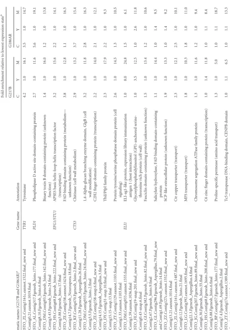
TABLE
1
(Continued)
Systematic
names
(G217B
and
G186AR)
a
Gene
name
Annotation
Fold
enrichment
relative
to
lowest
expression
state
b
G217B
G186AR
CY
M
C
Y
M
HISTO_ZL.Contig1161c.eannot.1522.final_new
and
Contig0.21.eannot.1019.final
TYR1
Tyrosinase
4.2
1.0
16.1
3.5
1.0
14.7
HISTO_AL.Contig317.Fgenesh_histo.177.final_new
and
Contig8.10.Fgenesh_histo.6.final
PLD1
Phospholipase
D
active
site
domain-containing
protein
2.7
1.0
11.6
5.6
1.0
19.1
HISTO_DM.Contig93.Fgenesh_histo.142.final_new
and
Contig3.43.Fgenesh_histo.24.final
Binary
toxin
B
domain-containing
protein
(unknown
function)
1.4
1.0
16.0
1.2
1.0
13.8
HISTO_HS.Contig68.Fgenesh_histo.222.final_new
and
Contig3.88.Fgenesh_histo.17.final
EFG1/STU1
APSES
basic
helix-loop-helix
transcription
factor
(transcription)
3.2
1.0
15.6
2.2
1.0
14.1
HISTO_ZE.Contig158.eannot.1424.final_new
and
Contig14.7.Fgenesh_Neurospora.18.final
FAD
binding
domain-containing
protein
(metabolism—
unknown
function)
3.8
1.0
12.8
1.1
1.0
16.5
HISTO_KF.Contig470.Fgenesh_histo.2.final_new
and
Contig11.28.Fgenesh_Aspergillus.26.final
CTS3
Chitinase
(cell
wall
metabolism)
2.9
1.0
13.2
3.7
1.0
15.4
HISTO_DM.Contig933.Fgenesh_histo.24.final_new
and
Contig3.5.Fgenesh_histo.2.final
1,4-Alpha-glucan
branching
enzyme
domain,
GlgB
(cell
wall
modification)
3.2
1.0
11.8
2.8
1.0
16.3
HISTO_ZE.Contig158-snap.6.final_new
and
Contig14.15.Fgenesh_Aspergillus.3.final
C2H2
finger
domain-containing
protein
(transcription)
2.2
1.0
14.0
2.1
1.0
12.1
HISTO_DY.Contig31-snap.10.final_new
and
Contig15.15-snap.13.final
ThiJ/PfpI
family
protein
2.3
1.0
17.9
2.2
1.0
9.3
HISTO_ZT.Contig1129.eannot.1103.final_new
and
Contig1.33.eannot.1103.final
Protein
tyrosine/serine
phosphatase
domain
protein
(cell
signaling)
2.6
1.0
14.5
1.3
1.0
10.5
HISTO_DM.Contig933.eannot.1555.final_new
and
Contig1.59.eannot.1036.final
ELI1
ELI-ag1-like
protein,
expression
library
immunization
antigen
1
(host
interaction)
1.0
8.0
24.8
1.5
1.0
6.1
HISTO_FE.Contig19-snap.201.final_new
and
Contig0.43-snap.6.final
Glycosylphosphatidylinositol
(GPI)-anchored
serine-threonine
rich
protein
(cell
wall)
1.0
3.5
12.5
1.0
2.6
11.8
HISTO_GI.Contig382.Fgenesh_histo.82.final_new
and
Contig2.67.Fgenesh_histo.9.final
Fasciclin
domain-containing
protein
(unknown
function)
1.5
1.0
13.4
1.2
1.0
10.6
HISTO_GL.Contig296.Fgenesh_Aspergillus.79.final_new
and
Contig1.69.Fgenesh_histo.14.final
Salicylate
hydroxylase,
FAD
binding
domain-containing
protein
1.1
1.0
14.4
1.0
1.4
9.5
HISTO_ZZ.Contig127c.eannot.1552.final_new
and
Contig13.21.eannot.1014.final
SCP-like
extracellular
protein
(unknown
function)
1.9
1.0
13.3
1.0
1.4
9.2
HISTO_ZL.Contig1161c.eannot.1487.final_new
and
Contig0.12.Fgenesh_histo.21.final
Ctr
copper
transporter
(transport)
2.3
1.0
12.1
2.5
1.0
10.1
HISTO_LG.Contig392.eannot.1695.final_new
and
Contig22.3.Fgenesh_Aspergillus.4.final
MFS
transporter
(transport)
1.8
1.0
10.3
1.8
1.0
11.0
HISTO_JG.Contig207.Fgenesh_histo.89.final_new
and
Contig6.30.Fgenesh_histo.55.final
Chromosome
segregation
ATPase
family
protein
1.3
1.0
11.6
1.2
1.0
9.4
HISTO_HS.Contig68.Fgenesh_histo.200.final_new
and
Contig3.88.Fgenesh_histo.6.final
C6
zinc
finger
domain-containing
protein
(transcription)
1.0
1.4
11.8
1.0
1.1
8.4
HISTO_BP.Contig457.Fgenesh_histo.177.final_new
and
Contig7.4.Fgenesh_Aspergillus.2.final
Proline-specific
permease
(amino
acid
transport)
1.3
1.0
5.0
1.0
1.1
18.7
HISTO_ZT.Contig174.eannot.1389.final_new
and
Contig6.15.eannot.1092.final
Tc5
transposase
DNA-binding
domain,
CENPB
domain
(Tn
transcription)
1.0
1.1
6.5
1.0
1.1
13.3
on September 8, 2020 by guest
http://ec.asm.org/
HISTO_BP.Contig457.Fgenesh_Aspergillus.216.final_new
and
Contig7.4.Fgenesh_Aspergillus.58.final
CCC2
Calcium
transporter
2.1
1.0
8.0
1.9
1.0
9.9
HISTO_ZE.Contig158.Fgenesh_Aspergillus.25.final_new
and
Contig14.7-snap.12.final
SAM-dependent
methyltransferase
(metabolism)
1.3
1.0
9.6
1.2
1.0
7.8
HISTO_ZL.Contig1161a.eannot.1162.final_new
and
Contig0.35.eannot.1060.final
MFS
monocarboxylate
transporter
(transport)
1.8
1.0
12.1
1.1
1.0
6.1
HISTO_ZE.Contig158.eannot.1426.final_new
and
Contig14.7.eannot.1095.final
MFS
multidrug
transporter
(transport—drug
resistance)
2.1
1.0
7.7
3.1
1.0
9.6
HISTO_BP.Contig459.Fgenesh_histo.160.final_new
and
Contig30.15.fgenesh_plus.20.final
C2H2
finger
domain-containing
protein
(transcription)
1.0
1.3
5.2
1.7
1.0
14.0
HISTO_ZL.Contig1161c.Fgenesh_histo.47.final_new
and
Contig0.19.Fgenesh_histo.2.final
Morphogenesis-related
protein,
Rho-GAP
superfamily
domain
(cell
signaling)
1.4
1.0
8.3
1.0
1.2
8.5
HISTO_BP.Contig457.Fgenesh_Aspergillus.17.final_new
and
Contig7.18.genewise.32.final
RNA-binding
protein
Nrd1,
negative
regulator
of
differentiation
(asexual
development)
1.0
1.6
7.8
1.0
1.8
9.1
HISTO_ZL.Contig658.Fgenesh_Aspergillus.66.final_new
and
Contig8.3.Fgenesh_Aspergillus.24.final
NIT88
PT
repeat
family
protein,
RNase
E
domains
1.0
1.2
7.0
2.8
1.0
9.7
HISTO_FE.Contig19.eannot.1909.final_new
and
Contig0.49.eannot.1124.final
FBC1
C2H2
finger
domain-containing
protein
FlbC
(transcription)
1.0
1.1
6.3
1.4
1.0
10.4
HISTO_ZT.Contig174.Fgenesh_histo.17.final_new
and
Contig1.50.Fgenesh_histo.1.final
PUM2
mRNA-binding
protein
Pumilio
2
(RNA
metabolism)
1.0
2.0
10.6
1.6
1.0
5.7
REPEAT
NAA.Contig17f.Fgenesh_histo.4.final_new
and
Contig11.4.Fgenesh_histo.7.final
Bifunctional
P-450:NADPH-P450
reductase
(metabolism)
1.8
1.0
5.6
1.4
1.0
10.4
HISTO_HS.Contig68-snap.7.final_new
and
Contig3.49.Fgenesh_histo.5.final
Protein
kinase
(protein
phosphorylation—cell
signaling)
1.0
1.2
5.1
2.8
1.0
11.5
HISTO_ZL.Contig1131-snap.217.final_new
and
Contig10.27-snap.8.final
MSB2
Mucin
family
signaling
protein,
putative
(cell
signaling)
1.0
1.0
7.5
1.0
1.0
7.6
HISTO_IAH.Contig38.eannot.1006.final_new
and
Contig20.3-snap.8.final
FDH1
Formate
dehydrogenase
(metabolism)
1.7
1.0
6.2
1.4
1.0
8.9
HISTO_BP.Contig457-snap.10.final_new
and
Contig7.4-snap.21.final
BSP1
PBSP
domain-containing
protein,
plant
basic
secretory
protein
1.2
1.0
5.6
1.0
1.0
8.8
HISTO_DY.Contig31.Fgenesh_Aspergillus.39.final_new
and
Contig15.6.eannot.1059.final
Secretory
phospholipase
A2
(lipid
metabolism)
1.0
1.3
4.9
2.1
1.0
10.0
HISTO_ZU.Contig153f.Fgenesh_histo.5.final_new
and
Contig17.12-snap.1.final
AT
DNA-binding
protein
(transcription)
1.1
1.0
6.3
2.6
1.0
7.7
HISTO_DM.Contig933.Fgenesh_histo.74.final_new
and
Contig3.2.Fgenesh_histo.1.final
Basic
region
leucine
zipper,
bZIP
transcription
factor
(transcription)
1.0
1.0
8.6
1.0
1.1
4.8
HISTO_KK.Contig134.Fgenesh_Aspergillus.17.final_new
and
Contig1.3-snap.11.final
Phosphoribulokinase/uridine
kinase
family
protein
(metabolism)
1.0
1.2
4.3
1.9
1.0
8.7
HISTO_ZL.Contig1161d-snap.23.final_new
and
Contig0.5-snap.37.final
Lactone
hydrolase
(metabolism—hydrolysis)
1.1
1.0
3.6
1.3
1.0
10.2
HISTO_DA.Contig93.Fgenesh_histo.176.final_new
and
Contig0.98.Fgenesh_histo.19.final
Calcium/calmodulin-dependent
protein
kinase
1.3
1.0
5.6
1.7
1.0
6.3
HISTO_ZZ.Contig127a.Fgenesh_histo.112.final_new
and
Contig9.8.Fgenesh_Neurospora.18.final
Asparagine
synthase
(metabolism)
1.3
1.0
4.0
2.0
1.0
8.5
HISTO_ZL.Contig1161b.eannot.1356.final_new
and
Contig0.29.eannot.1064.final
ESDC
GTP-binding
protein
(sexual
development)
1.2
1.0
7.6
1.0
1.0
4.4
(Continued
on
following
page)