ESSENTIAL ROLES FOR POLYMERASE THETA-MEDIATED END JOINING IN REPAIR OF CHROMOSOME BREAKS
David William Wyatt
A dissertation submitted to the faculty at the University of North Carolina at Chapel Hill in partial fulfillment of the requirements for the degree of Doctor of Philosophy in the
Curriculum in Genetics and Molecular Biology in the College of Arts and Sciences.
Chapel Hill 2016
Approved by:
ABSTRACT
David William Wyatt: Essential Roles for Polymerase Theta-Mediated End Joining in Repair of Chromosome Breaks
(Under the direction of Dale Ramsden)
DNA double strand breaks (DSBs) constitute a rare but lethal class of genomic
damage that must be efficiently repaired. Deficiencies in DSB repair pathways manifest
themselves as severe phenotypes including cancer predisposition, accelerated aging,
and immunodeficiency. On the cellular level, failure to repair DSBs can result in
genomic abnormalities, chromosomal rearrangements, and apoptosis.In mammalian
cells, repair of DSBs proceeds by classical nonhomologous end joining (NHEJ),
homologous recombination (HR), or by a third loosely described non-canonical repair
pathway, termed alternative end joining (Alt-EJ). Amongst the most clearly defined
characteristics of Alt-EJ is the presence microhomology, or small patches of
complementary DNA sequence flanking a chromosome break, in the repair junction.
3’ resected ends) or replication fork collapse. It typically does not compete with canonical repair pathways, but in NHEJ-deficient cells is engaged more frequently and protects against translocation. Cell viability is also severely impaired upon combined deficiency in Pol qand a factor that antagonizes end resection (Ku or 53BP1). TMEJ thus employs a flexible mechanism to help sustain cell viability and genome stability by rescuing chromosome break repair when resection is misregulated or NHEJ is
ACKNOWLEDGEMENTS
I would like to thank my advisor, Dale, for his guidance and support over the course of my doctoral work. Dale possess an incredible passion for science, an even more incredible wealth of knowledge, and has been integral in my development as a scientist. I would also like to thank my committee members Cyrus Vaziri, Jeff Sekelsky, Mike Emanuele, and Shawn Ahmed for both their time and interest in my work.
I am very grateful for all the Ramsden lab members that I have worked with over the past five years. Without the training, insights, and thoughtful discussion that they have provided, this work would not have been possible. In particular, I would like to thank Dr. John Pryor. His friendship and advice in navigating all aspects of the doctoral process has been invaluable.
I would not have arrived to this point without the support of my family and I am thankful for the opportunities that they have provided over the years. Finally, I would like to thank my fiancée Elizabeth Mutter-Rottmayer who has been an endless source of support, patience, and motivation.
Additional acknowledgments specific to each chapter Chapter 1
Chapter 2
This chapter was modified from its original version appearing in Molecular Cell in 2016. I have divided the publication here for the purpose of logical flow and have
modified figure layouts for this dissertation. Supplemental figures and 2 tables were included here. I have additionally included one piece of unpublished data (Figure 2.4). D.A.R., R.D.W, and D.W.W. designed the studies and analyzed the data. D.W.W. and M.J.Y. generated and characterized cell lines deficient in Pol q and Ku70 and variants. D.W.W. performed all experiments presented in this chapter. D.W.W. and S.A.R. performed bioinformatic analyses. D.A.R. and D.W.W. wrote the manuscript with input from all authors.Studies presented in this chapter were supported by NCI grant
CA097096 (D.A.R), AHA grant 14PRE20380258 (D.W.W), RP130297 and RP150102 from the Cancer Prevention and Research Institute of Texas and the Grady F. Saunders Research Professorship (R.D.W.), T32 CA09480 (MJY), DOD grant BC141727 and NIEHS grant R00ES02633 (S.A.R.), and the Burroughs Welcome Fund Career Award for Medical Scientists (G.P.G.). I thank Luis Blanco for the gift of Polm-/-Pol-/- MEFs.
Chapter 3
This chapter was modified from its original version appearing in Molecular Cell in 2016. I have divided the publication here for the purpose of logical flow and have
and variants. D.W.W. performed chromosomal end joining assays, resection assays, sequencing library preparation, translocation, and cell proliferation assays. W.F. performed immunofluorescence and colony formation assays. M.P.C. performed homologous recombination assays. D.W.W. and S.A.R. performed bioinformatic
analyses. D.A.R. and D.W.W. wrote the manuscript with input from all authors. Studies presented in this chapter were supported by NCI grant CA097096 (D.A.R), AHA grant 14PRE20380258 (D.W.W), RP130297 and RP150102 from the Cancer Prevention and Research Institute of Texas and the Grady F. Saunders Research Professorship
(R.D.W.), T32 CA09480 (MJY), DOD grant BC141727 and NIEHS grant R00ES02633 (S.A.R.), and the Burroughs Welcome Fund Career Award for Medical Scientists
TABLE OF CONTENTS
LIST OF TABLES ... xi
LIST OF FIGURES ... xii
LIST OF ABBREVIATIONS AND SYMBOLS ... xiii
CHAPTER 1: INTRODUCTION ... 1
1.1 DNA double strand break repair ... 1
1.2 Nonhomologous End Joining ... 2
1.3 DNA end resection ... 4
1.3.1 Initiation of homologous recombination ... 5
1.4 Alternative end joining ... 5
1.5 DNA Polymerase q ... 6
1.5.1 In vitro characterization of Polymerase q ... 7
1.5.2 Polymerase q and double strand break repair ... 8
1.6 Polymerase q and cancer……….9
CHAPTER 2: MECHANISM OF POLYMERASE q-MEDIATED END JOINING ... 13
2.1 Introduction ... 13
2.2 Methods ... 14
Cell lines ... 14
Sequencing ... 16
Gel shift assay ... 17
Strand displacement assay ... 18
2.3 Results ... 18
Distinct substrates for NHEJ and Pol q-mediated end joining……….18
Mechanism of Pol q-mediated end joining………..………20
2.4 Discussion ... 23
Identification of biologically relevant substrates and mechanism………...24
CHAPTER 3: ROLE OF TMEJ IN CHROMOSOMAL DOUBLE STRAND BREAK REPAIR ... 40
3.1 Introduction ... 40
3.2 Methods ... 41
Cell lines ... 41
Chromosomal double strand break repair assays ... 42
Sequencing ... 44
Cell cycle analysis... 45
Chromosomal aberrations ... 45
Immunofluorescence ... 45
Cell proliferation assays ... 46
Resection assay ... 46
3.3 Results………...47
Role of TMEJ in double strand break repair ... 53
TMEJ and genome instability ... 55
CHAPTER 4: DISCUSSION ... 74
4.1 Identification of biological substrates ... 75
4.2 Microhomology search mechanism ... 76
4.3 Other TMEJ factors ... 78
4.4 Relationships between TMEJ and canonical double strand break repair pathways ... 79
4.5 Polymerase q and genome stability ... 81
4.6 Polymerase q as a therapeutic target ... 82
4.6 Concluding remarks ... 83
LIST OF TABLES
Table 2.1 – Reagents used during experiments……… 37 Table 2.2 – Inferred alignment structures and recovery frequencies
LIST OF FIGURES
Figure 1.1 – DNA double strand break repair pathways ... 11
Figure 1.2 – Hypersensitivity of Polq-/- bone marrow stromal cells to DNA strand breaking agents ... 12
Figure 2.1 – Validation of NHEJ deficient cells and complementation ... 26
Figure 2.2 – Effect of end joining deficiencies on repair of pre-resected ends ... 28
Figure 2.3 – Mechanism of Pol q-mediated end joining ... 30
Figure 2.4 – Pol q is a strand displacing polymerase ... 32
Figure 2.5 – Proposed model of Pol q-mediated end joining ... 33
Figure 2.6 – Effect of varied overhang length and polarity on end joining ... 34
Figure 2.7 – Separation of function in Pol q and role of terminal microhomology ... 36
Figure 3.1 – Effect of end joining deficiencies on cell proliferation ... 57
Figure 3.2 – Effect of end joining deficiencies on repair of Cas9-induced chromosome breaks ... 58
Figure 3.3 – Effect of aberrant end resection on repair and viability ... 60
Figure 3.4 – Direct measurement of resected double strand breaks ... 61
Figure 3.5 – Effect of end joining deficiencies on repair of chromosomal breaks by homologous recombination and translocation ... 62
Figure 3.6 – Mechanism and cellular roles of Pol q-mediated end joining ... 64
Figure 3.7 – Characterization of Ku70/Pol q synthetic sickness ... 65
Figure 3.8 – Supplemental characterization of effects of end joining deficiencies on repair of Cas9-induced chromosome breaks ... 67
LIST OF ABBREVIATIONS AND SYMBOLS
Alt-EJ, alternate end joining bp, base pair
BRCA1/2, breast cancer 1/2 BRCT, BRCA1 Carboxy-Terminal
CRISPR, Clustered Regularly Interspaced Short Palindromic Repeats CtIP, CtBP Interacting Protein
ddPCR, digital droplet polymerase chain reaction
DNA-PKcs, DNA dependent Protein Kinase catalytic subunit dRP, deoxy ribose phosphate
ds, double stranded DSB, double strand break
EMSA, electrophoretic mobility shift assay HR, homologous recombination
IR, ionizing radiation kDa, kilo Dalton
MEF, mouse embryo fibroblast
MMBIR, microhomology-mediated break induced replication MMEJ, microhomology-mediated end joining
MRN, Mre11-Rad50-Nbs1
PAGE, polyacrylamide gel electrophoresis PARPi, Poly(ADP)ribose polymerase inhibitor PBS, phosphate buffered saline
Pol, polymerase
PCR, polymerase chain reaction
qPCR, quantitative polymerase chain reaction SNP, single nucleotide polymorphism
ss, single stranded
V(D)J, Variable, Diversity, Joining ul, microliter
XLF, XRCC4-like factor/Cernunnos
XRCC4, X-ray cross-complementarity gene 4 g, gamma
k, kappa
l, lambda
µ, mu n, nu
q, theta
CHAPTER 1: INTRODUCTION
1.1 DNA double strand break repair
DNA double strand breaks (DSBs) are a rare but potentially lethal class of genomic
damage. DSBs are generated by both exogenous agents (e.g. ionizing radiation (IR)
and chemical mutagens) and during endogenous processes (e.g. meiotic recombination
and V(D)J recombination). Efficient DSB repair is essential for maintaining genome
integrity and cell viability, as unrepaired DSBs can induce apoptosis. On the organismal
level, deficiencies in DSB repair display as phenotypes including accelerated aging,
immunodeficiency, and cancer predisposition1-5. Induction of DSBs is the basis of many
frontline cancer treatments, including IR therapy and chemotherapeutic drugs such as
etoposide and bleomycin. These therapies rely on overwhelming the capacity of DSB
repair processes or exploiting repair deficiencies in the tumor cell, ultimately resulting in
cell death.
There are two major pathways for resolving DSBs: nonhomologous end joining
(NHEJ) and homologous recombination (HR)6 (Figure 1.1). In mammals, repair of DSBs
proceeds predominantly by NHEJ7. NHEJ is a ligation pathway in which DSBs are
resolved with little to no reliance on sequence homology at the breakpoint. This property
DSB termini and to be robustly active throughout the cell cycle, at the expense of repair
fidelity8. Frequently, repair by NHEJ results in small insertion/deletion events at the
repair junction. Inaccurate repair of DSBs can induce mutations and chromosomal
rearrangements that harbor the propensity to become oncogenic. In contrast to NHEJ,
the other major repair pathway, HR, performs high fidelity repair of DSBs by using a
sister chromatid or homologous chromosome as a template to direct repair. As such,
HR is active only during S and G2 phases of the cell cycle when an undamaged
template is available9,10.
While NHEJ and HR are the predominant DSB repair pathways, a third, loosely
described repair option termed both alternative end joining (Alt-EJ) and
microhomology-mediated end joining (MMEJ) has been observed, primarily in systems that are deficient
in classical NHEJ11-15 (Figure 1.1). Alt-EJ/MMEJ is thought to be a “back-up” repair
pathway, that while minor, may have significant effects on genome integrity that are not
yet understood.
1.2 Nonhomologous end joining
The NHEJ core complex consists of the Ku heterodimer (Ku70:Ku80),
DNA-PKCS, XLF, and the DNA Ligase IV:XRCC4 complex16. NHEJ is initiated by the loading
of Ku onto exposed DSB termini. The Ku heterodimer forms a ring structure that binds
DNA with high affinity through a central channel and has the ability to translocate on
DNA and peel DNA from chromatin17-22. In addition to direct roles in end processing,
including 5’dRP lyase activity for the removal of nucleotide damage at the break termini,
holoenzyme. This large kinase forms a synaptic complex that bridges and aligns the
broken ends26. Additionally, DNA-PK regulates access of processing factors such as
polymerases and nucleases to the break ends, with autophosphorylation of DNA-PKcs
mediating the remodeling of the synaptic complex to allow access27,28. Finally, the ligase
complex consisting of DNA Ligase IV, with XRCC4 and XLF forming a stabilizing
filament, ligates the aligned double strand break ends, thus restoring genome integrity
29-34. DNA Ligase IV is a flexible translesion ligase that can tolerate mismatch distortions
and some damage such as 8-oxo guanine nucleotides at the repair junction35.
The termini of DSBs are rarely fully complementary, and frequently have
associated nucleotide damage. Common damages include base mismatches, oxidized
nucleotides, abasic sites (where the nitrogenous base is lost and only the
sugar-phosphate backbone remains), and bulky adducts such as alkyl groups or covalently
linked proteins36-38. A host of accessory processing factors associate with the NHEJ
core complex to aide in the resolution of these damages. These factors include
nucleases (e.g. Artemis), specialized factors for removing protein adducts (e.g. Tdp2),
and X-family DNA polymerases (e.g. Pol l and Pol µ)39-42. Of particular interest, these
polymerases associate with the NHEJ core complex via BRCT domains and allow for
alignment and gap filling synthesis of breaks that would otherwise require more
extensive deletion to repair43. Pol l and Pol µ are each individually active on cognate
substrates and single loss of either enzyme is well tolerated by cells; yet double
deficiency in both polymerases results in a sensitivity to ionizing radiation on-par with
extensive nucleolytic processing of the DSB and resolution by a different pathway is
preferred.
1.3 DNA end resection
DNA end resection is the 5’ to 3’ degradation of DSB termini, resulting in the
generation of long 3’ single-stranded DNA (ssDNA) tails. Resection is a tightly regulated
process with a host of both pro- and anti-resection factors that control the fate of the
DSB. Pro-resection factors include the MRN complex (MRE11, Rad50, and Nbs1), CtIP,
and BRCA145-47. A general model for resection involves the recognition of a break by
the MRN complex, followed by nicking of the DNA by the endonuclease activity of
MRE11 and 3’-5’ degradation towards the break termini by the exonuclease activity of
MRE1148-52. CtIP is recruited to the MRN complex by the Nbs1 component and is an
essential accessory factor for Mre11 nuclease activity in this process53-55. BRCA1,
though not essential for resection, is thought to increase the initial rate of resection
through CtIP56,57. This initial processing results in relatively short 3’ overhangs, on the
order of 25-50 base pairs (bp). Subsequently, exonucleases and RecQ helicases such
as ExoI, DNA2, and BLM are recruited to processively unwind and resect much larger
tracts of DNA58.
Proper regulation of resection is crucial for optimal repair pathway choice.
Anti-resection factors include 53BP1, RIF1, PTIP, and Ku59-64. 53BP1 is recruited to DSBs
through the reading of both methyl and ubiquitin marks on histones at sites of damage
These factors suppress resection by physically blocking the accessibility of the DNA
ends to pro-resection nucleases, thus promoting repair by NHEJ.
1.3.1 Initiation of homologous recombination
Repair by HR is essential for organismal development and is a major mediator of
genetic diversity through meiotic recombination. The ssDNA tails exposed by end
resection are an essential structural intermediate in the initiation of DSB repair by HR67.
As the resection machinery exposes DNA tails, they are quickly coated by the trimeric
ssDNA binding protein RPA and are protected from degradation by cellular nucleases
and the formation of secondary DNA structures68. Subsequently, RPA is displaced from
the ssDNA tails by the Rad51 recombinase. This exchange is promoted through BRCA2
and DSS1, which bind Rad51 monomers and destabilize RPAs affinity for ssDNA
respectively, allowing Rad51 to properly bind and nucleate a protein:DNA filament along
resected 3’ ends69-74. The Rad51-coated nucleofilament then proceeds to invade the
sister chromatid and search for homology between the resected end and the template
chromosome75. Subsequently, a host of late-stage HR factors use the ssDNA tail as a
primer to direct high-fidelity DNA synthesis off the intact template chromosome, and
resolve the break via mechanisms that can result in either a crossover or non-crossover
of genetic information between chromosomes.
1.4 Alternative end joining
both NHEJ and HR. This pathway has been defined primarily in the background of
NHEJ deficiency and initially manifested as the observation that DSB repair junctions in
the absence of NHEJ tended to join using small patches of complementary DNA
("microhomology”) derived from sequence flanking the DSB76,77. Genetic analysis of this
class of repair has provided evidence that Alt-EJ is dependent on the MRN complex and
CtIP (and their orthologs MRX and Sae2, respectively, in S. cerevisiae), suggesting that
like HR, Alt-EJ acts downstream of resection78-80.
Very little is known about the cellular roles, context, and factors that are involved
in the repair by of Alt-EJ. Alt-EJ appears to have some contribution to class switch
recombination, where microhomology usage can occur during heavy chain
rearrangement to generate different antibody isotypes, though a defined role in normal
cellular repair remains elusive81,82. It has been suggested that Alt-EJ is dependent on
the activity of Poly(ADP)ribose polymerase 1 (PARP1), as well as DNA Ligase I and
DNA Ligase III83-86. Specifically, depletion of Ligase III leads to a decrease in
microhomology-mediated chromosomal translocation junctions. These studies have
tended to rely on the indirect output of reporter assays and bear further investigation, as
there appear to be discrepancies between reporters both within and across species.
Recently, studies have proposed a role for DNA Polymerase q (Pol q, Polq) in
Alt-EJ/MMEJ.
1.5 DNA Polymerase q
evolutionarily conserved domain structure consisting of a superfamily II helicase-like
domain in the N-terminal third of the protein, a disordered central domain of unknown
function, and an A-family polymerase domain in the C-terminal third87. The helicase-like
domain does not appear to have any DNA unwinding activity, but does harbor an active
ATPase motif88. Additionally, this domain harbors several putative Rad51 interaction
motifs89.Recent structural studies of the helicase-like domain have proposed that the
domain may serve as a ssDNA binding module, in which 3’ overhang structures are
channeled through a central region in the domain90. At the C-terminal end of the protein,
the polymerase domain shares sequence identity with the other mammalian A-family
polymerases, Pol n and Pol g, yet structurally diverges by the acquisition of 3 loop
inserts91. Crystallization of this domain bound to DNA shows that these loop inserts
make novel stabilizing contacts with DNA near the polymerase active site92.
1.5.1 In vitro characterization of Polymerase q
Pol q has activity in vitro that is consistent with a relatively low fidelity DNA
polymerase. The enzyme displays moderate processivity (up to 75 bp in a single round
of extension), a single-base error rate (2.4x10⁻³) similar to Y-family translesion
polymerases such as Pol k, and does in fact display some translesion synthesis
activity93. For example, Pol q can bypass thymine-glycol adducts and incorporate
nucleotides across from abasic sites94,95. Further, Pol q has the ability to extend
following this base incorporation event, which could be biologically useful in the repair of
is far from duplexed DNA (at least 14 bp from duplex)96. This is opposite of X-family
polymerases, which can perform template independent synthesis only when overhangs
are short (< 4 nt). Finally, though the protein contains the evolutionary remnants of a
3’-5’ exonuclease “proofreading” domain, it maintains no nucleolytic activity in vitro92.
1.5.2 Polymerase q and DSB repair
Several in vivo studies are strongly suggestive of a role for Pol q in protecting
genome stability, though there do appear to be subtle variations in Pol q activities
depending on species. The earliest studies of Pol q as a mediator of genome stability
came from a screen in which the chaos1 allele, a mutation in mouse chromosome 16
that correlated with increased levels of micronuclei in blood samples (an indicator of
chromosomal fragmentation), was determined to be a point mutation in Pol q97. Further
investigation of this phenotype by the generation of a full knockout mouse confirmed
that loss of Pol q was the causative agent of this instability. Of interest, mice made
doubly deficient in Pol q and ATM, a master regulator kinase of repair by HR, die during
embryonic development, suggesting that Pol q is involved in maintaining genome
stability in a manner distinct from classical HR repair98.
Pioneering studies in three model organisms (D. melanogaster, C. elegans, and M.
musculus) have reached the accord that Pol q mediates a non-canonical DSB repair
pathway, though with some evolutionary differences in cellular roles. The first direct
reports of Pol q involvement in a DSB repair pathway came from D. melanogaster,
where end joining junctions following p-element excision only contained
junctions frequently contained templated insertion events that could be attributed to one
or more microhomology dependent intra-molecular priming events100. Drosophila
studies have also identified a role for Mus308 in the repair of inter-strand crosslinks
(ICLs); a class of repair that typically involves a DSB intermediate101.
Similarly, work in C. elegans has shown a crucial role for Pol q in repair of complex
replication blocks such as G-quadraplex DNA structures, CRISPR/Cas9 breaks, and
damage induced by crosslinking agents102-105. Further, Pol q appears to be a mediator
of C. elegans germline mutagenesis and evolution through a mechanism that utilizes
exactly one nucleotide of microhomology at DSB repair junctions106.
Studies in mammalian systems have further confirmed a role for Pol q in DSB repair,
while highlighting evolutionary differences. Treatment of both WT and Pol q-deficient
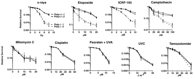
murine bone marrow stromal cells with a variety of damaging agents showed that Pol q
confers a specific resistance to classical DSB inducing agents such as ionizing radiation
and etoposide, but not to ICL inducing agents (Figure 1.2)107. These studies in
mammalian systems have also addressed the broad role of Pol q in genome stability
through translocation formation. Translocations result from the aberrant joining of two
DSBs on different chromosomes and are a common rearrangement in many cancers.
Work in murine and human cells has suggested opposing roles for Pol q in translocation
formation (a suppressive role in mice compared with a promoting role in human), though
the methods in each study varied substantially (spontaneous IgH:Myc translocation in
1.6 Polymerase q and cancer
Several types of cancer including stomach, lung, colorectal, and breast cancer have
been associated with elevated Pol q expression. Further, cancers overexpressing Pol q
are significantly associated with poor patient survival109. The relationship between Pol q
and breast cancer is currently the most explored association and in fact, expression
levels of Pol q and a cohort of its neighboring genes appear to be diagnostic for triple
negative breast cancer110. Two single nucleotide polymorphisms (SNPs) in or near the
POLQ gene have also been identified and associated with hereditary breast cancer in a
case-control study of patients with either hereditary or sporadic breast cancers111. A
SNP in the promoter region of the gene was most strongly associated, though the
mechanistic implications of this SNP are unknown.
Recently, studies have suggested that Pol q may be be a promising target in battling
chemotherapeutic resistance. Co-inhibition of Pol q and some HR-involved genes
re-sensitized cisplatin-resistant lung cancer cells to the drug112. Similarly, inhibition of Pol q
greatly sensitized HR deficient tumor cell lines to a variety of cytotoxic agents including
PARP inhibitors (PARPi). Most strikingly, relative tumor volume, clonogenic survival,
and organismal survival were all positively impacted by depletion of Pol q combined with
PARPi therapy in a murine FANCD2-null xenograft model89. These results rationalize
the exploration of Pol q as a therapeutic candidate for the treatment of some cancers
and highlight the need for a deeper mechanistic understanding of the enzyme and its
Figure 1.1: DNA double strand break repair pathways.
Figure 1.2: Hypersensitivity of Polq-/- bone marrow stromal cells to DNA strand-breaking agents.
BMSCs were exposed to x-rays or UVC at the indicated doses, and to etoposide, ICRF-193, camptothecin, olaparib, temozolomide, mitomycin c, cisplatin, and HMT
psoralen+UVA at the indicated concentrations and plated in triplicate. Two isogenic bone marrow stromal cell lines were used of each genotype, Polq+/+ or Polq-/-. Colonies
were stained and counted seven to ten days later.
The DNA polymerase activity of POLQ is required to confer resistance to DNA damaging agents
We sought next to investigate which catalytic activities of POLQ are necessary to confer resistance to DNA damaging agents. Lentiviral-delivered expression vectors were constructed to express wild-type or mutant versions of POLQ in immortalized MEFs, in order to test for functional complementation (Figure 3A). A tandem (D2330A,Y2331A) mutation was introduced into the DNA polymerase domain (POL); mutation of the corresponding residues in other DNA polymerases completely inactivates polymerase activity [27]. In a separate construct, a mutation was introduced into the conserved ATP-binding site of the Walker A motif (K121M) in the helicase-like domain (HLD). An equivalent mutation eliminates DNA helicase activity in related enzymes, including HELQ [28]. A third construct (DM) was made harboring mutations in both domains. These vectors expressed full-length recombinant POLQ as tested in transfected 293T cells (Figure 3B and C).
The mutant cDNAs were tested for their ability to genetically complement the bleomycin sensitivity ofPolq-null MEFs. Stable clones with each of the constructs were generated and analyzed for expression of POLQ (Figure 3D). Independent clones of knockout MEFs expressing wild-type recombinant POLQ (WT4 and WT6) were able to rescue bleomycin hypersensitivity (Figure 3E) as an antibody that recognizes endogenous POLQ does not yet exist. Neither the polymerase domain mutant (POL) nor the polymer-ase-helicase double mutant (DM) restored bleomycin sensitivity
(Figure 3E, Figure S1B). Expression of a construct with a mutation only in the helicase-like domain (HLD) was, however, still able to restore resistance to bleomycin. These data indicate that POLQ polymerase activity is essential for conferring resistance to DNA damage, while the ATPase activity of the helicase-like domain is not necessary. Similarly reintroduction of polymerase activity of POLQ into Polq-deficient MEFs was able to rescue chromosomal instability (micronuclei and DNA DSBs, as measured by 53BP1 andcH2AX colocalization (Figure 3F and 3G, Figure S2).
Mice with an S1932P mutation inPolq(the ‘‘chaos1’’ allele) have an increased spontaneous frequency of micronuclei [13]. We generated a humanPOLQ cDNA mimicking thechaos1mutation (S1977P), but attempted expression of POLQ with this mutation in 293T cells did not yield detectable protein (Figure S3). This suggests that thechaos1-encoded mutant protein is unstable, consistent with thefinding thatchaos1mice have a phenotype essentially indistinguishable fromPolqknockout mice [13].
POLQ operates in a pathway of altEJ during mouse Ig class-switching
Immunoglobulin class-switch recombination (CSR) uses DNA end joining to exchange one constant region of an antibody gene for another constant region. CSR can occur by both Ku-dependent classical non-homologous end joining and Ku-inde-pendent altEJ [20]. The overall frequencies of CSR are similar in
Polq-defective mice [29] and cultured B cells [30]. To determine whether POLQ is involved in a mechanistically distinct subset of
Figure 1. Hypersensitivity of Polq2/2 bone marrow stromal cells to DNA strand-breaking agents. BMSCs were exposed to x-rays or UVC at the indicated doses, and to etoposide, ICRF-193, camptothecin, olaparib, temozolomide, mitomycin c, cisplatin, and HMT psoralen+UVA at the indicated concentrations and plated in triplicate. Two isogenic bone marrow stromal cell lines were used of each genotype,Polq+/+orPolq2/2. Colonies were crystal violet stained and counted seven to ten days later. Experiments were repeated three times. Circles,Polq+/+clone 1; Squares,
Polq+/+clone 1; Triangles,Polq2/2 clone 1; inverted triangles,Polq2/2 clone 3. doi:10.1371/journal.pgen.1004654.g001
CHAPTER 2: MECHANISM OF POLYMERASE q-MEDIATED END JOINING
2.1 Introduction
DNA double-strand breaks (DSBs) can arise via a number of different exogenous
exposures (e.g. ionizing radiation or chemotherapeutics), as well as through
endogenous processes such as V(D)J recombination during immune system
diversification and during normal cellular duplication cycles, when DNA replication stalls
at naturally occurring structures or at sites of internally-generated DNA damage.
Because DSBs are toxic and/or mutagenic if not repaired, organisms have multiple
mechanisms for DSB repair113,114. The primary strategies are nonhomologous end
joining (NHEJ), which processes and rejoins DSBs independent of sequence homology,
and homologous recombination (HR) which employs an undamaged copy of the DNA
for high fidelity repair115. In addition to these canonically studied repair processes, one
or more “alternative” end joining pathways (Alt-EJ) also exist, which are independent of
these factors116,117.
Alt-EJ in eukaryotic cells has been linked to activity of DNA polymerase theta
(Pol q; encoded by Polq)107. This large, 290 kDa protein has an unusual bipartite
structure with an N-terminal helicase-like domain and a C-terminal DNA polymerase
domain88. This domain arrangement and the Pol q protein sequence is highly conserved
Several functions have been suggested for Pol q, including bypass of template
DNA lesions such as abasic sites and thymine glycols95,118, a backup role in DNA base
excision repair119,120, and influencing the timing of DNA replication origin firing121. Loss
of Pol q homologs in Drosophila and C. elegans causes hypersensitivity to DNA
interstrand crosslink (ICL)-forming agents122,123 such as nitrogen mustards or cisplatin,
while mammalian cells lacking Pol q are uniquely sensitive to DSB inducing agents,
such as ionizing radiation.
The mechanistic constraints on Pol q activity in cellular repair are still unclear. We
investigated these questions through use of extrachromosomal assays and a panel of
mouse embryo fibroblast lines (MEF) cell lines deficient in Polq and Ku70. Our results
provide insight into how Pol q activity contributes to cellular DSB repair. We show Pol q
activity is facilitated by an ability to couple microhomology primed synthesis to a
“microhomology search” and removal of non-homologous tails. We further identify an
essential role for this enzyme in repair of DSBs in contexts where ends are inaccessible
to canonical NHEJ, as is the case when resected ends exceed 45 nt in length, or at “Y”
structures expected during replication fork collapse.
2.2 Methods
Cell lines
Experiments varying Polq status employed SV-40 Large T antigen transformed
MEFs recovered from mice where a Polq null allele was generated by conventional
complemented by lentiviral delivery of cDNA coding wild type human Polq. Deficiency or
complementation was assessed by RT-qPCR for mRNA expression relative to a
housekeeping control (GAPDH) (Figure 2.1A).
Variants of the above WT and Polq-/- lines deficient in the Ku70 gene were made
following transient expression of Cas9 and sgRNA targeting exon 3 of Ku70. Following
Cas9 mutagenesis, cells were subcloned and screened for biallelic frameshift
mutations. Complemented variants of these Ku70 deficient lines were made with
lentivirus delivering mouse Ku70 cDNA. Ku70 deficiency or complementation was
validated by western analysis (Figure 2.1B) and a functional assay for NHEJ activity
(Figure 2.1C). All cell lines were cultured in DMEM with 10% FBS (Sigma), 2 mM
L-Glutamine (Gibco), 1x penicillin-streptomycin, and 2 ug/ml puromycin, 4 ug/ml
blasticidin, or 0.5 mg/ml G418 as necessary for transgene maintenance.
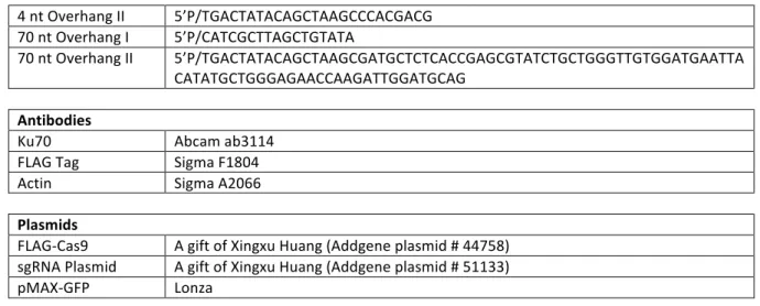
Substrate preparation
Substrates were prepared using a variation of the golden gate cloning method. A
596 base pair common core DNA segment was amplified by PCR and digested with
BsaI-HF (NEB) to yield 4 nucleotide overhangs where head and tail overhang had
distinct sequence. Caps with a common 15 bp double stranded region and overhangs
specific for core head and tail ends were assembled by annealing of oligonucleotides
(IDT), then ligating the caps to the core fragment, generate the linear fragments with
head and tail end structures varied as noted in Table 2.1. Excess cap was removed
using the QIAquick PCR purification kit (Qiagen) and substrate purity validated by
Extrachromosomal end joining assay
DNA substrates (75 ng) of the noted substrate and pMAX-GFP (1 ug, Lonza)
were introduced into 2x10⁵ cells by electroporation (Neon, Invitrogen) using a 1350 V,
30 ms pulse in a 10 μL chamber and incubated for 1 hour. Cells were washed,
incubated with 12.5U of Benzonase (Sigma) to digest extracellular substrate, and
cellular DNA purified (QIAamp DNA mini kit, Qiagen) to determine substrate joining
efficiency and junction character by sequencing. Joining efficiency of extrachromosomal
substrates introduced into cells was quantified by qPCR using either primers Sub
Duplex Fwd/Rev or primers Sub Overhang Fwd/Rev, depending on the experiment.
Total substrate recovery (both joined and unjoined) was determined as a validation of
transfection efficiency across genotypes by qPCR using primers Sub Recovery
Fwd/Rev. All PCRs were confirmed to be >98% efficient over the range relevant to
these experiments by parallel analysis of a standard curve generated by serial dilution
of model amplicons. Product structures were characterized by polyacrylamide gel
electrophoresis on an 8% gel with SYBR green staining or sequencing.
Sequencing
Template DNA for each sequencing library was from independent
electroporations performed across three days. 1x10⁵ input junction molecules were
amplified using Phusion DNA polymerase (NEB) and variants of the qPCR primers
noted above, with the addition of 6 nt barcode sequences appended to their 5’ ends for
21 cycles. 15.5 ng of amplified DNA from each library was pooled in groups of ~10
libraries, phosphorylated with T4 PNK (NEB), treated with dATP and Klenow Exo –
appended to the amplicons by ligation, and free adaptor removed by gel purification.
Gel purified pooled libraries were subjected to a 10 cycle enrichment amplification with
adaptor-specific primers and purified with Ampure XP beads (Agencourt, Beckman
Coulter). Equal amounts of DNA from each pooled library set were combined and
submitted for a 2x150 paired end sequencing run (MiSeq, Illumina), with a PhiX174
DNA spike.
After sequencing, reads of PhiX174 DNA were removed. Remaining reads were
then trimmed for quality, paired ends were merged, and libraries were de-indexed using
Genomics workbench v8.0 (CLC-Bio). Characterization of substrate repair junctions was
performed by alignment of reads to a reference junction, export in SAM format, and
deconstruction of the CIGAR string in Excel (Microsoft) to yield parameters including
flanking deletion, length of ssDNA tails generated, and microhomology content at the
junction.
Gel shift assay
Cy-5 labeled DNA substrates were generated by annealing of oligonucleotides to
generate a 20 bp duplex core. Subsequent ligation of cap structures generated
substrates with either 4 nt or 70 nt 3’ ssDNA overhangs. Resulting substrates (5 nM)
were incubated with 500 nM Streptavidin for 10 minutes at room temperature, then
incubated with 5nM Ku for 5 minutes at 37°C in 1x EMSA buffer (20 mM Tris pH 8.0, 90
mM NaCl, 25 mM KCl, and 10% glycerol) at 37°C before addition of a 10-fold excess of
Strand displacement assay
Substrates bearing either 4 nt or 45 nt 3’ overhangs were prepared as described
above, with the variation that substrates now contained a mispaired BamHI restriction
endonuclease site embedded 15 bp into the duplex DNA adjacent to the 3’ overhangs.
Substrate variations also contained either a 5’ phosphate or 5’ THF. The restriction site
was mispaired such that 4 out of 6 positions of the BamHI recognition site were
mispaired. Substrates were introduced into WT MEFs by electroporation as described
above and harvested after 1 hr. Repair junctions were digesting with BamHI overnight at
37°C in a 20 ul reaction volume then used as template in a PCR amplification using
primers Sub Duplex Fwd/Rev. Products were analyzed by PAGE electrophoresis on a
6% gel and qPCR.
2.3 Results
Distinct substrates for NHEJ and Pol q-mediated end joining
We previously showed that Pol q is required for a cellular end joining pathway –
Polymerase q mediated end joining, or TMEJ – that is independent of Ku, and uniquely
able to join ends with extended 3’ssDNA tails107. To better address the relationship
between TMEJ and classically defined NHEJ we sought to make cell lines deficient in
one or the other pathway, or both. We consequently employed CRISPR/Cas9 to
generate variants of existing isogenic wild type and Polq-/- MEF lines107(Figure 2.1A)
that do not express significant Ku70 protein (Figure 2.1B). These lines are deficient in
NHEJ, and can be complemented by expression of introduced Ku70 cDNA (Figure
The progressive 5’ to 3’ resection of DSB termini is a necessary pre-requisite for
repair of DSBs by homologous recombination (HR). To address how resection affects
the deployment of different end joining pathways we introduced a panel of linear double
stranded DNA substrates with “pre-resected” ends, where 3’ ssDNA overhangs ranged
from 4-70 nucleotides (nt) (Figure 2.2A, Figure 2.6A), into the cell lines described
above. Head and tail overhang sequence were designed such that the terminal 4 nt
were complementary, i.e. contain a microhomology. The impact of differing location and
length of microhomologies is discussed in greater detail below.
Overall joining efficiency was reduced 5-10 fold for overhang lengths in excess of 10
nucleotides, and two populations of end joining products were readily evident; one
where most of both overhangs had been removed, and another where most of both
overhangs had been retained (Figure 2.2B-D). As overhang length is increased, joining
is progressively more reliant on products where most of both overhangs are retained
(Figure 2.6A, 2.6B). In all contexts joining after overhang loss was largely dependent on
Ku70/NHEJ, while joining associated with retention of 3’ overhangs was dependent on
Pol q (bottom panels, Figure 2.2B-D). Pol q additionally was not required for either
product class (overhangs retained and overhangs lost) using a substrate where the
overhang polarity was reversed (5’ overhang), but which was otherwise equivalent
(Figure 2.6C). TMEJ is thus uniquely employed for repair of products of 5’>3’ resection.
When the 3’ overhang was only 45 nt, overall joining efficiency was reduced 3-fold in
Ku70 deficient cells and not affected by Pol q deficiency (Figure 2.2B, top panel). In
substrate variants in cells deficient in both Pol q and Ku70 (Figure 2.2B, 2.2C, top
panels). The two pathways thus together account for most of the cellular capacity to
repair pre-resected end structures, but TMEJ assumes prominence as the extent of
5’>3’ resection exceeds 45 nt (see also Figure 2.6B).
We reasoned that this increased reliance on Pol q-dependent repair as the ssDNA
tails are extended reflects a reduced ability of the Ku heterodimer to load on substrates
with long ssDNA tails (Figure 2.6C). To further investigate this possibility, we generated
a variant of the shorter 45 nt 3’ overhang with a “Y” or forked end structure, by including
a 5’ streptavidin-blocked flap (Figure 2.2D). Such a substrate is expected to completely
block loading of Ku, and is analogous to products of replication fork collapse (“one
ended breaks”). Accordingly, we observed no significant impact of Ku-dependent NHEJ
when using this substrate. Repair was instead Pol q-dependent (Figure 2.2D, top
panel), consisted almost entirely of products that retained the majority of both 3’
overhangs (Figure 2.2D, bottom panel), and thus can be readily defined as TMEJ. We
conclude Pol q-dependent repair is most important when considering end structures
where loading of Ku is impaired. This includes those end structures expected after
aborted HR – ends with extended 3’ ssDNA tails - or after replication fork collapse.
Mechanism of Pol q-mediated end joining
Products that retain significant amounts of overhang are mostly dependent on Pol q;
conversely, Pol q deficiency had little impact on either the efficiency or structure of those
products generated after overhang loss (Figure 2.2). Relocation of qPCR primers to
require that end joining products retain at least 10 nt of each overhang is thus sufficient
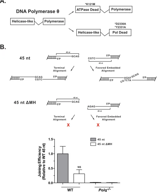
Pol q is a multi-domain protein, with an N-terminal helicase-like domain and a
C-terminal polymerase domain (Figure 2.7A). To investigate the contributions of each
activity we compared the ability to complement Polq-/- cells for TMEJ capacity by
expression of the wild type Polq cDNA, a variant cDNA with a mutation inactivating
ATPase activity of the helicase domain (K121M), or a variant cDNA with mutations that
inactivate the polymerase domain (D2330A/Y2331A; Motif A)107. Levels of TMEJ in cells
expressing the D2330A/Y2331A construct were indistinguishable from cells with an
empty vector control (Figure 2.3A); polymerase activity is thus essential for this
pathway. In contrast, loss of ATPase activity in the helicase-like domain had no
significant impact on joining efficiency, but did affect product spectra (discussed below).
We also assessed a possible role for Pol µ and Pol l, which have previously been
associated with an ability to direct intermolecular DNA synthesis, i.e. can prime
synthesis from a 3’ tail from one DSB end, and use a second DSB end as template44.
Both of these polymerases are dispensable for cellular end joining using pre-resected
substrates (Figure 2.3A), with levels of TMEJ instead increased in cells deficient in both
Pol µ and Pol l. This may reflect the important role of these polymerases in NHEJ, and
increased TMEJ in NHEJ-deficient contexts.
The substrates described above were designed such that the terminal 4 nt were
complementary, to allow for alignment-directed synthesis. Surprisingly, products
consistent with synthesis from these terminal alignments accounted for only 56% of
TMEJ when using the 45 nt 3’ overhang substrate, and 20% of TMEJ when using the 70
alignments). For example, repair of the 70 nt overhang substrate frequently employed
all four of the alignments that are greater than 3 bp and can also be found within 25 nt
of the 3’ ends. Taken together, these 4 products account for 2/3rds of all products
recovered (Figure 2.3B), with the three embedded alignments used almost twice as
often as the terminal alignment. Moreover, there were only mild effects on the efficiency
of repair when using a substrate where both the terminal and most favored embedded
alignments were disrupted (Figure 2.7B). We additionally did not see significant
recovery of products where an embedded alignment directed synthesis from a
mispaired tail (Figure 2.3B). We can therefore infer synthesis by Pol q using embedded
alignments is efficiently coupled to a prior step where the unpaired tail is removed.
Notably, the ability to use embedded alignments was modestly reduced in cells
expressing the Pol q variant with an ATPase inactivating mutation (K121M; Figure
2.3C). This suggests TMEJ is facilitated by an active search for complementary
sequence alignments (i.e. microhomology in finished product) that involves ATPase
activity in the N-terminal helicase-like domain. Characterization of products also clarifies
the constraints on this putative microhomology search mechanism. Considering first the
amount of complementary sequence, only those products with alignments > 2bp were
significantly enriched (Figure 2.3D). However, the length of nonhomologous tails
impacts the ability to use such alignments. For example, a product generated from a 6
bp alignment, but which would generate long nonhomologous tails (52 nt and 22 nt), is
observed 20-fold less frequently than a 4 bp alignment with short tails (9 nt and 2 nt,
Figure 2.3B). Alignments resulting in tails with one long and one short tail (e.g. 51 nt
termini. In sum, 91% of products recovered using the 70 nucleotide overhang substrate
(140 nt of ssDNA) have deletion less than 50 bp (Table 2.2).
Moreover, we show here that the synthesis activity of TMEJ does not terminate
at a downstream 5’ phosphate and instead involves a strand displacement intermediate.
Repair of substrate containing a BamHI site on the template strand that is mispaired
and buried 15 bp into the substrate duplex is not sensitive to restriction enzyme digest
when overhang length is 4 nt and thus repaired by NHEJ (Figure 2.4A, 2.4B). Joining of
this substrate is reduced when the 5’ phosphate is replaced with a 5’ abasic site mimetic
(e.g. THF). In contrast, when the overhang length is extended to 45 nt, the substrate is
repaired by TMEJ and junctions are rendered sensitive to pre-amplification digestion by
BamHI (Figure 2.4B). Further, joining of this substrate not influenced by the presence of
a 5’ abasic site in lieu of a 5’ phosphate. This result confirms that the synthesis step of
TMEJ preferentially displaces the mispaired strand and uses the intact
BamHI-containing strand as template in a “long patch” DSB repair process.
2.4 Discussion
Our results indicate that in cells, Pol q has a central role in an end joining pathway –
TMEJ – that is uniquely able to repair substrates where classically defined NHEJ is
ineffective (Figure 2.2). The effects of Pol q deficiency on both which substrates can be
joined (Figure 2.2) and product spectra (Figure 2.3, 2.4) indicates that in mammals, Pol
q/TMEJ is required for most of what had previously been defined as MMEJ or Alt-NHEJ.
synthesis by Pol q is coupled to two prior steps, including a search for microhomology
and removal of nonhomologous tails (Figure 2.3).
Identification of biologically significant substrates and mechanism
We show there are marked differences between cellular TMEJ and the in vitro
activities of the truncated Pol q polymerase domain. TMEJ is uniquely required as
ssDNA extensions exceed 45 nt (Figure 2.2), as well as forked ends (i.e. when a paired
5’ terminus is unavailable); by comparison, in vitro studies124 indicate activity was limited
to extensions 15 nt or shorter. We suggest full length Pol q may be intrinsically more
processive93, Pol q may employ a processivity clamp (PCNA) during cellular TMEJ, or
there may be polymerase switching after synthesis initiation. The unique activity on
forked structures is consistent with studies identifying an important role for Pol q in
replication fork re-start after fork collapse (“one-ended” chromosome breaks)89,103.
Indeed, this result predicts TMEJ will be critical in joining any pair of ends where either
of the 5’ termini is inaccessible to Ku or “unligatable”, i.e. has irreversible bulky adducts
or is associated with extensive base damage. In accord with this prediction, we show
that TMEJ is not impeded by a 5’ abasic site – a lesion that ablates joining by NHEJ
(Figure 2.4).
Cellular TMEJ is also rarely associated with synthesis from terminal mispairs (Figure
2.3), as can be observed in in vitro studies of truncated Pol q activity124. This limitation
could dramatically restrict the fraction of ends that could be joined (<1/64), given the
requirement for annealing at significant microhomologies (3-6 bp) for synthesis. Our
results indicate cellular TMEJ instead efficiently samples all of the useable alignments
of over 99% of ends of random sequence. The spectrum of cellular TMEJ products also
emphasizes another advantage of a homology-search mechanism, its ability to mitigate
deletion. 91% of TMEJ-dependent products of an extrachromosomal substrate with 140
nucleotides of ssDNA have less than 50 bp (2x25) of deletion.
Cellular TMEJ is thus made flexible, and deletion constrained, through an ability to
tightly couple together three steps (Figure 2.5): 1) a microhomology search that usually
generates nonhomologous tails, 2) excision of these tails, and 3) strand-displacement
synthesis from the newly annealed 3’ terminus. The search for microhomology might be
dependent on the Pol q N-terminal helicase-like domain, as suggested by a structural
model for an oligomer of this domain that juxtaposes a pair of 3’ ssDNA termini90. In
accord with this model, the ability to use embedded alignments is specifically impaired
in cells expressing a Pol q mutation that inactivates this domain’s ATPase activity.
Effects of ATPase activity on microhomology search could plausibly be explained if it
drove adjustment of the relative position of the two aligned termini. With regard to the
next step, removal of nonhomologous 3’ tails, Pol q has no intrinsic nuclease activity87.
However, such an activity is present as a contaminant of Pol q partially purified from
HeLa cells125, and results described here suggest this activity is tightly coupled to Pol q
activity. Mre11 and XPF:ERCC1 are both attractive candidates for this nuclease activity,
as they have appropriate substrate specificities and have already been implicated in
Alt-NHEJ/MMEJ77. The third step, synthesis from 3’ termini with 3-6 bp annealed, is
facilitated by the tight grasp Pol q maintains on the primer termini92. Altogether, Pol q is
Figure 2.1: Validation of NHEJ deficient cells and complementation
A.
0.0 0.5 1.0 1.5Polq: +/+
-/-hPolq hPolq (K121M)
hPolq (D2330A) (Y2331A)
mRNA Expression (R
e la tive to WT ) <0.0001
B.
Ku70 ActinWT Ku70 -/- Polq -/- Polq
-/-Ku70
-/-FLAG Ku70 Actin Ku70 -/-+mKu70 Polq -/-Ku70 -/-+mKu70 Polq -/-Ku70 -/-Ku70 -/-4 nt 5’P3’OH J oining E ff ic ie n cy (R e la tive to WT ) ** **
WT Polq-/- Polq
-/-Ku70
-/-Polq
-/-Ku70 -/-+mKu70 Ku70-/- Ku70
Figure 2.1: Validation of NHEJ deficient cells and complementation
Figure 2.2: Effect of end joining deficiencies on repair of pre-resected ends
B.
45 nt 5’P 3’OH 70 nt 5’P 3’OH 20 nt Bio-Strep45 nt 3’OH
C.
D.
200 bp 150 bp 75 bp 200 bp 150 bp 75 bp 200 bp 150 bp 75 bpWT Polq-/- Ku70-/- Polq
-/-Ku70 -/-0.0 0.5 1.0 1.5 J oining E ff ic ie n cy (R e la tive to WT ) 0.0 0.5 1.0 1.5 J oining E ff ic ie n cy (R e la tive to WT ) 0.0 0.5 1.0 1.5 J oining E ff ic ie n cy (R e la tive to WT ) NS ** ** *** * **** ** NS ** ✂ ✂ ✂ ✂ ✂ ✂
A.
MEFs, + SV40Tag
G CGA
GC GA
1 hour
G CGA
GC GA Electroporate Efficiency (qPCR) Product structure (gel, sequence) 5’P 5’P
qPCR PAGE Analysis Sequencing Incubate 1 hr.
Harvest Electroporate MEFs 5’P 3’OH 5’P 3’OH N nt ssDNA
WT Polq-/- Ku70-/- Polq
-/-Ku70
-/-WT Polq-/- Ku70-/- Polq
-/-Figure 2.2: Effect of end joining deficiencies on repair of pre-resected ends
(A) Linear substrates with varied end structures were introduced into noted cell types and end joining products characterized by quantitative polymerase chain reaction (top panels) or electrophoresis of amplified products (bottom panels); Data shown below are the mean +/- SEM, n=3. Statistical significance was assessed by one-way ANOVA with Bonferoni correction of p values to account for multiple comparisons. NS not significant, *p<0.05, **p<0.01, ***p<0.001, ****p<0.0001. (B) A substrate with symmetrical 45 nt 3’ single stranded DNA overhangs was introduced into the noted cell types, and the mean efficiency of end joining determined by qPCR and expressed as a fraction of that
observed in WT cells. Amplified products generated in a parallel fixed-cycle number PCR were also characterized by electrophoresis; products of size consistent with joining after overhang clipping vs. overhang retention are noted. (C) A substrate with 70 nt 3’ overhangs was introduced into the noted cell lines and joining characterized as
Figure 2.3: Mechanism of Pol q mediated end joining
A.
*qPCR primers sit in ssDNA overhang
GCAG CGTC 5’P 5’P 45 nt
C.
D.
J oining E ff ic ie n cy (R e la tive to WT ) 0.0 0.5 1.0 1.5 2.0 3.0 4.0Polq: +/+
-/-hPolqhPolq
K121M
hPolq
D2330A Y2331A
WT Polm
-/-Poll
-/-B.
GCAG CGTC 5’P ssDNA Tails ATCT TAGA 5’P CTCTT GAGAA 5’P 0/0% of Total Junctions Recovered (70 nt substrate)
9/2 22/10 16/13 52/22 51/4 20.0% 33.4% 8.3% 5.8% 1.3% 0.1% CAGC GTCG GATACG CTATGC 5’P AGAT TCTA 5’P
0 10 20 30 40
0.04% MH C G 5’P 4/0 4 4 4 5 6 4 1 Alignment of termini
Embedded alignments
-/-hPolq hPolq
K121M
Polq: Alignment of termini
All embedded alignments
Microhomology Usage by Pol θ
freq MHN(obs/exp)
No MH 1 bp 2 bp 3 bp 4 bp 5 bp 6 bp ATCT TAGA
5’P 4 9/2
Synthesis from mispaired tail
0.008%
NS NS
*
* ***
% of all products recovered
25 50 75 100 45 nt 5’P 3’OH 0 70 nt Empty Vector 3
10-2 10-1 100 101 102 10
5’P
Figure 2.3:Mechanism of Pol q mediated end joining
(A) The 45 nt 3’ overhang substrate described in Fig. 2B was introduced into cells that were WT, Polq-/-, and Polq-/-engineered to express wild type human Polq or variants deficient in helicase-like domain ATPase activity (K121M), or polymerase activity
(D2330A+Y2331A), and joining efficiency assessed by qPCR using primers that require retention of at least 10 nt of each 3’ overhang sequence in junctions. Joining efficiency was also characterized in Poll-/- Polm-/- MEFs, relative to matched WT control MEFs.
Data shown are the mean +/- SEM, n=3. Statistical significance was assessed by one-way ANOVA with Bonferoni correction. NS =not significant, *p<0.05, **p<0.01,
***p<0.001. (B) Observed frequency of different joining products of the 70 nt overhang substrate (Fig. 2C) recovered from wild type cells, including the 4 most common
products (above dashed line) and 4 other representative examples (see also Table S1). The first 3 columns summarize the structures of the inferred intermediates. (C) For experiments described in Fig. 3A (using the 45 nt overhang) we report the fraction of products directed by the terminal 4 bp microhomology (shown in panel B), as
Figure 2.4: Pol q is a strand displacing polymerase
(A) Schematic showing the possible Pol q repair fates and the impact of each fate on digestion with BamHI and subsequent PCR amplification. (B) qPCR quantification (top panel) and 6% PAGE gel analysis (bottom panel) of noted substrates, either treated as a mock sample or BamHI digested, followed by PCR amplification.
BamHI: - + - + - + - +
3’ 4 nt 5’ Phos
3’ 4 nt 5’ Abasic
3’ 45 nt 5’ Abasic 3’ 45 nt
5’ Phos TMEJ NHEJ 100 bp 200 bp 0.0 0.5 1.0 Joining Efficiency
(Relative to 3’
4 nt 5’
Phos)
CCTAGG CGGAAC 5’ P
or 5’ THF
CCTAGG CGGAAC
Dissociation at 5’ phosphate Ligation No BamHI digestion
Amplification CCTAGG CG GAA C GGATCC Strand displacement BamHI digestion No amplification
Pol θ
Pol θ
Pol θ
Or
A.
Figure 2.5: Proposed model of Pol q-mediated end joining
Figure 2.6: Effect of varied overhang length and polarity on end joining
WT Polq -/- Ku70-/- Polq -/- Ku70-/-0.0 0.5 1.0 1.5 2.0
45 nt 5’P
3’OH 200 bp 150 bp 75 bp J oining E ff ic ie n cy (R e la tive to WT )
C.
B.
A.
Joining Efficiency (Relative to 4 nt Overhang)20 nt 3’OH
10 nt 3’OH 4 nt 3’OH 30 nt 3’OH 45 nt 3’OH 70 nt 3’OH *qPCR primers sit
in duplex flank
GCAG CGTC 5’P
3’ Overhang
Length (nt) 4 10 20 30 45 70 75 bp 150 bp 200 bp 5’P ✂ ✂ Overhang Clipping Overhang Retention 4-70 nt
0.0 0.5 1.0
Product Ratio (Retention:
Clipping) 0 0.40 0.72 1.32 1.61 6.48
**
**** ****
D.
Ku: - 5 nM - 5 nM
38 bp duplex
4 nt 3’ overhang 70 nt 3’ overhang38 bp duplex
Substrate +Streptavidin Substrate +Streptavidin +Ku 4 nt 3’OH
Figure 2.6: Effect of varied overhang length and polarity on end joining
(A) Substrates with 3’ single stranded DNA overhangs ranging from 4-70 nt were introduced into WT MEFs. Joining efficiency was quantified as described in Figure 2. Data are the mean +/- SEM, n=3. (B) Joined products from panel A were characterized by amplification of end joining products and polyacrylamide gel electrophoresis.
Products of size consistent with joining after overhang clipping vs. joining with overhang retained are noted, and their relative abundance (determined after correction for size) noted below. (C) A substrate with head and tail 45 nt 5’ overhangs was introduced into the noted cell types and the efficiency (top panel) and character (bottom panel) of end joining were assessed as described in Figure 2. Data are the mean +/- SEM, n=3. Statistical significance assessed by one-way ANOVA with Bonferoni correction. (D)
Figure 2.7: Separation of function in Pol q and role of terminal microhomology
(A) Domain structure of Pol q. Experiments performed in Figure 3 use Polq-/- MEFs expressing WT human Polq cDNAs, as well as Polq cDNAs with an ATPase defective mutation (K121M) or mutations that ablate polymerase activity (D2330A, Y2331A). (B)
The 45 nt 3’ overhang substrate described in Fig. 2B was transfected into WT and Polq -/-MEFs. A variant of this substrate with no ability to align either at the extreme 3’ termini,
or the second most favored alignment (shown in the cartoon) was transfected in parallel; joining efficiency was assessed using overhang specific primers as described in Figure 3. Data are the mean +/- SEM, n=3. Statistical significance assessed one-way ANOVA with Bonferoni correction.
A.
Helicase-like Polymerase
Polymerase ATPase Dead
*K121M
Helicase-like Pol Dead *D2330A *Y2331A DNA Polymerase θ
0.0 0.5 1.0 1.5 WT Polq-/-NS J oining E ff ic ie n cy (R e la tive
to WT 45 nt
) 45 nt GCAG 45 nt 5’P GCAG 5’P
B.
GCAG CGTC5’P 5’P 5’P AGATTCTA 5’P
GCAG CG CGTC 5’P 45 nt Favored Embedded Alignment Terminal Alignment 45 nt
45 nt ΔMH
Favored Embedded Alignment Terminal Alignment X X 45 nt
45 nt ΔMH
AGAG 5’P
45 nt
Extrachromosomal Substrate: Left Caps
4 nt 5’P/AGTCTGAGATGGGTGCCACGACG
10 nt 5’P/AGTCTGAGATGGGTGTGAGAGTGATCTGC
20 nt 5’P/AGTCTGAGATGGGTGTGAGAGTGAAGATCCTCGATCTGC
30 nt 5’P/AGTCTGAGATGGGTGTGAGAGTGAAGATCCTCACCTTCGGACGATCTGC
45 nt 5’P/AGTCTGAGATGGGTGTGAGAGTGAAGATCCTCACCTTCGGAGTACTCCTTCTTTTGAGATCTGC
70 nt 5’P/AGTCTGAGATGGGTGTGAGAGTGAAGATCCTCACCTTCGGAGTACTCCTTCTTTTGACCATTGATACGATACTTCTCAGCCGAGATCTGC
45 nt DMH 5’P/AGTCTGAGATGGGTGTGAGAGTGAAGATCCTCACCTTCGGAGTACTCCTTCTTTTGACTAGAGA
45 nt 5’ Overhang 5’P/GCAGATCTCAAAAGAAGGAGTACTCCGAAGGTGAGGATCTTCACTCTCACACCCATCTCA
Annealing Strand 5’P/CTCACACCCATCTCA
5’ Biotin Flap 5’Biotin/TTTTTTTTTTTTTTTTTTTTCTCACACCCATCTCA
Annealing Strand for
5’ Overhang 5’P/AGTCTGAGATGGGTGTGAG
Extrachromosomal Substrate: Right Caps
4 nt 5’P/TGACTATACAGCTAAGCCCACGACG
10 nt 5’P/TGACTATACAGCTAAGCGATGCTCGATGCAG
20 nt 5’P/TGACTATACAGCTAAGCGATGCTCTCACCGAGCGGATGCAG
30 nt 5’P/TGACTATACAGCTAAGCGATGCTCTCACCGAGCGTATCTGCTGTGATGCAG
45 nt 5’P/TGACTATACAGCTAAGCGATGCTCTCACCGAGCGTATCTGCTGTGTTGTGGATGAATTAGATGCAG
70 nt 5’P/TGACTATACAGCTAAGCGATGCTCTCACCGAGCGTATCTGCTGGGTTGTGGATGAATTACATATGCTGGGAGAACCAAGATTGGATGCAG
45 nt DMH 5’P/TGACTATACAGCTAAGCGATGCTCTCACCGAGCGTATCTGCTGTGTTGTGGATGAATTAGATGCAG
45 nt 5’ Overhang 5’P/CTGCATCTAATTCATCCACAACACAGCAGATACGCTCGGTGAGAGCATCGCTTAGCTGTATA
Annealing Strand 5’P/CATCGCTTAGCTGTATA
5’ Biotin Flap 5’Biotin/TTTTTTTTTTTTTTTTTTTTCATCGCTTAGCTGTATA
Annealing Strand for
5’ Overhang 5’P/TGACTATACAGCTAAGCGATG
Primers/Probes
Sub Duplex Fwd 5’CTTACGTTTGATTTCCCTGACTATACAG
Sub Duplex Rev 5’GCAGGGTAGCCAGTCTGAGATG
Sub Overhang Fwd 5’TAAGCGATGCTCTCACCGAG
Sub Overhang Rev 5’GATGGGTGTGAGAGTGAAGATC
Sub Recovery Fwd 5’GGCACTCTCCAAGGCAAAGA
Sub Recovery Rev 5’ACATGTCTAGCCTATTCCCGGCTT
sgRNA Sequences
Ku70 Exon 3 5’TCTTACTGGTGTACACACTC
Gel Shift Assay Oligos (Fig. S2)